. 2022 Mar 9;96(5):e0199021.
doi: 10.1128/jvi.01990-21. Epub 2022 Jan 12.

Influenza A Virus Uses PSMA2 for Downregulation of the NRF2-Mediated Oxidative Stress Response

Affiliations

Influenza A Virus Uses PSMA2 for Downregulation of the NRF2-Mediated Oxidative Stress Response

Mahamud-Ur Rashid et al. J Virol. .

Abstract

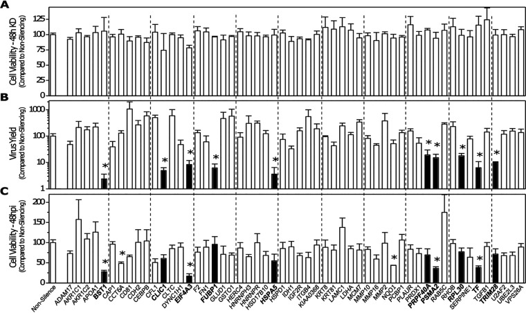
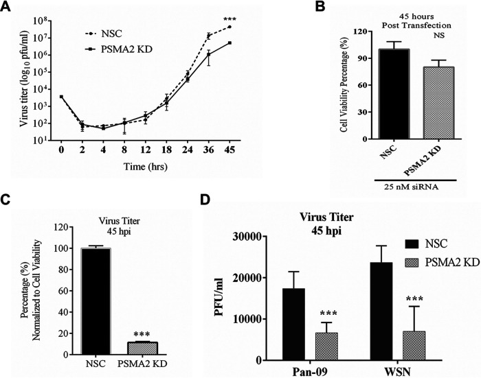
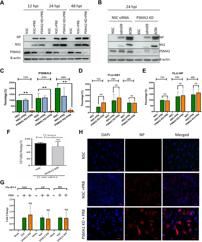
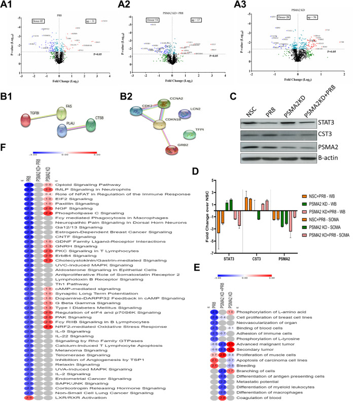
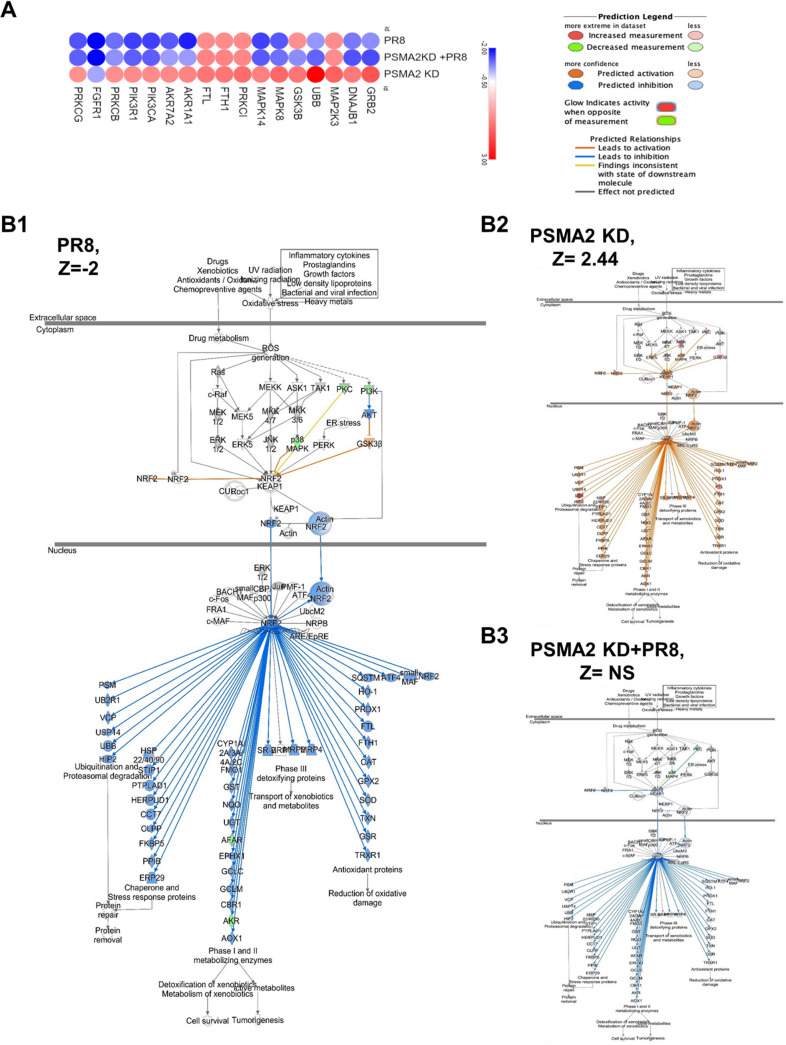
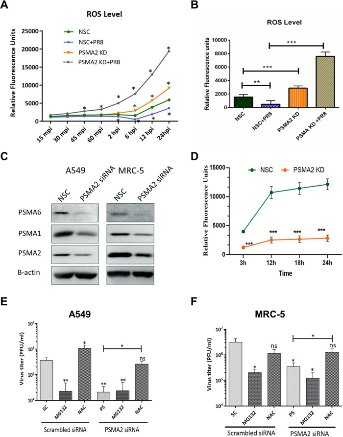
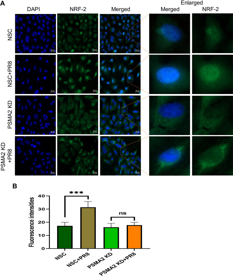
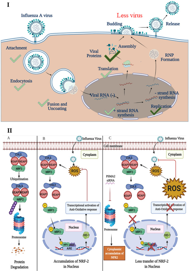
Influenza A virus (IAV), an obligatory intracellular parasite, uses host cellular molecules to complete its replication cycle and suppress immune responses. Proteasome subunit alpha type 2 (PSMA2) is a cellular protein highly expressed in IAV-infected human lung epithelial A549 cells. PSMA2 is part of the 20S proteasome complex that degrades or recycles defective proteins and involves proteolytic modification of many cellular regulatory proteins. However, the role of PSMA2 in IAV replication is not well understood. In this study, PSMA2 knockdown (KD) in A549 cells caused a significant reduction in extracellular progeny IAV, but intracellular viral protein translation and viral RNA transcription were not affected. This indicates that PSMA2 is a critical host factor for IAV maturation. To better understand the interplay between PSMA2 KD and IAV infection at the proteomic level, we used the SomaScan 1.3K version, which measures 1,307 proteins to analyze alterations induced by these treatments. We found seven cellular signaling pathways, including phospholipase C signaling, Pak signaling, and nuclear factor erythroid 2p45-related factor 2 (NRF2)-mediated oxidative stress response signaling, that were inhibited by IAV infection but significantly activated by PSMA2 KD. Further analysis of NRF2-mediated oxidative stress response signaling indicated IAV inhibits accumulation of reactive oxygen species (ROS), but ROS levels significantly increased during IAV infection in PSMA2 KD cells. However, IAV infection caused significantly higher NFR2 nuclear translocation that was inhibited in PSMA2 KD cells. This indicates that PSMA2 is required for NRF2-mediated ROS neutralization and that IAV uses PSMA2 to escape viral clearance via the NRF2-mediated cellular oxidative response. IMPORTANCE Influenza A virus (IAV) remains one of the most significant infectious agents, responsible for 3 million to 5 million illnesses each year and more than 50 million deaths during the 20th century. The cellular processes that promote and inhibit IAV infection and pathogenesis remain only partially understood. PSMA2 is a critical component of the 20S proteasome and ubiquitin-proteasome system, which is important in the replication of numerous viruses. This study examined host protein responses to IAV infection alone, PSMA2 knockdown alone, and IAV infection in the presence of PSMA2 knockdown and determined that interfering with PSMA2 function affected IAV maturation. These results help us better understand the importance of PSMA2 in IAV replication and may pave the way for designing additional IAV antivirals targeting PSMA2 or the host proteasome for the treatment of seasonal flu.

Keywords: NRF-2; PSMA2; host protein; influenza A virus; oxidative stress; proteasome.


Conflict of interest statement

The authors declare no conflict of interest.

Figures

FIG 1
FIG 1
siRNA array screen of selected fibronectin-interacting proteins shown previously to be up- or downregulated by IAV infection. (A) Viability of cells transfected with a 100 nM concentration of the indicated siRNA was determined at 48 h posttransfection (hpt) by WST-1 assay. (B) Indicated 48-h-knockdown cells were infected with PR8 at an MOI of 0.02, supernatants were harvested at 42 hpi, and viral progeny replication was determined by plaque assay on MDCK cells. (C) Viability of the 48-h-transfected/48-h-infected cells determined by WST-1 assay. All assays performed in triplicate, with average values compared to the cognate values of cells transfected with a scrambled siRNA control (nonsilencing). Error bars represent SEM, and values determined to be statistically significantly altered (P < 0.05) are indicated with an asterisk.
FIG 2
FIG 2
PSMA2 is required for replication of IAV. A549 cells were treated with either nonsilencing siRNA (NSC) or PSMA2 siRNA (PSMA2 knockdown [KD]) for 48 h and infected with IAV PR8 at an MOI of 0.01. Supernatants from the infected cells were collected at 0, 2, 4, 8, 12, 18, 24, 36, and 45 h postinfection (hpi). Similarly, NSC and PSMA2 KD cells were infected with IAV strains pdm09 and WSN and supernatants were collected at 45 hpi. Virus titers were determined by plaque assay. (A) Influenza A virus (PR8 strain) titer in the PSMA2 KD cell supernatant compared to the control (NSC) over time. (B) Viability of cells measured by WST-1 assay at 45 h post-siRNA transfection. (C) Percentage of virus titer in PSAM2 KD cell supernatant at 45 hpi compared to the control and normalized with cell viability. (D) Impact of PSMA2 KD on IAV pdm09 and WSN strains. NS, not significant. ***, P < 0.001.
FIG 3
FIG 3
PSMA2 KD does not impact translation of viral proteins and transcription of vRNAs but impacts maturation. A549 cells were treated with either nonsilencing siRNA (NSC) or PSMA2 siRNA (PSMA2 KD) for 48 h and infected with IAV PR8, pdm09, or WSN at an MOI of 3. Cell lysates were collected at 12, 24, and 48 hpi from PR8-infected cells and at 24 hpi from pdm09-infected and WSN-infected cells for analyzing the expression of viral proteins by Western blotting. After 24 hpi, cells were fixed on slides to measure viral protein localization by immunofluorescence microscopy. Viral RNAs were collected at 24 hpi, and the comparative vRNA transcripts were determined by qRT-PCR. (A) Expression of IAV PR8 NP and NS1 proteins in PSMA2 cells at 12, 24, and 48 hpi. (B) Expression of viral proteins in PSMA2 KD at 24 hpi after infection with pdm09 and WSN strains. (C to E) Quantitative densitometry analysis of Western blot images to determine knockdown of PSMA2 expression (C), IAV NS1 protein expression (D), and Flu-NP protein expression (E). (F) Impact of PSMA2 KD on cell viability measured by WST-1 assay at 72 h after transfection by PSMA2 siRNA. (G) IAV NS1, NP, and HA vRNA transcripts in PSMA2 KD cells compared to mock-infected cells and NSC control. (H) Immunofluorescence images showing the expression of IAV NP protein in infected PSMA2 KD cells. *, P < 0.05; **, P < 0.01; ***, P < 0.001.
FIG 4
FIG 4
Proteomic analysis to delineate the impact of PSMA2 KD on IAV replication. NSC and PSMA2 KD cells were infected with IAV PR8 at an MOI of 3. Cell lysates were collected from uninfected NSC and PSMA2 KD cells and after infection with PR8 at 24 hpi. Uninfected NSC and PSMA2 KD cells were used as controls. Cell lysates were analyzed by the SomaScan platform, which can detect >1,300 predefined proteins simultaneously from each sample. The protein expression values were compared between the groups to determine whether the protein dysregulation was an experimental condition. PSMA2 KD versus NSC, NSC infected with PR8 versus NSC, and PSMA2 KD infected with PR8 versus PSMA2 KD comparisons were made to determine the impact of PSMA2 knockdown (PSMA2 KD), PR8 infection (PR8) and impact of PR8 infection in PSMA2 KD cells (PSMA2 KD+PR8), respectively. (A) Volcano plot of proteins dysregulated in IAV PR8-infected (A1), PSMA2 KD (A2), and IAV PR8-infected PSMA2 KD (A3) cells. (B1) Proteins significantly dysregulated by PR8 infection but with an opposite trend of expression in PSMA2 KD+PR8 cells. (B2) Proteins significantly dysregulated by PSMA2 KD+PR8 infection but with an opposite trend of expression in PR8-infected cells. (C) Validation SomaScan data by Western blot detection of STAT3, CST3, and PSMA2 proteins. (D) Quantitative densitometry of Western blot images and comparison with SomaScan data for data validation. (E) Heat map of the disease and functions significantly dysregulated by either PR8 or PSMA2 KD+PR8 but not by the others. (F) Heat map of significantly dysregulated canonical pathways in PR8-infected cells; significance could not be predicted by IPA in PSMA2 KD+PR8 cells.
FIG 5
FIG 5
Influenza A virus uses PSMA2 for downregulation of NRF2-mediated oxidative stress response. (A) Proteins associated with NRF2-mediated oxidative stress response pathway dysregulated by PR8 infection, PSMA2 KD, and PSMA2 KD+PR8. Red, upregulated; blue, downregulated. (B) NRF2-mediated oxidative stress response signaling pathway activation by PR8 infection (B1), PSMA2 KD (B2), and PR8 infection with PSMA2 KD (B3). Orange and blue indicate IPA-predicted activation and inactivation, respectively. mpi, minutes postinfection.
FIG 6
FIG 6
PSMA2 KD reduces proteasome activity but does not affect IAV replication in the presence of NAC. (A and B) Change in reactive oxygen species (ROS) levels over time (A) and at 6 hpi (B) by PR8 infection, PSMA2 KD, and PR8 infection in A549 cells. (C) Expression of PSMA1 and PSMA6 in A549 and MRC-5 cells after PSMA2 knockdown. (D) 20S proteasome activity in PSMA2 KD cells. (E) Impact of MG132 and NAC on IAV replication in wild-type cells and after PSMA2 KD in A549 cells. (F) Impact of MG132 and NAC on IAV replication in wild-type MRC-5 cells and after PSMA2 KD. All significance levels were calculated in comparison with NSC/SC, without the bars compared with the horizontal lines. *, P < 0.05; **, P < 0.01; ***, P < 0.001.
FIG 7
FIG 7
PSMA2 is required for nuclear translocation of NRF2. (A) Immunofluorescence images showing the impact of PSMA2 KD on NRF2 nuclear translocation in influenza A virus-infected A549 cells. (B) Quantitative fluorescence intensity of NRF2 in the nucleus determined by ImageJ. The nonsilencing scrambled siRNA control shows the distribution of NRF2 in noninfected cells treated with nontargeted siRNAs. NSC+PR8 indicates the nuclear translocation of NRF2 in IAV-infected cells. PSMA2 KD shows the distribution of NRF2 in noninfected, PSMA2-depleted cells. PSMA2 KD+PR8 shows the impact of PSMA2 KD on translocation of NRF2 in IAV-infected cells.
FIG 8
FIG 8
Proposed model showing the role of PSMA2 in IAV replication cycle and NRF2-mediated oxidative response pathway during IAV infection in human lung epithelial cells. (I) The translation of viral proteins and transcription of vRNAs were not affected in PSMA2 KD cells during IAV infection. This indicates that earlier steps in the IAV replication cycle (i.e., attachment, entry, nuclear transport, and mRNA synthesis) were unaffected. Significantly fewer virus progeny were detected in the supernatant of PSMA2 KD cells compared to the control. Furthermore, although viral protein expression was not affected by PSMA2 KD, higher intracellular intensities of NP proteins suggest that PSMA2 is involved in a maturation step of IAV replication. (II) (A) Normally in cells, NRF2 is located in the cytoplasm in a complex form bound with KEAP1 and CUL3. NRF2-KEAP1 complex gets recycled by ubiquitination and frequent degradation by the proteasome. (B) Viral infection and any other stress condition cause increase of the cellular ROS level. The ROS induce the NRF2-KEAP1 complex to dissociate. Then NRF-2 gets phosphorylated and translocates into the nucleus. In the nucleus, it works as a transcription activator and activates expression of antioxidant proteins. The antioxidant proteins translocate to the cytoplasm and reduce ROS levels to protect the cell from ROS-mediated cell injury. (C) IAV infection of PSMA2 KD cells causes an increase in ROS levels and subsequent dissociation of the NRF2-KEAP1 complex. But PSMA2 KD causes a significant reduction in 20S proteasome activity. Inactivation of proteasome activity may cause NRF2 accumulation in the cytoplasm and is required for nuclear translocation of the protein. Thus, the transcriptional activation of antioxidant response may not be activated, which may result in a higher level of ROS accumulation in the cells. The ROS may act on the virus and inactivate it.

Similar articles

Cited by

References

    1. Taubenberger JK, Morens DM. 2010. Influenza: the once and future pandemic. Public Health Rep 125:15–26. 10.1177/00333549101250S305. - DOI - PMC - PubMed
    1. Johnson NP, Mueller J. 2002. Updating the accounts: global mortality of the 1918–1920 “Spanish” influenza pandemic. Bull Hist Med 76:105–115. 10.1353/bhm.2002.0022. - DOI - PubMed
    1. Glezen WP. 2008. Prevention and treatment of seasonal influenza. N Engl J Med 359:2579–2585. 10.1056/NEJMcp0807498. - DOI - PubMed
    1. Shaw ML, Palese P. 2013. Orthomyxoviridae, p 1151–1184. In Knipe DM, Howley PM, Cohen JI, Griffin DE, Lamb RA, Martin MA, Racaniello VR, Roizman B (ed), Fields virology, 6th ed, vol 1. Lippincott Williams & Wilkins, Philadelphia, PA.
    1. Tong SX, Li Y, Rivailler P, Conrardy C, Castillo DAA, Chen LM, Recuenco S, Ellison JA, Davis CT, York IA, Turmelle AS, Moran D, Rogers S, Shi M, Tao Y, Weil MR, Tang K, Rowe LA, Sammons S, Xu XY, Frace M, Lindblade KA, Cox NJ, Anderson LJ, Rupprecht CE, Donis RO. 2012. A distinct lineage of influenza A virus from bats. Proc Natl Acad Sci USA 109:4269–4274. 10.1073/pnas.1116200109. - DOI - PMC - PubMed

Publication types

MeSH terms

Substances

Grants and funding