. 2022 Mar 9;96(5):e0214921.
doi: 10.1128/jvi.02149-21. Epub 2022 Jan 12.

Mutations at the Alphavirus E1'-E2 Interdimer Interface Have Host-Specific Phenotypes

Affiliations

Mutations at the Alphavirus E1'-E2 Interdimer Interface Have Host-Specific Phenotypes

Sophia C Ren et al. J Virol. .

Abstract

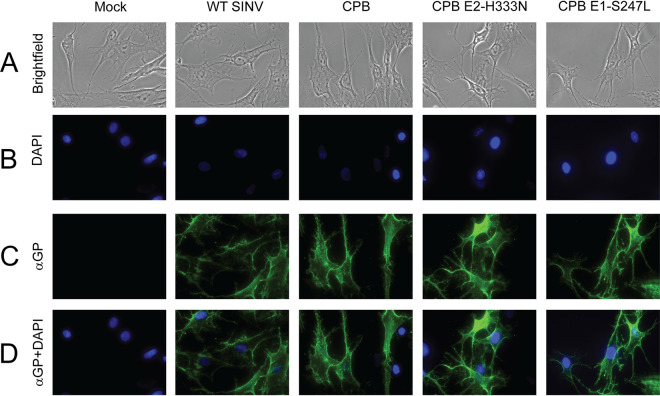
Alphaviruses are enveloped viruses transmitted by arthropod vectors to vertebrate hosts. The surface of the virion contains 80 glycoprotein spikes embedded in the membrane, and these spikes mediate attachment to the host cell and initiate viral fusion. Each spike consists of a trimer of E2-E1 heterodimers. These heterodimers interact at the following two interfaces: (i) the intradimer interactions between E2 and E1 of the same heterodimer and (ii) the interdimer interactions between E2 of one heterodimer and E1 of the adjacent heterodimer (E1'). We hypothesized that the interdimer interactions are essential for trimerization of the E2-E1 heterodimers into a functional spike. In this work, we made a mutant virus (chikungunya piggyback [CPB]) where we replaced six interdimeric residues in the E2 protein of Sindbis virus (wild-type [WT] SINV) with those from the E2 protein from chikungunya virus and studied its effect in both mammalian and mosquito cell lines. CPB produced fewer infectious particles in mammalian cells than in mosquito cells, relative to WT SINV. When CPB virus was purified from mammalian cells, particles showed reduced amounts of glycoproteins relative to the capsid protein and contained defects in particle morphology compared with virus derived from mosquito cells. Using cryo-electron microscopy (cryo-EM), we determined that the spikes of CPB had a different conformation than WT SINV. Last, we identified two revertants, E2-H333N and E1-S247L, that restored particle growth and assembly to different degrees. We conclude the interdimer interface is critical for spike trimerization and is a novel target for potential antiviral drug design. IMPORTANCE Alphaviruses, which can cause disease when spread to humans by mosquitoes, have been classified as emerging pathogens, with infections occurring worldwide. The spikes on the surface of the alphavirus particle are absolutely required for the virus to enter a new host cell and initiate an infection. Using a structure-guided approach, we made a mutant virus that alters spike assembly in mammalian cells but not mosquito cells. This finding is important because it identifies a region in the spike that could be a target for antiviral drug design.

Keywords: Glycoprotein spike; alphaviruses; assembly; host-range.


Conflict of interest statement

The authors declare no conflict of interest.

Figures

FIG 1
FIG 1
Interdimer interface residues in Domain C of E2 may be important for spike assembly. (A) Crystal structure of CHIKV glycoproteins E2 (blue) and E1 (yellow) (PDB: 3N40) (29) fit into the cryo-EM map of SINV (PDB: 1Z8Y) (29). One trimeric spike is outlined. (B) During assembly, E1 (shades of yellow) forms heterodimers with E2 (shades of blue), and these dimers then trimerize. A top view of one of these trimeric spikes, with the individual E2 (in teal, light blue, and blue) and E1 (in gold, light yellow, and saffron) proteins is shown here. A total of 80 spikes are on the surface of the alphavirus particle. (C) The trimer is rotated 90 degrees for a side view. The light-blue E2 and light-yellow E1 are a heterodimer. Intradimer contacts occur between Domains I and II of E1 with Domains A, B, and C of E2. The teal E2’ and gold E1’ form another heterodimer, with similar intradimer contacts. The last dimer in the spike is shown in gray. (D) A 60-degree rotation of C highlights an interdimer interface in the trimer. Interdimer contacts are between the gold E1’ of one heterodimer and the light-blue E2 in the adjacent dimer. Domain C of E2 (red dashed circle) is sandwiched between the adjacent E1’ (gold) and its cognate E1 (light yellow), forming interdimer and intradimer contacts, respectively. (E) The same proteins, namely, E1’, E2, and E1, shown in D are color coded by the domain here. Domain III of E1' and E1 is dark green, Domain I of E1' and E1 is yellow green, and Domain II of E1' and E1 is yellow. Domain C of E2 is in dark purple and Domains A and B are in light purple. The other monomers are colored gray for clarity. (F) Ribbon diagram of the interdimer of E1’-E2. Residues in E1’ that contact E2 are in yellow residues in E2 that contact E1’ are in blue spheres (light and dark). The residues mutated in this study are in dark-blue spheres. (G) Amino acid alignment of CHIKV and SINV E2 in the interdimer region; SINV residues shown in black and CHIKV residues are shown in red. The CHIKV piggyback (CPB) chimera was generated by substituting the nonhomologous E2 CHIKV residues for the corresponding SINV E2 residues (highlighted in red in CPB) in this region.
FIG 2
FIG 2
CPB grows slower than WT SINV in mammalian cells than in mosquito cells. Cells were infected at an MOI of 1 PFU/cell. At the indicated time points, the medium was collected and replaced with fresh media. The titers of the collected samples were determined by standard plaque assay on BHK cells. Results are shown for one representative experiment (n = 5). (A) Growth kinetics of infectious virus released from BHK cells at 37°C show that CPB was attenuated by 1 to 1.5 logs relative to WT SINV. (B) Growth kinetics of virus grown in C6/36 cells at 28°C show CPB releases infectious particles at the same rate as WT SINV. (C) Growth kinetics of virus grown in BHK cells at 28°C show that CPB growth is still attenuated by 1 to 1.5 logs relative to WT SINV.
FIG 3
FIG 3
Two CPB revertants map close to the interdimer interface and grow similarly to WT SINV. (A) CPB collected 20 to 40 hours postelectroporation have smaller plaques than WT SINV plaques (P < 0.0015, see 3C). When CPB is harvested later, or as it is passaged in BHK cells, larger plaques are seen in addition to the small plaques. Five of these larger plaques were isolated, the RNA was sequenced, and two independent second-site revertants were found. (B) The locations of 2 second-site revertant sites, namely, H333N in E2 (cyan) and S247L in E1 (orange), are shown in the E1’-E2 dimer. E1’ and E2 contact residues are colored in yellow and blue, respectively, as they were in Fig. 1F; mutated sites in CPB are in dark blue. (C) CPB E2-H333N and CPB E1-S247L were introduced independently into CPB. The plaque size of CPB E2-H333N and CPB E1-S247L in BHK cells was larger than that of CPB and was not significantly (ns) different from that of the WT. (D and E) Growth kinetics of the two revertants show they have comparable titers as WT SINV in BHK (D) and C6/36 (E) cells. Representative curves are shown (n = 3). Cells were infected at an MOI of 1 PFU/cell, medium was collected at the indicated time points, and the titers of samples were determined on BHK cells.
FIG 4
FIG 4
Particle morphology and composition are restored in CPB revertants to various degrees. Cells were infected at an MOI of 0.1 PFU/cell for 1 hour, after which serum-free medium was overlaid. Infected BHK medium was collected after 24 hours, and infected C6/36 medium was collected after 120 hours. The medium was clarified and purified through a sucrose cushion at 140,000 × g for 2.5 h. (A and B) To determine the composition of the virions, purified virus was solubilized in reducing SDS sample buffer, run on an 8% SDS-PAGE gel, and imaged by stain-free 2,2,2-trichloroethanol (TCE). CPB particles have a reduced level of E2 and E1 glycoproteins in particles purified from BHK cells. The E1 deglycosylation control is SINV E1-N245/246Q, which has one of its two E1 glycosylation sites deglycosylated resulting in a faster migrating E1 protein band, and the E1/E2 deglycosylation control is E1-N139Q/E2-N318Q and has a deglycosylation site in both E1 and E2 resulting in faster migration in each of those protein bands. (C and D) Purified particles were applied to a Formvar- and carbon-coated 400-mesh copper grid, stained with 2% uranyl acetate, and imaged at ×20,000 magnification with the JEOL 1010 transmission electron microscope. Scale bar for TEM, 200 nm.
FIG 5
FIG 5
CPB glycoprotein spikes are transported to the plasma membrane of BHK cells in approximately equal amounts compared with WT. BHK cells were infected at an MOI of 2 PFU/cell. Ten hours postinfection, the cells were fixed with 4% EM-grade paraformaldehyde and probed for SINV E1/E2. Images were taken at 40× magnification on a Nikon Ni-E microscope. Representative images are shown (n = 4). (A and B) Brightfield images (A) and DAPI staining (B) show cells in field of view and their nuclei in blue. (C) Cell surface E2 and E1 expression is shown in green, and Alexa Fluor 488 was the secondary antibody. (D) Anti-E2 and E1+DAPI merge shows green fluorescence from the surface glycoprotein expression relative to the cells present.
FIG 6
FIG 6
Cryo-EM 3D reconstructions show that CPB spikes from mammalian cells have an altered interdimer organization. (A to F) Radially colored isosurface rendering of WT SINV, CPB, and SINV E1-S247L from BHK cells (A to C, top row) and C6/36 cells (D to F, bottom row). All views are at the 5-fold axis. All six maps have a similar outer appearance with 80 spikes decorating a fenestrated surface. In A, the white triangle shows one asymmetric unit. The filled oval, triangle, and pentagon indicate locations of 2-fold, 3-fold, and 5-fold axes, respectively. The dotted circle shows one of the spikes on the quasi-three axis. (G to L) Enlarged views of SINV spikes at the quasi-3-fold location, and all images are shown at a 10-Å resolution for comparison purposes. The spike of CPB from BHK (H) shows a distinct organization from other SINV spikes. Black arrowheads indicate the difference in the electron density between each interdimer interface. Red arrowheads show glycan modification at E2-196.

Similar articles

Cited by

References

    1. Griffin DE. 2013. Chapter 23: alphaviruses, p 652–770. In Knipe DM, Howley PM (ed), Fields’ virology. Lippincott Williams & Wilkins, Philadelphia, PA.
    1. Kuhn RJ. 2013. Chapter 22: Togaviradae, p 629–650. In Knipe DM, Howley PM (ed), Fields’ virology. Lippincott Williams & Wilkins, Philadelphia, PA.
    1. Hermanns K, Marklewitz M, Zirkel F, Overheul GJ, Page RA, Loaiza JR, Drosten C, van Rij RP, Junglen S. 2020. Agua Salud alphavirus defines a novel lineage of insect-specific alphaviruses discovered in the New World. J Gen Virol 101:96–104. 10.1099/jgv.0.001344. - DOI - PMC - PubMed
    1. Forrester NL, Palacios G, Tesh RB, Savji N, Guzman H, Sherman M, Weaver SC, Lipkin WI. 2012. Genome-scale phylogeny of the alphavirus genus suggests a marine origin. J Virol 86:2729–2738. 10.1128/JVI.05591-11. - DOI - PMC - PubMed
    1. Firth AE, Chung BY, Fleeton MN, Atkins JF. 2008. Discovery of frameshifting in Alphavirus 6K resolves a 20-year enigma. Virol J 5:108. 10.1186/1743-422X-5-108. - DOI - PMC - PubMed

Publication types

LinkOut - more resources