

Acute effects of ketamine on the pregenual anterior cingulate: linking spontaneous activation, functional connectivity, and glutamate metabolism
- PMID: 35020021
- PMCID: PMC9095553
- DOI: 10.1007/s00406-021-01377-2
Acute effects of ketamine on the pregenual anterior cingulate: linking spontaneous activation, functional connectivity, and glutamate metabolism
Abstract
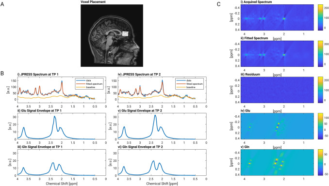
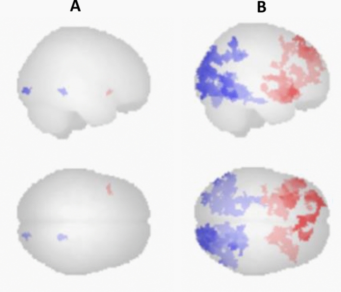
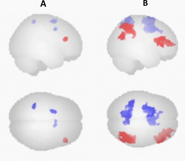
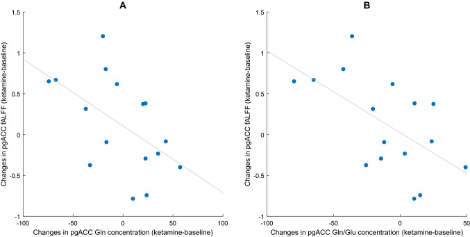
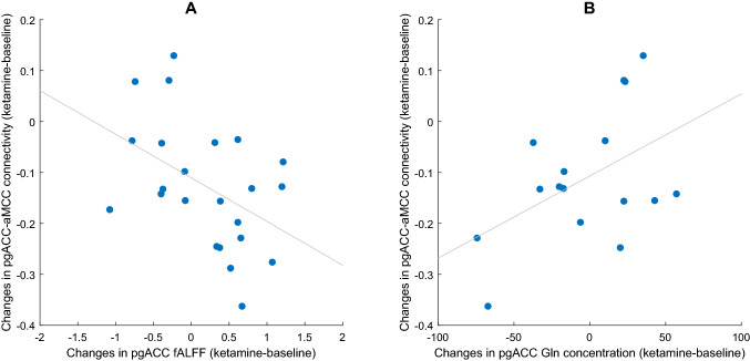
Ketamine exerts its rapid antidepressant effects via modulation of the glutamatergic system. While numerous imaging studies have investigated the effects of ketamine on a functional macroscopic brain level, it remains unclear how altered glutamate metabolism and changes in brain function are linked. To shed light on this topic we here conducted a multimodal imaging study in healthy volunteers (N = 23) using resting state fMRI and proton (1H) magnetic resonance spectroscopy (MRS) to investigate linkage between metabolic and functional brain changes induced by ketamine. Subjects were investigated before and during an intravenous ketamine infusion. The MRS voxel was placed in the pregenual anterior cingulate cortex (pgACC), as this region has been repeatedly shown to be involved in ketamine's effects. Our results showed functional connectivity changes from the pgACC to the right frontal pole and anterior mid cingulate cortex (aMCC). Absolute glutamate and glutamine concentrations in the pgACC did not differ significantly from baseline. However, we found that stronger pgACC activation during ketamine was linked to lower glutamine concentration in this region. Furthermore, reduced functional connectivity between pgACC and aMCC was related to increased pgACC activation and reduced glutamine. Our results thereby demonstrate how multimodal investigations in a single brain region could help to advance our understanding of the association between metabolic and functional changes.
Keywords: Glutamate; Ketamine; MR Spectroscopy; Resting state fMRI.
© 2022. The Author(s).
Conflict of interest statement
The authors declare no conflict of interest.
Figures
Similar articles
-
Default mode network connectivity change corresponds to ketamine's delayed glutamatergic effects.Eur Arch Psychiatry Clin Neurosci. 2020 Mar;270(2):207-216. doi: 10.1007/s00406-018-0942-y. Epub 2018 Oct 23. Eur Arch Psychiatry Clin Neurosci. 2020. PMID: 30353262 Clinical Trial.
-
Temporal Dynamics of Antidepressant Ketamine Effects on Glutamine Cycling Follow Regional Fingerprints of AMPA and NMDA Receptor Densities.Neuropsychopharmacology. 2017 May;42(6):1201-1209. doi: 10.1038/npp.2016.184. Epub 2016 Sep 8. Neuropsychopharmacology. 2017. PMID: 27604568 Free PMC article. Clinical Trial.
-
The relationship between aberrant neuronal activation in the pregenual anterior cingulate, altered glutamatergic metabolism, and anhedonia in major depression.Arch Gen Psychiatry. 2009 May;66(5):478-86. doi: 10.1001/archgenpsychiatry.2009.39. Arch Gen Psychiatry. 2009. PMID: 19414707
-
A Meta-Analysis of Functional Neuroimaging Studies of Ketamine Administration in Healthy Volunteers.J Psychoactive Drugs. 2024 Apr-Jun;56(2):211-224. doi: 10.1080/02791072.2023.2190758. Epub 2023 Mar 15. J Psychoactive Drugs. 2024. PMID: 36921026 Review.
-
The potential of 1H-MRS in CNS drug development.Psychopharmacology (Berl). 2021 May;238(5):1241-1254. doi: 10.1007/s00213-019-05344-7. Epub 2019 Sep 5. Psychopharmacology (Berl). 2021. PMID: 31486875 Free PMC article. Review.
Cited by
-
Ketamine and Zinc: Treatment of Anorexia Nervosa Via Dual NMDA Receptor Modulation.CNS Drugs. 2023 Feb;37(2):159-180. doi: 10.1007/s40263-022-00984-4. Epub 2023 Jan 22. CNS Drugs. 2023. PMID: 36681939 Free PMC article. Review.
-
Brain Networks, Neurotransmitters and Psychedelics: Towards a Neurochemistry of Self-Awareness.Curr Neurol Neurosci Rep. 2024 Aug;24(8):323-340. doi: 10.1007/s11910-024-01353-y. Epub 2024 Jul 9. Curr Neurol Neurosci Rep. 2024. PMID: 38980658 Free PMC article. Review.
-
Association of the delayed changes in glutamate levels and functional connectivity with the immediate network effects of S-ketamine.Transl Psychiatry. 2023 Feb 16;13(1):60. doi: 10.1038/s41398-023-02346-0. Transl Psychiatry. 2023. PMID: 36797238 Free PMC article.
References
MeSH terms
Substances
Grants and funding
LinkOut - more resources
Full Text Sources
