. 2022 Jan 25;94(3):1594-1600.
doi: 10.1021/acs.analchem.1c03503. Epub 2022 Jan 12.

"Fix and Click" for Assay of Sphingolipid Signaling in Single Primary Human Intestinal Epithelial Cells

Affiliations

"Fix and Click" for Assay of Sphingolipid Signaling in Single Primary Human Intestinal Epithelial Cells

Luke A Gallion et al. Anal Chem. .

Abstract

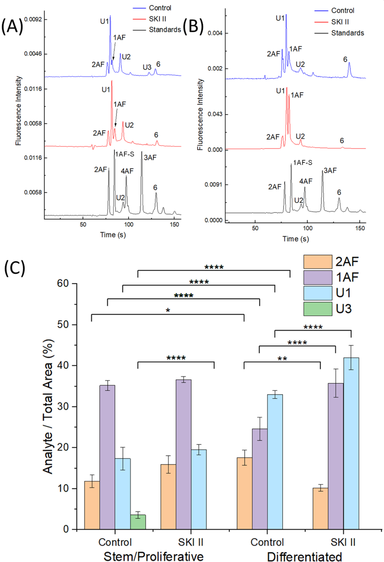
Capillary electrophoresis with fluorescence detection (CE-F) is a powerful method to measure enzyme activation in single cells. However, cellular enzymatic assays used in CE-F routinely utilize reporter substrates that possess a bulky fluorophore that may impact enzyme kinetics. To address these challenges, we describe a "fix and click" method utilizing an alkyne-terminated enzyme activation reporter, aldehyde-based fixation, and a click chemistry reaction to attach a fluorophore prior to analysis by single-cell CE-F. The "fix and click" strategy was utilized to investigate sphingolipid signaling in both immortalized cell lines and primary human colonic epithelial cells. When the sphingosine alkyne reporter was loaded into cells, this reporter was metabolized to ceramide (31.6 ± 3.3% peak area) without the production of sphingosine-1-phosphate. In contrast, when the reporter sphingosine fluorescein was introduced into cells, sphingosine fluorescein was converted to sphingosine-1-phosphate and downstream products (32.8 ± 5.7% peak area) without the formation of ceramide. Sphingolipid metabolism was measured in single cells from both differentiated and stem/proliferative human colonic epithelium using "fix and click" paired with CE-F to highlight the diversity of sphingosine metabolism in single cells from primary human colonic epithelium. This novel method will find widespread utility for the performance of single-cell enzyme assays by virtue of its ability to temporally and spatially separate cellular reactions with alkyne-terminated reporters, followed by the assay of enzyme activation at a later time and place.


Conflict of interest statement

Notes

Y.W. and N.L.A. disclose a financial interest in Altis Biosystems, Inc. L.A.G., A.M., M.Y., B.V.P., Q.Z., W.H., A.J.C., and Q.Z. declare no financial conflict.

Figures

Figure 1:
Figure 1:
Schematic of the “fix and click” method and sphingosine reporters. (A) Schematic of “fix and click” reaction for substrate and product assay. (i) Cells are loaded with 2A, which is converted to metabolites. (ii) Cells are fixed to halt enzyme activity with retention of the reporter and its product forms within the cell. (iii) Alkyne-terminated sphingolipids within the cell undergo a click reaction to attach a fluorophore. (iv) Cells are washed to remove excess click reagent 6. (v) Cell contents are electrophoretically separated to display migration time (X) vs fluorescence intensity (Y). (B) Sphingosine reporters (2F and 2A) and the fluorescent product yielded by the click reaction between 2A and 6. (C) Effect of fixation method. Glyoxal indicates the glyoxal-based Prefer fixative while PFA indicates paraformaldehyde. Shown is total cellular fluorescence vs fixative. p = 0.0024. (D) Post-click cellular fluorescence stability. Cells loaded with SA and fixed and clicked were stored at 4°C for varying times. Shown is total cellular fluorescence vs time.
Figure 2.
Figure 2.
2F and 2A reporter metabolism in Caco-2 cells. (A) Representative electropherograms of standards and Caco-2 cells incubated with 10 μM 2F with and without exposure to SKI II. Cells were then fixed, and methanol-soluble lipids extracted from the cells. (B) Representative electropherograms of standards and Caco-2 cells incubated with 10 μM 2A with and without exposure to SKI II. Cells were then fixed and clicked, and methanol-soluble lipids extracted from the cells. (C) Quantification of metabolites formed from cells loaded with 2F or 2A. The y axis displays the percentage of the analyte peak area divided by the total area of the peaks on the electropherogram. n=3 biological replicates for each condition. “Reporter” corresponds to 2AF or 2F + 2F-G, “SphK Products” refers to the sum of 3F + 5F (no SphK products observed with 2A reporter), and “Ceramide” refers to 1AF (no ceramide observed with 2F reporter).
Figure 3:
Figure 3:
2A reporter metabolism by primary human colonic epithelial cells. (A) Representative electropherograms of standards and stem/proliferative cells incubated with 1 μM 2A with and without exposure to SKI II. Cells were then fixed and clicked, and methanol-soluble lipids extracted from the cells. (B) Representative electropherograms of standards and differentiated cells incubated with 2A with and without exposure to SKI II. Cells were then fixed and clicked, and methanol-soluble lipids extracted from the cells. (C) Quantification of metabolites formed from cells loaded with 2A. The y-axis displays the percentage of the analyte peak area divided by the total area of the peaks on the electropherogram. n=3 biological replicates for each condition.
Figure 4:
Figure 4:
2A reporter metabolism by single primary human colonic epithelial cells. (A) Representative electropherograms of two cells from a stem/proliferative cell culture and standards. (B) Representative electropherograms of two cells from a differentiated cell culture and standards. (C) Quantification of the peak areas observed in individual cells. n = 13 stem/proliferative cells, 11 differentiated cells. No significant difference was observed between stem/proliferative cells and differentiated cells for any analytes.

References

    1. Maceyka M; Harikumar KB; Milstien S; Spiegel S Sphingosine-1-Phosphate Signaling and Its Role in Disease. Trends Cell Biol. 2012, 22 (1), 50–60. DOI: 10.1016/j.tcb.2011.09.003. - DOI - PMC - PubMed
    1. Morad SAF; Cabot MC Ceramide-Orchestrated Signalling in Cancer Cells. Nat. Rev. Cancer 2013, 13 (1), 51–65. DOI: 10.1038/nrc3398. - DOI - PubMed
    1. Evangelisti C; Evangelisti C; Buontempo F; Lonetti A; Orsini E; Chiarini F; Barata JT; Pyne S; Pyne NJ; Martelli AM Therapeutic Potential of Targeting Sphingosine Kinases and Sphingosine 1-Phosphate in Hematological Malignancies. Leukemia 2016, 30 (11), 2142–2151. DOI: 10.1038/leu.2016.208. - DOI - PubMed
    1. Paugh SW; Paugh BS; Rahmani M; Kapitonov D; Almenara JA; Kordula T; Milstien S; Adams JK; Zipkin RE; Grant S; Spiegel S A Selective Sphingosine Kinase 1 Inhibitor Integrates Multiple Molecular Therapeutic Targets in Human Leukemia. Blood 2008, 112 (4), 1382–1391. 10.1182/blood-2008-02-138958. - DOI - PMC - PubMed
    1. Siegel RL; Miller KD; Fuchs HE; Jemal A Cancer Statistics, 2021. CA: A Cancer Journal for Clinicians 2021, 71 (1), 7–33. DOI: 10.3322/caac.21654. - DOI - PubMed

Publication types