. 2022 Jan 14;8(2):eabi8683.
doi: 10.1126/sciadv.abi8683. Epub 2022 Jan 12.

Imaging and tracing the pattern of adult ovarian angiogenesis implies a strategy against female reproductive aging

Affiliations

Imaging and tracing the pattern of adult ovarian angiogenesis implies a strategy against female reproductive aging

Xueqiang Xu et al. Sci Adv. .

Abstract

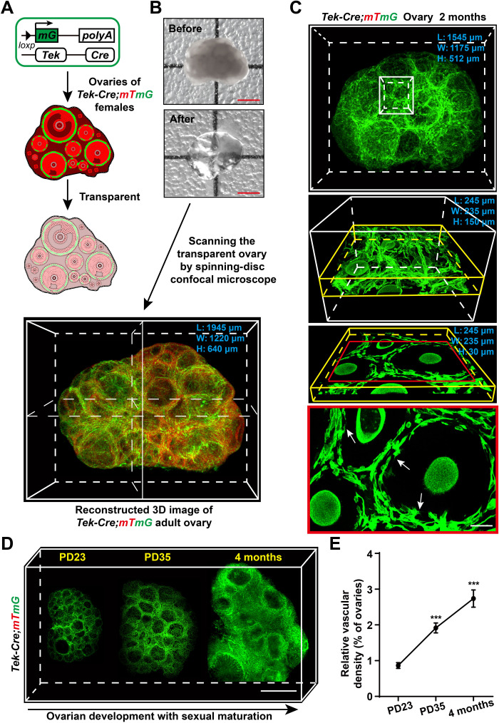
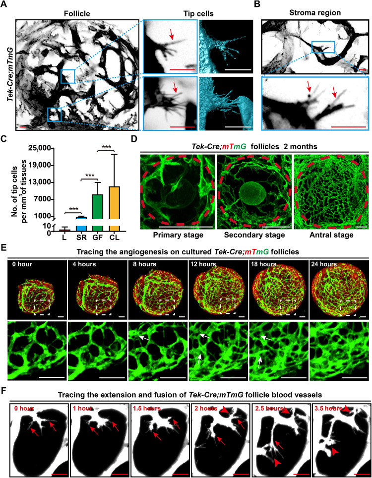
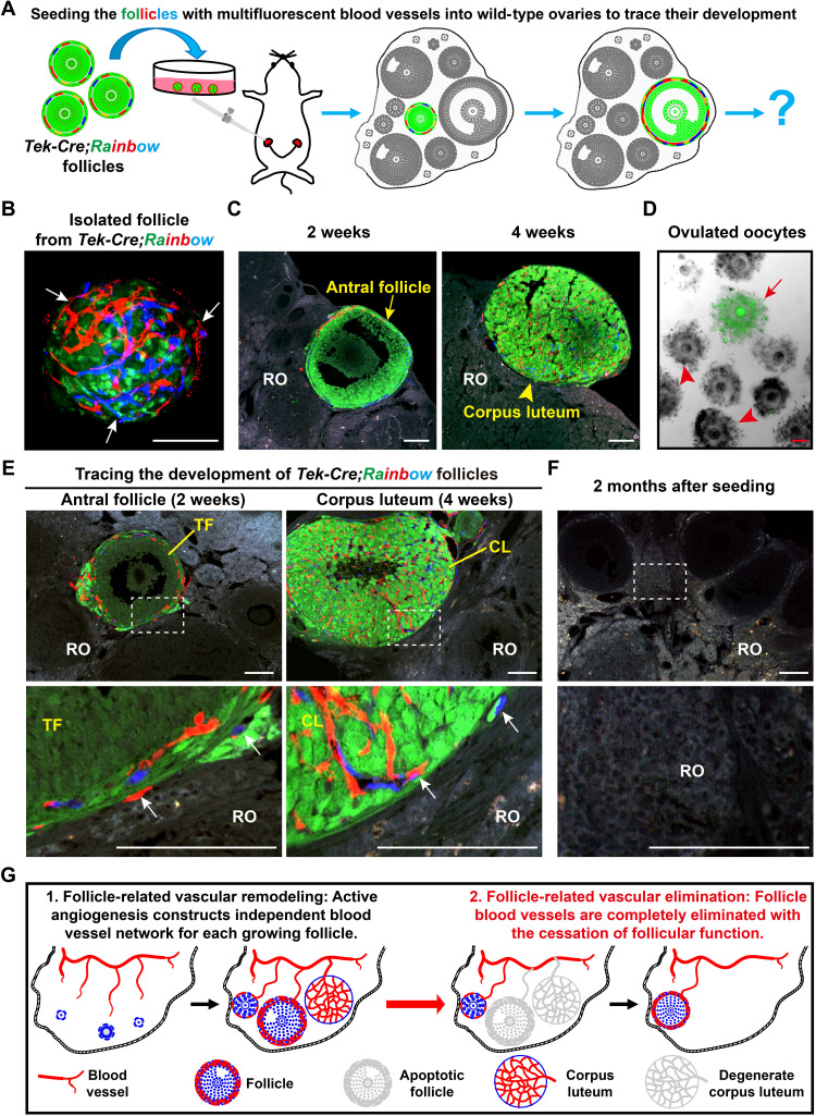
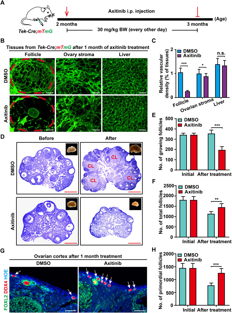
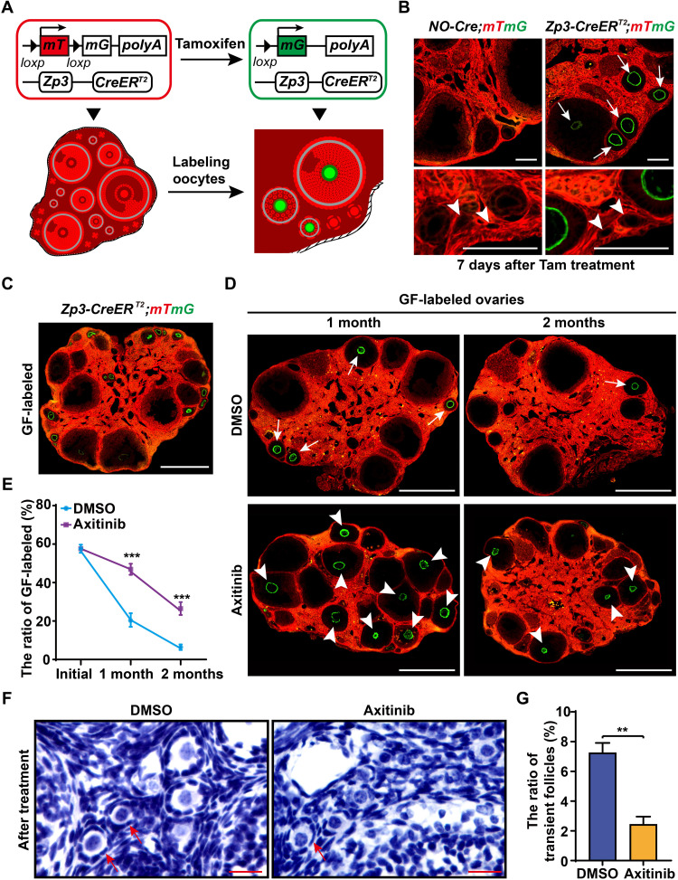
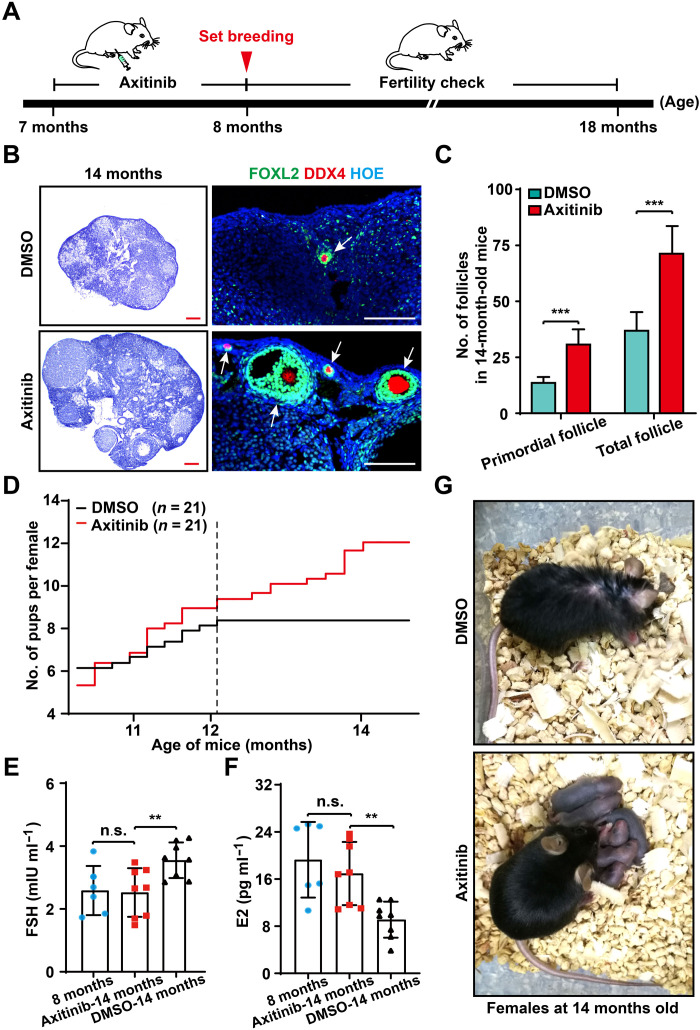
Robust angiogenesis is continuously active in ovaries to remodel the ovary-body connections in mammals, but understanding of this unique process remains elusive. Here, we performed high-resolution, three-dimensional ovarian vascular imaging and traced the pattern of ovarian angiogenesis and vascular development in the long term. We found that angiogenesis was mainly active on ovarian follicles and corpus luteum and that robust angiogenesis constructs independent but temporary vascular networks for each follicle. Based on the pattern of ovarian angiogenesis, we designed an angiogenesis-blocking strategy by axitinib administration to young females, and we found that the temporary suppression of angiogenesis paused ovarian development and kept the ovarian reserve in the long term, leading to postponed ovarian senescence and an extension of the female reproductive life span. Together, by uncovering the detailed model of physiological ovarian angiogenesis, our experiments suggest a potential approach to delay female reproductive aging through the manipulation of angiogenesis.


Figures

Fig. 1.
Fig. 1.. High-resolution 3D imaging to reconstruct adult ovarian blood vessels in mice.
(A and B) Schematic diagram showing the strategy used to achieve high-resolution 3D imaging of ovarian blood vessels. In Tek-expressing endothelial cells, Cre recombinase mediated a switch from mTomato (red) to mGFP (green). The Tek-Cre;mTmG ovaries with the mG-labeled endothelium were fixed and cleared (A), and the transparent ovaries were scanned by high-resolution confocal microscopy to reconstruct a 3D image of the ovarian blood vessels (B). (C) The reconstructed 3D images of Tek-Cre;mTmG ovaries were kept at a high resolution and were available for different analysis scales from the whole-organ level (white box) and single-follicle level (yellow box) to the subcellular resolution (red frame). The subcellular structures (arrows) are shown in the image. (D) Reconstructed ovarian blood vessels at the organ level showing a significant accumulation of blood vessels in mouse ovaries with sexual maturation, from puberty onset at PD23 to adulthood at 4 months. (E) Quantification of the relative changes in ovarian vessel density showing a significant increase in ovarian vessel density from PD23 to 4 months (n = 9 ovaries per group). The data are presented as means ± SD. The data were analyzed by a two-tailed unpaired Student’s t test and ***P < 0.001. Scale bars, 500 μm (B and D) and 25 μm (C).
Fig. 2.
Fig. 2.. Imaging and tracing angiogenesis in adult ovaries.
(A and B) Analysis of the 3D-reconstructed ovarian images at the single-cell level to reveal the pattern of angiogenesis in the adult ovaries. Tip cells with filopodia (arrows) were mainly observed on the endothelium of GFs (A), and a few tip cells were randomly detected in the ovarian stromal region (B). (C) Quantification of the tip cell number in the liver (L), stromal region (SR), growing follicles (GFs), and CL of ovaries (n = 20 for each group) in adult mice (2 months), showing the abundance of tip cells in the adult ovaries, especially on GFs and CL. (D) Reconstructed images of ovarian follicles at different stages, showing a significantly increased density of blood vessels from primary follicles to antral follicles. (E) Live-cell imaging analysis recorded a marked growth of blood vessels on the surface of Tek-Cre;mTmG follicles. During 24 hours of in vitro culture, branching and extension were observed on the membrane GFP–labeled follicle endothelium (see movie S3). (F) High-magnification time-lapse imaging showing the behaviors of tip cells on follicle blood vessels (white). The extension of filopodia (arrows) on tip cells and the fusion of different tip cells (arrowheads) were recorded (see movie S4). GFP was converted to black and white in (A) and (B) to highlight the cell details. The data are presented as means ± SD. The data were analyzed by a two-tailed unpaired Student’s t test and ***P < 0.001. Scale bars, 10 μm (A and B), 50 μm (D and E), and 25 μm (F).
Fig. 3.
Fig. 3.. Angiogenesis established an independent and temporary follicle vascular network in the adult ovaries.
(A) Illustration of the strategy used to trace the development of follicle blood vessel endothelial cells in vivo. GFs with fluorescent-labeled blood vessels were isolated from Tek-Cre;Rainbow females and were surgically seeded into the ovaries of wild-type recipient females for long-term tracing. (B) Before seeding, a single follicle from Tek-Cre;Rainbow ovaries showed red or blue fluorescent blood vessels (arrows) surrounding the GFP follicle. (C and D) The transplanted Tek-Cre;Rainbow follicles successfully developed to the antral stage and formed a CL in the recipient ovaries (ROs) and also ovulated the GFP oocytes from recipient females, showing the normal development of the seeded follicles. (E) Tracing the developmental fate of follicle blood vessels after transplantation. All fluorescent blood vessels (arrows) were limited to the surface of transplanted follicles and the CL, which were derived from transplanted follicles. (F) At 2 months after seeding, all fluorescent blood vessels were eliminated because of the exhaustion of transplanted follicles in the ROs (0 of 97). The transplanted window is indicated by the white frame. (G) Schematic of the fate of new follicular blood vessels formed via angiogenesis, showing the formation of an independent blood vessel network for each GF in the ovaries, and a complete elimination of follicle blood vessels with the cessation of follicle function. RO, recipient ovary; TF, transplanted follicle; CL, corpus luteum. Scale bars, 100 μm (B to E).
Fig. 4.
Fig. 4.. Axi treatment disrupted follicle blood vessel construction but kept the ovarian reserve in adult females.
(A) The strategy of Axi treatment in females. Females at 2 months were treated with Axi (30 mg/kg BW, every other day) for 1 month to temporarily block angiogenesis. i.p., intraperitoneal. (B) After 1 month of Axi treatment, a significantly decreased vascular density was observed on the follicles of Axi-treated ovaries compared with those in the DMSO group (n = 8), but no marked effect was found for the stable blood vessels of the ovarian stromal region and liver. (C) Quantification of vascular density in the follicles and stromal region of ovaries and the liver after Axi treatment (n = 9 ovaries per group). (D) Histological analysis showed the decrease in ovarian size and in the number of GFs in the Axi group compared with the controls. (E) Follicle counts showed a significant decrease in the number of GFs in the Axi-treated ovaries (n = 6). (F) The total follicle number in the Axi-treated and control ovaries, showing that the total number of follicles in the Axi group was significantly greater than that in the controls (n = 6). (G) Immunostaining results revealed more primordial follicles (arrows) in the cortex of Axi-treated ovaries. Green: anti-FOXL2/GCs. Red: anti-DDX4/oocytes. Blue: Hoechst33342. (H) Follicle count results of primordial follicles (PFs), showing an increased number of PFs in Axi-treated ovaries compared with the controls (n = 6). The data are presented as means ± SD. The data were analyzed by a two-tailed unpaired Student’s t test; ***P < 0.001, **P < 0.01, *P < 0.05, n.s. P ≥ 0.05. Scale bars, 50 μm (B and G) and 500 μm (D).
Fig. 5.
Fig. 5.. Blocking angiogenesis paused ovarian development.
(A and B) Illustration of tamoxifen (Tam)–induced labeling of growing oocytes in the Zp3-CreERT2;mTmG mice. CreERT2 recombinase is active upon Tam induction, which mediates the switch from mTomato (red) to mGFP (green) in Zona pellucida protein 3 (Zp3)–expressing oocytes, resulting in the labeling of GFs with green oocytes. (B) Validating the labeling specificity and efficiency of Zp3-CreERT2;mTmG females. After 7 days of Tam treatment, numerous oocytes in GFs were labeled with mG (arrows), whereas none of the dormant oocytes (arrowheads) were labeled in the Zp3-CreERT2;mTmG ovaries. (C) The 2-month Zp3-CreERT2;mTmG females after 7 days of Tam treatment are referred to as “GF-labeled” females. (D) Tracing the developmental dynamics of GFs in GF-labeled females with or without Axi treatment. GFP oocytes (arrowheads) were widely distributed in GF-labeled ovaries at either 1 or 2 months in Axi-treated females but were exhausted in control ovaries after 2 months (arrows). (E) Quantification of the labeling ratio of the GF showed a significant extension of GF life span in Axi-treated ovaries (n ≥ 6). (F) Histological identification of the transient follicles (arrows) in the controls and Axi-treated ovaries. (G) Counting the transient follicles showed a significantly decreased ratio of transient follicles in the Axi-treated ovaries compared with the controls (n = 5). The data are presented as means ± SD. The data were analyzed by a two-tailed unpaired Student’s t test; ***P < 0.001, **P < 0.01. Scale bars, 100 μm (B), 500 μm (C and D), and 25 μm (F).
Fig. 6.
Fig. 6.. Appropriate inhibition of adult angiogenesis extended the reproductive life span of aged females.
(A) The strategy of Axi treatment in females of late reproductive life span. After 1 month of Axi or vehicle treatment, females at 8 months were mated to check their fertility and were used to detect other reproductive-related indices at different time points. (B) Histological analysis showing Axi-treated ovaries exhibited a clear younger status at 14 months with many follicles (arrows) compared with the control ovaries. (C) Follicle counting results showing that more follicles survived in the Axi-treated ovaries compared with the controls at the age of 14 months (n = 6). (D) Fertility test results showed a significant extension of the female reproductive life span after Axi treatment (n = 21). (E and F) The sex-related hormones, including follicle-stimulating hormone (FSH) and estrogen (E2), were found to be comparable in untreated females at 8 months and Axi-treated females at 14 months of age, whereas significantly increased levels of FSH and decreased levels of E2 were detected in the 14-month-old females of the DMSO group (n = 8 per group). (G) The general appearance of Axi-treated and the control females at 14 months, showing healthy pups with Axi-treated mothers (Photo credit: Xueqiang Xu, China Agricultural University). The data are presented as means ± SD. The data were analyzed by a two-tailed unpaired Student’s t test; ***P < 0.001, **P < 0.01, n.s. P ≥ 0.05. Scale bars, 100 μm (B).

References

    1. Hsueh A. J. W., Kawamura K., Cheng Y., Fauser B. C. J. M., Intraovarian control of early folliculogenesis. Endocr. Rev. 36, 1–24 (2015). - PMC - PubMed
    1. Carmeliet P., Angiogenesis in life, disease and medicine. Nature 438, 932–936 (2005). - PubMed
    1. Cao Y. H., Adipose tissue angiogenesis as a therapeutic target for obesity and metabolic diseases. Nat. Rev. Drug Discov. 9, 107–115 (2010). - PubMed
    1. Cao Y., Angiogenesis and vascular functions in modulation of obesity, adipose metabolism, and insulin sensitivity. Cell Metab. 18, 478–489 (2013). - PubMed
    1. Robinson R. S., The critical importance of ovarian angiogenesis. Reprod. Fertil. Dev. 25, iii–v (2013). - PubMed

LinkOut - more resources