. 2023 Jan 18;118(17):3386-3400.
doi: 10.1093/cvr/cvac007.

Smooth muscle mineralocorticoid receptor as an epigenetic regulator of vascular ageing

Affiliations

Smooth muscle mineralocorticoid receptor as an epigenetic regulator of vascular ageing

Jaime Ibarrola et al. Cardiovasc Res. .

Abstract

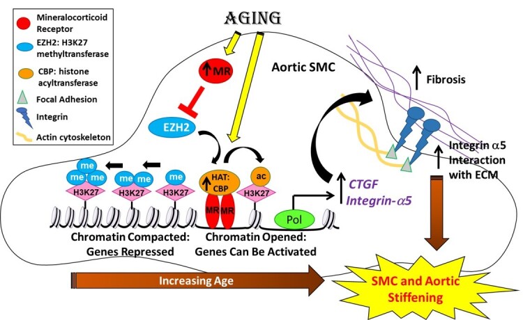
Aims: Vascular stiffness increases with age and independently predicts cardiovascular disease risk. Epigenetic changes, including histone modifications, accumulate with age but the global pattern has not been elucidated nor are the regulators known. Smooth muscle cell-mineralocorticoid receptor (SMC-MR) contributes to vascular stiffness in ageing mice. Thus, we investigated the regulatory role of SMC-MR in vascular epigenetics and stiffness.

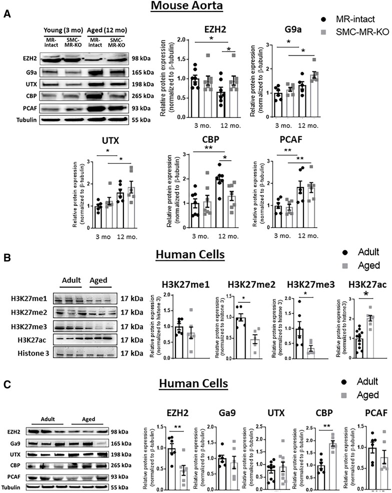
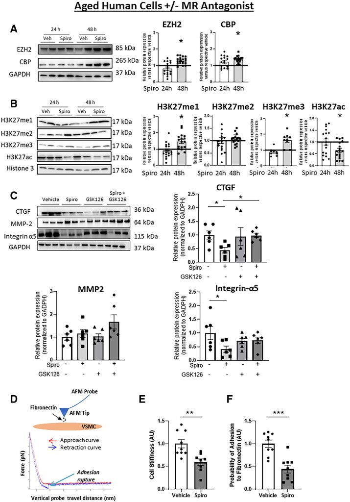
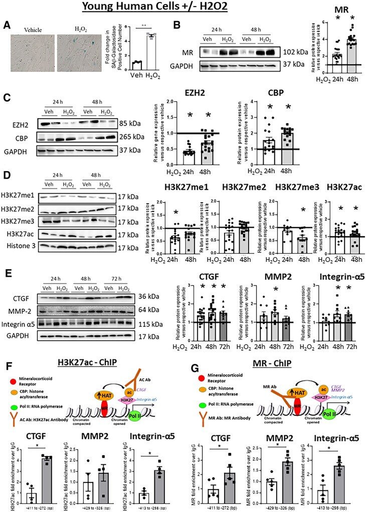
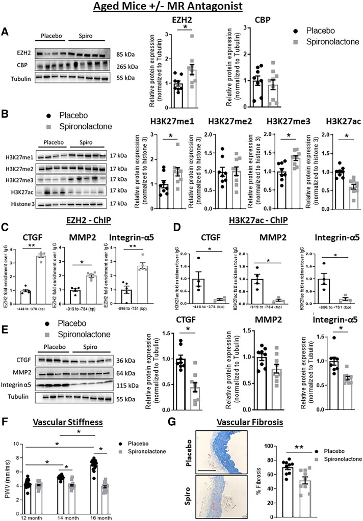
Methods and results: Mass spectrometry-based proteomic profiling of all histone modifications completely distinguished 3 from 12-month-old mouse aortas. Histone-H3 lysine-27 (H3K27) methylation (me) significantly decreased in ageing vessels and this was attenuated in SMC-MR-KO littermates. Immunoblotting revealed less H3K27-specific methyltransferase EZH2 with age in MR-intact but not SMC-MR-KO vessels. These ageing changes were examined in primary human aortic (HA)SMC from adult vs. aged donors. MR, H3K27 acetylation (ac), and stiffness gene (connective tissue growth factor, integrin-α5) expression significantly increased, while H3K27me and EZH2 decreased, with age. MR inhibition reversed these ageing changes in HASMC and the decline in stiffness genes was prevented by EZH2 blockade. Atomic force microscopy revealed that MR antagonism decreased intrinsic stiffness and the probability of fibronectin adhesion of aged HASMC. Conversely, ageing induction in young HASMC with H2O2; increased MR, decreased EZH2, enriched H3K27ac and MR at stiffness gene promoters by chromatin immunoprecipitation, and increased stiffness gene expression. In 12-month-old mice, MR antagonism increased aortic EZH2 and H3K27 methylation, increased EZH2 recruitment and decreased H3K27ac at stiffness genes promoters, and prevented ageing-induced vascular stiffness and fibrosis. Finally, in human aortic tissue, age positively correlated with MR and stiffness gene expression and negatively correlated with H3K27me3 while MR and EZH2 are negatively correlated.

Conclusion: These data support a novel vascular ageing model with rising MR in human SMC suppressing EZH2 expression thereby decreasing H3K27me, promoting MR recruitment and H3K27ac at stiffness gene promoters to induce vascular stiffness and suggests new targets for ameliorating ageing-associated vascular disease.

Keywords: EZH2; Epigenetics; Vascular ageing; Vascular stiffness; Mineralocorticoid receptor.


Conflict of interest statement

Conflict of interest: none declared. JDJ is currently employed by Odyssey Therapeutics. His contribution to the manuscript was completed while he was at the Broad Institute. There is no overlap between the subject of the manuscript and the role of JDJ at Odyssey Therapuetics.

Figures

Graphical Abstract
Graphical Abstract
Figure 1
Figure 1
Mineralocorticoid receptor (MR) and stiffness gene expression increase with age in primary human aortic smooth muscle cells (HASMC). Primary HASMC were compared from adult (average age 40s) vs. aged (average age late 70s) donors. (A) Representative immunoblots and quantification of MR protein expression by immunoblotting. n = 9 for each age group. (B) Representative HASMC images of senescence-associated (SA) β-galactosidase staining and quantification of SAβ-galactosidase staining. n = 4 of each age group. (C) Representative immunoblots and quantification of protein expression of select stiffness genes: connective tissue growth factor (CTGF), matrix metalloproteinase 2 (MMP2), and integrin-α5. n = 7 for each age group. Dot plots show the individual data points and bars indicate the mean with error bars indicating the SEM for relative protein expression normalized to GAPDH. Results were analysed using Student’s t-test. *P < 0.05, **P < 0.01.
Figure 2
Figure 2
Proteomic profiling of all aortic histone modifications from young and old mouse aortas distinguishes old from young vessels. Mass spectrometry-based profiling of histone modifications was performed on mouse aortic chromatin from 3 month and 12 month old mineralocorticoid receptor (MR)-intact and smooth muscle cells (SMC)-MR knock out littermates. (A) Principal component analysis (PCA) comparing the histone proteomic data and plotting by co-ordinates for principal Components 1 and 2, colour-coded by the age and genotype of each vessel. Principal Component 2 separates all vessels by age and principal Component 1 further separates most of the old MR-intact from old SMC-MR-KO vessels. (B) Hierarchical cluster analysis of global histone modification patterns in MR-intact and SMC-MR-KO mice young and old. Red indicates an increase and blue a decrease in the amount of each histone modification relative to the mean for all vessels. (C) Marker selection analysis identifies specific histone modifications that significantly distinguish young from old vessels. Histone modifications with a FDR < 0.05 are listed. ac, acetyl; me, methyl.
Figure 3
Figure 3
Ageing-associated changes in histone modifications and histone-modifying enzymes in mouse aortas and primary human aortic SMC (HASMC). (A) Representative immunoblots and quantification of protein expression of enzymes involve in the methylation (me; EZH2, G9a, UTX) and acetylation (ac; CBP, PCAF/P300) of histones in aortas from mice with MR-intact vs. SMC-MR-KO. n = 6–7 of each age group. (B) Representative immunoblots and protein expression of mono (me1), di (me2), and tri (me3)-methylation and acetylation of histone 3 (H3) lysine 27 (H3K27) in primary HASMC from adult and aged donors. n = 6–12 of each age group. (C) Representative immunoblots and quantification of protein expression of enzymes involve in the methylation (EZH2, G9a, UTX) and acetylation (CBP, PCAF) of histone H3 in HASMC from adult and aged donors. Dot plots show the individual data points and bars indicate the mean with error bars indicating the SEM of protein expression normalized to β-tubulin or histone modifications normalized to total histone H3. n = 6–8 of each age group *P < 0.05; **P < 0.01. Mouse aorta results were analysed using two-way ANOVA with Tukey’s post hoc test. Human cell results were analysed using Student’s t-test.
Figure 4
Figure 4
MR inhibition in aged human cells reverses the impact of ageing on histone modifications, stiffness gene expression, and SMC stiffness. HASMCs from aged human donors were treated with vehicle or the MR antagonist spironolactone for 24, 48, and 72 h. Protein was quantified by immunoblotting relative to vehicle treatment at each time and representative immunoblots are shown for: (A) histone-modifying enzymes (EZH2 and CBP). n = 17 of each age group; (B) histone modifications [mono- (me1), di- (me2), tri (me3)-methylation, and acetylation (ac)] of H3K27. n = 10–17 of each age group. (C) HASMCs from aged human donors were treated with vehicle, MR antagonist spironolactone ± EZH2 inhibitor GSK128 for 48 h. Representative immunoblots and protein was quantification for stiffness genes (CTGF, MMP2, Integrin-α5) n = 6. (D) Schematic of a fibronectin (FN) functionalized AFM tip and probe interacting with the surface of a single SMC. Representative force curve as the probe is lowered (red) to indent the cell surface and induce upward deflection of the probe (indentation force) and is then retracted (blue) to rupture adhesions formed between fibronectin on the AFM tip and integrins on the SMC. AFM was performed on HASMC from aged donors treated with vehicle or spironolactone for 48 h. (E) SMC stiffness was calculated from the rising portion of the approach curve, n = 9 of each age group and (F) SMC adhesion to fibronectin was quantified by determining the probability of adhesion events (# of force curves with at least one adhesion event/total # of force curves collected) in HASMC. Dot plots show the individual data points and bars indicate the mean with error bars indicating the SEM in relative protein expression normalized to GAPDH (A and C) or histone modifications normalized to total histone H3 (B), n = 9 of each age group. Results were analysed using Student’s t-test (A, B, E, and F) or two-way ANOVA (C) with Tukey’s post hoc test. (A and B) *P < 0.05. vs. respective vehicle at each time (CF) *P < 0.05, ** P < 0.01, *** P < 0.001.
Figure 5
Figure 5
Induction of an ageing phenotype in young HASMC drives changes in MR, histone modifications, and stiffness gene expression. Primary human aortic SMC (HASMC) from adult donors (age 40s) were treated with hydrogen peroxide (H2O2) for the indicated time (24–72 h) to induce an ageing phenotype. (A) Representative HASMC images of SAβ-galactosidase staining and quantification of SAβ-galactosidase staining in HASMCs treated with vehicle or H2O2 for 24 h to confirm induction of cell senescence, n = 4. Protein was quantified by immunoblotting and representative immunoblots are shown for: (B) MR, n = 16; (C) histone-modifying enzymes EZH2 and CBP, n = 16; (D) histone modifications [mono- (me1), di- (me2), tri (me3)-methylation and acetylation (ac)] of H3K27, n = 12–19. (E) and stiffness genes (CTGF, MMP2, Integrin-α5), n = 12–16. (F and G) Chromatin immunoprecipitation (ChIP)-qPCR was performed in chromatin isolated from HASMC treated with vehicle or H2O2 for 24 h followed by PCR with primers specific to the CTGF, MMP2, and Integrin-α5 promoter gene loci. ChIP was performed in cells treated with vehicle (white bar) or H2O2 (grey bar) compared to control IgG with antibodies specific for: (F) H3K27 acetyl to indicate open chromatin, n = 4 experiments; or (G) MR to indicate the degree of MR enrichment at the promoter, n = 4 experiments. Dot plots show the individual data points and bars indicate the mean with error bars indicating the SEM in relative protein expression normalized to GAPDH (B, C, and E) or to total histone H3 (D). Protein results were analysed using two-way ANOVA (treatment, time) with Tukey’s post hoc test. ChIP results were analysed using Student’s t-test. *P < 0.05 vs. vehicle.
Figure 6
Figure 6
Mineralocorticoid receptor antagonism attenuates epigenetic and gene expression changes with decreased vascular fibrosis and stiffness in aged mice. Twelve-month-old mice were treated with placebo (black) or MR antagonist (spironolactone, grey) for 4 months and aortas were isolated for protein quantification and ChIP (AE). Representative immunoblots and protein quantification are shown for: (A) histone-modifying enzymes (EZH2 and CBP), n = 8–9; (B) histone modifications [mono- (me1), di- (me2), tri (me3)-methylation and acetylation (ac)] of H3K27, n = 8–9; and (E) and stiffness genes, CTGF, MMP2, Integrin-α5, n = 8–9. Relative protein expression is normalized to tubulin (A and E) or to total histone H3 (B). ChIP-qPCR data quantifies enrichment of (C) EZH2, n = 5 or (D) H3K27ac, vs. control IgG at the CTGF, MMP2, and Integrin-α5 promoter gene loci, n = 5. (F) Aortic stiffness measured by abdominal aortic pulse wave velocity (PWV) at randomization (12 months) and again after 2 (14 months) and 4 (16 months) months of treatment with placebo or spironolactone (n = 15–19 mice per group). (G) Vascular fibrosis quantified in aorta sections stained with Masson’s trichrome after the 4 months treatment with spironolactone or placebo. Representative images are displayed and scale bar = 10 μm, n = 9. Dot plots show the individual data points and bars indicate the mean with error bars indicating the SEM. (AE and G) Results were analysed using Student’s t-test. (F) PWV results were analysed using two-way ANOVA with Tukey’s post hoc test, n = 4–8 of each age group. *P < 0.05, **P < 0.01.
Figure 7
Figure 7
Correlations between age and protein levels of mineralocorticoid receptor (MR), histone-modifying enzymes, histone modifications, and stiffness marker genes in human aortic tissue. Immunoblotting was performed in aortic tissue from adult donors from a range of ages. (A) Correlation between increasing age and MR, CTGF, MMP2, and Integrin-α5 protein in human aortic tissue. (B) Correlations between age and H3K27 modifications normalized to total H3 in human aortic tissue. (C) Correlation of aortic EZH2 protein level with age of the donor or with MR expression in human aortic tissue, n = 11. Pearson correlation coefficients and the degree of significance are indicated in each graph.

References

    1. North BJ, Sinclair DA. The intersection between aging and cardiovascular disease. Circ Res 2012;110:1097–1108. - PMC - PubMed
    1. Mitchell GF, Parise H, Benjamin EJ, Larson MG, Keyes MJ, Vita JA, Vasan RS, Levy D. Changes in arterial stiffness and wave reflection with advancing age in healthy men and women: the Framingham Heart Study. Hypertension 2004;43:1239–1245. - PubMed
    1. Lakatta EG, Wang M, Najjar SS. Arterial aging and subclinical arterial disease are fundamentally intertwined at macroscopic and molecular levels. Med Clin North Am 2009;93:583–604. - PMC - PubMed
    1. Cecelja M, Shanahan CM. Targeting cell stiffness: a paradigm shift in the treatment of aortic stiffness. Circ Res 2021;128:769–771. - PubMed
    1. Harvey A, Montezano AC, Lopes RA, Rios F, Touyz RM. Vascular fibrosis in aging and hypertension: molecular mechanisms and clinical implications. Can J Cardiol 2016;32:659–668. - PMC - PubMed

Publication types

Substances