Review
. 2022 Jan 11;55(1):14-30.
doi: 10.1016/j.immuni.2021.12.012.

Metabolic adaptation of lymphocytes in immunity and disease

Affiliations
Review

Metabolic adaptation of lymphocytes in immunity and disease

Nicole M Chapman et al. Immunity. .

Abstract

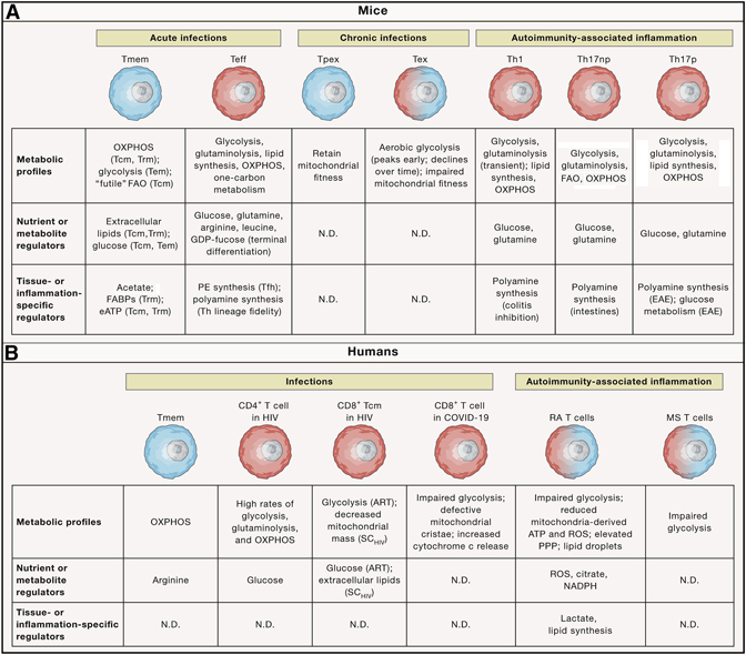
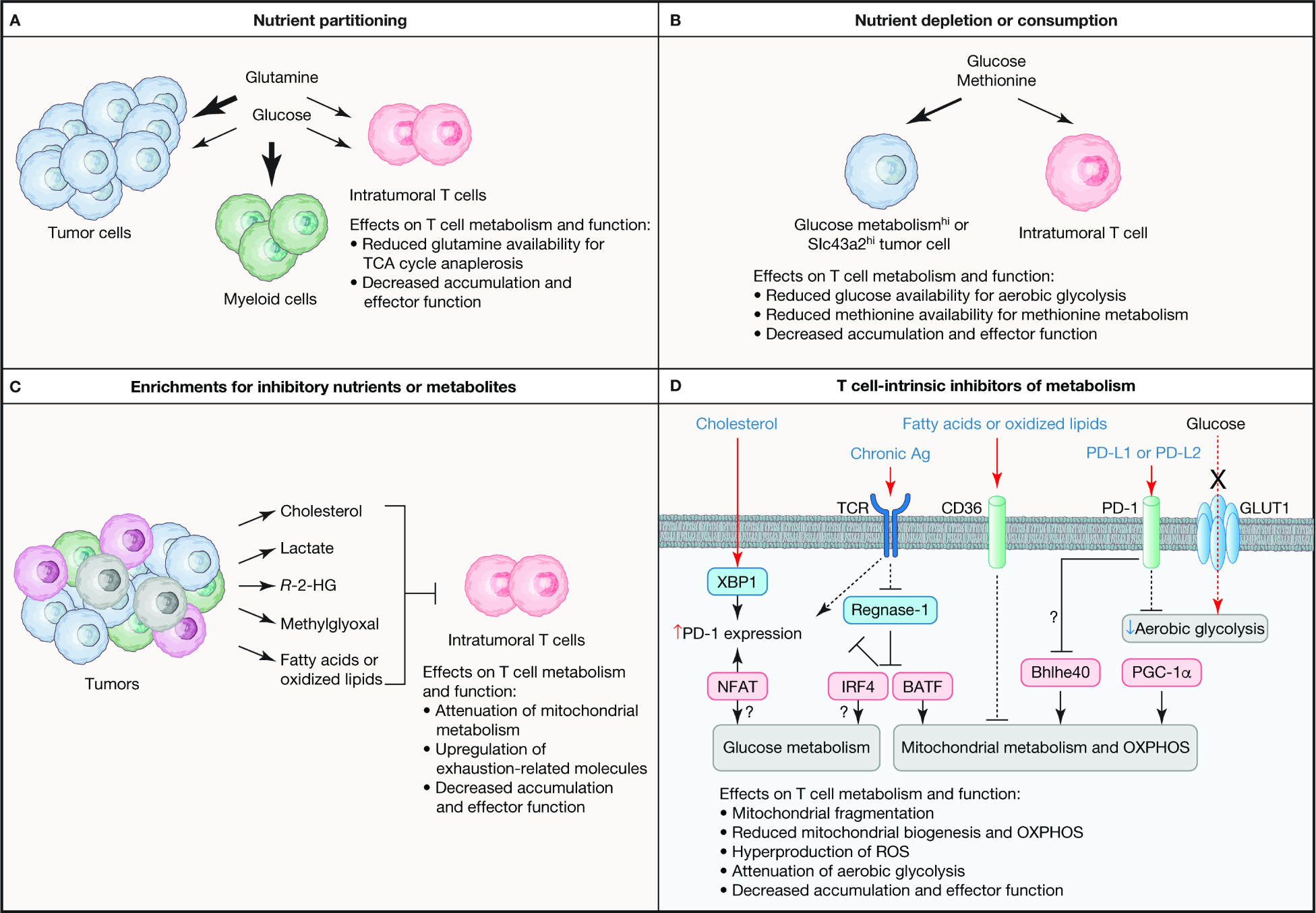
Adaptive immune responses mediated by T cells and B cells are crucial for protective immunity against pathogens and tumors. Differentiation and function of immune cells require dynamic reprogramming of cellular metabolism. Metabolic inputs, pathways, and enzymes display remarkable flexibility and heterogeneity, especially in vivo. How metabolic plasticity and adaptation dictate functional specialization of immune cells is fundamental to our understanding and therapeutic modulation of the immune system. Extensive progress has been made in characterizing the effects of metabolic networks on immune cell fate and function in discrete microenvironments or immunological contexts. In this review, we summarize how rewiring of cellular metabolism determines the outcome of adaptive immunity in vivo, with a focus on how metabolites, nutrients, and driver genes in immunometabolism instruct cellular programming and immune responses during infection, inflammation, and cancer in mice and humans. Understanding context-dependent metabolic remodeling will manifest legitimate opportunities for therapeutic intervention of human disease.


Conflict of interest statement

Declaration of interests H.C. is a consultant for Kumquat Biosciences.

Figures

Figure 1.
Figure 1.. Signaling networks orchestrate metabolism for coordination of T cell fate
(A) Signaling networks regulate metabolic state to tune T cell fate decisions. Mechanistic target of rapamycin (mTOR) complexes 1 and 2 (mTORC1 and mTORC2, respectively) orchestrate signaling to induce anabolic metabolism in T cells. Mechanistically, mTORC1 activity regulates these programs, in part, by upregulating transcriptional programs mediated by MYC, sterol regulatory binding element proteins (SREBPs), and hypoxia inducible factor-1α (HIF-1α). Additional transcriptional networks regulated by NFAT, BATF, and IRF4 are also crucial to support certain anabolic processes. In contrast, AMP-dependent protein kinase (AMPK) activation promotes fatty acid oxidation (FAO), a catabolic program. The metabolic programs are shown in grey boxes and their impacts on the activation, proliferation, and differentiation of conventional CD4+ and CD8+ T cells are shown. (B) The interplay between the major metabolic pathways involved in T cell fate decisions are depicted. Key selective nutrients are shown in red. Note that serine can be de novo synthesized or acquired from extracellular sources. PPP, pentose phosphate pathway; Tcm, central memory T cell; Tem, effector memory T cell; Th, CD4+ T helper; Trm, tissue-resident T cell.
Figure 2.
Figure 2.. Metabolic reprogramming in response to infection or autoimmune inflammation
Metabolic adaptation occurs in acute or chronic infection and autoimmunity-associated inflammation. A summary of the metabolic profiles, nutrient or metabolic regulators, and tissue- or inflammation-specific regulators that orchestrate metabolic reprogramming in the representative murine (upper) and human (lower) T cell populations. Small, blue cells display more catabolic metabolism; large, red cells are anabolic; and intermediate-sized cells with blue–red gradient coloring represent cells that display some features of anabolism and catabolism. Labels in parentheses indicate the specific contexts (e.g. cell subset or disease) in which the metabolic pathways or regulators have been shown to be functionally important. ART, antiretroviral therapy; eATP, extracellular adenosine triphosphate; FAO, fatty acid oxidation; HIV, human immunodeficiency virus; MS, multiple sclerosis, N.D., not determined; OXPHOS, oxidative phosphorylation; PE, phosphatidylethanolamine; PPP, pentose phosphate pathway; RA, rheumatoid arthritis; ROS, reactive oxygen species; SCHIV, spontaneous HIV controller; Tcm, central memory T cell; Teff, effector T cell; Tem, effector memory T cell; Tex, terminally exhausted T cell; Th, CD4+ T helper; Th17np, non-pathogenic Th17; Th17p, pathogenic Th17; Tmem, memory T cell; Tpex progenitor of exhausted T cell; Trm, tissue-resident T cell.
Figure 3.
Figure 3.. Mechanisms for inhibition of T cell metabolism in the TME
Intratumoral T cells show defects in metabolic programs that are associated with their dysfunction, with both cell-extrinsic and -intrinsic mechanisms orchestrating metabolic adaptation in the TME. (A) Nutrient partitioning in the TME contributes to dysfunctional T cell responses. Specifically, glucose is preferentially consumed by tumor-infiltrated myeloid cells, which creates competition between tumor cells and intratumoral T cells for glutamine. In instances where tumor cells have elevated glutamine consumption as compared with T cells, glutamine-dependent regeneration of TCA cycle metabolites is impaired, leading to reduced T cell accumulation and function in the TME. (B) Certain tumor cells have higher rate of aerobic glycolysis (indicated as Glucose metabolismhi) or increased expression of the methionine transporter Slc43a2 (denoted as Slc43a2hi) than intratumoral T cells, resulting in depletion or consumption of these nutrients. Restriction of glucose or methionine reduces glucose and mitochondrial metabolism in intratumoral T cells, thereby limiting the accumulation and function of these cells. (C) The tumor is a source for nutrients and metabolites that restrain intratumoral T cell function, such as by suppressing mitochondrial metabolism or increasing the expression of exhaustion-related proteins. See the main text for more details. (D) T cell-intrinsic regulators also reprogram metabolism in the TME to impede T cell function. Mitochondrial metabolism and OXPHOS are antagonized by chronic antigen (Ag) stimulation of the T cell receptor (TCR) or extracellular fatty acids or oxidized lipids that are transported into cells via CD36. TCR signals induce glucose and mitochondrial metabolism, in part, by inactivating Regnase-1 that acts to restrain IRF4 and BATF activity, which suppresses PD-1 expression. However, chronic antigen stimulation can also promote upregulation of PD-1 via NFAT. PD-1–PD-L1 or PD-L2 signaling may inactivate the transcription factor Bhlhe40, which is important to upregulate mitochondrial metabolism in certain contexts, with PD-1 signaling also being further amplified by the cholesterol-dependent activation of XBP1 that can induce PD-1 expression. Intratumoral T cells also have low expression of the transcription factor PGC-1α that is important for mitochondrial biogenesis. Moreover, glucose metabolism is often inhibited in intratumoral T cells, which may be mediated by PD-1 signaling, as well as intrinsic defects in glucose metabolism, such as reduced expression of GLUT1 or glycolytic enzymes. R-2-HG, R-2-hydroglutarate.

References

    1. Angin M, Volant S, Passaes C, Lecuroux C, Monceaux V, Dillies MA, Valle-Casuso JC, Pancino G, Vaslin B, Le Grand R, et al. (2019). Metabolic plasticity of HIV-specific CD8(+) T cells is associated with enhanced antiviral potential and natural control of HIV-1 infection. Nat Metab 1, 704–716. - PubMed
    1. Araki K, Turner AP, Shaffer VO, Gangappa S, Keller SA, Bachmann MF, Larsen CP, and Ahmed R (2009). mTOR regulates memory CD8 T-cell differentiation. Nature 460, 108–112. - PMC - PubMed
    1. Araujo L, Khim P, Mkhikian H, Mortales CL, and Demetriou M (2017). Glycolysis and glutaminolysis cooperatively control T cell function by limiting metabolite supply to N-glycosylation. Elife 6, e21330. - PMC - PubMed
    1. Arthur L, Esaulova E, Mogilenko DA, Tsurinov P, Burdess S, Laha A, Presti R, Goetz B, Watson MA, Goss CW, et al. (2021). Cellular and plasma proteomic determinants of COVID-19 and non-COVID-19 pulmonary diseases relative to healthy aging. Nature Aging 1, 535–549. - PubMed
    1. Bachem A, Makhlouf C, Binger KJ, de Souza DP, Tull D, Hochheiser K, Whitney PG, Fernandez-Ruiz D, Dahling S, Kastenmuller W, et al. (2019). Microbiota-Derived Short-Chain Fatty Acids Promote the Memory Potential of Antigen-Activated CD8(+) T Cells. Immunity 51, 285–297 e285. - PubMed

Publication types