Review
. 2022 Mar;33(3):472-486.
doi: 10.1681/ASN.2021101293. Epub 2022 Jan 12.

Mechanisms and Models of Kidney Tubular Necrosis and Nephron Loss

Affiliations
Review

Mechanisms and Models of Kidney Tubular Necrosis and Nephron Loss

Francesca Maremonti et al. J Am Soc Nephrol. 2022 Mar.

Abstract

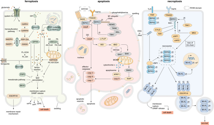
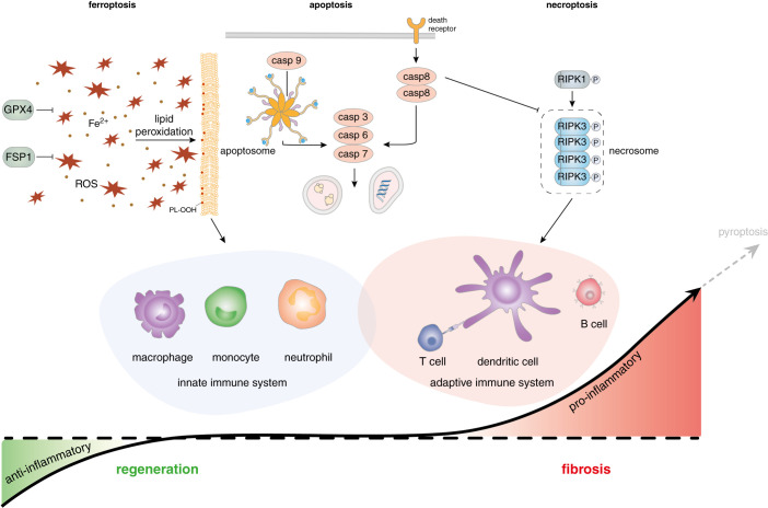
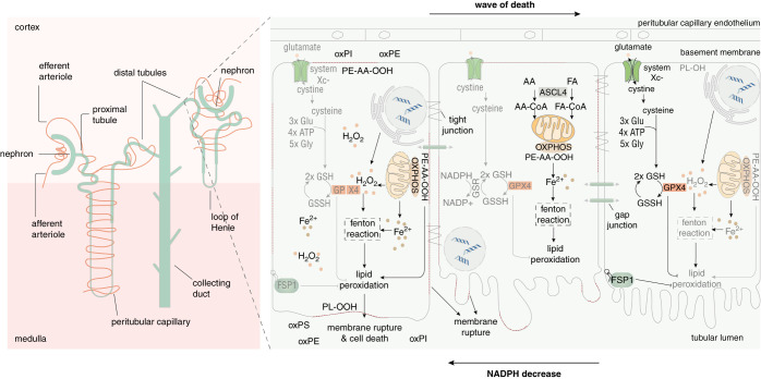
Understanding nephron loss is a primary strategy for preventing CKD progression. Death of renal tubular cells may occur by apoptosis during developmental and regenerative processes. However, during AKI, the transition of AKI to CKD, sepsis-associated AKI, and kidney transplantation ferroptosis and necroptosis, two pathways associated with the loss of plasma membrane integrity, kill renal cells. This necrotic type of cell death is associated with an inflammatory response, which is referred to as necroinflammation. Importantly, the necroinflammatory response to cells that die by necroptosis may be fundamentally different from the tissue response to ferroptosis. Although mechanisms of ferroptosis and necroptosis have recently been investigated in detail, the cell death propagation during tubular necrosis, although described morphologically, remains incompletely understood. Here, we argue that a molecular switch downstream of tubular necrosis determines nephron regeneration versus nephron loss. Unraveling the details of this "switch" must include the inflammatory response to tubular necrosis and regenerative signals potentially controlled by inflammatory cells, including the stimulation of myofibroblasts as the origin of fibrosis. Understanding in detail the molecular switch and the inflammatory responses to tubular necrosis can inform the discussion of therapeutic options.

Keywords: acute kidney injury; acute tubular necrosis; cell death; ferroptosis; necroinflammation; necroptosis; nephron loss.


Figures

Figure 1.
Figure 1.
Pathways of renal cell death. Ferroptosis (left) is a failsafe rather than a typical cell death pathway. In cellular homeostasis, H2O2 concentrations and iron-catalyzed Fenton reactions are limited by diverse cellular antiredox systems. The best studied system relies on GSH, which is generated intracellularly and depends on supply via system Xc-, a cys/glu antiporter in the plasma membrane, or products of the trans-sulfuration pathway. On sufficient GSH concentrations, GPX4 prevents lipid peroxidation that otherwise leads to plasma membrane rupture by unknown mechanisms. In contrast, the oxidoreductase FSP1 (also known as AIFM2) prevents lipid peroxidation on myristoylation-dependent recruitment to the plasma membrane in a GSH-independent manner. Ferroptosis occurs as a noncell-autonomous pathway in a process referred to as synchronized regulated necrosis (SRN). Apoptosis (center) represents a noninflammatory pathway that is mediated by caspases. Two distinct signaling pathways of apoptosis, extrinsic and intrinsic apoptosis, have been characterized. In extrinsic apoptosis, death receptors such as TNFR1, CD95 (Fas), and TRAIL-R, through the engagement of various intracellular adaptor proteins (Fas-associated protein with death domain, TRADD) lead to the activation of the master regulator caspase-8. On homodimerization, caspase-8 cleaves the effector caspases-3/-6/-7 to propagate the apoptosis program. On loss of mitochondrial outer membrane potential (MOMP) and the BAX-BAK–mediated release of cytochrome c from the mitochondria into the cytosol, the intrinsic apoptotic pathway is triggered. Within the cytosol, cytochrome c, APAF1 and caspase-9 form the apoptosome, which activates the effector caspases-3/-6/-7. The typical morphology includes nuclear condensation, early loss of cellular volume (shrinking), subsequent membrane blebbing, and exposure of PtdSer. Importantly, PtdSer exposure represents an eat-me signal to macrophages that eliminate apoptotic cells. Importantly, the plasma membrane does not lose its integrity during this process. Whereas apoptosis depends on the activation of caspases, necroptosis (right) is mediated by kinases. Depending on its RHIM domain, RIPK3 forms an amyloid-like structure referred to as the necrosome, the central signaling platform of necroptosis. Therein, RIPK3 phosphorylates the pseudokinase MLKL. By unknown mechanisms, pMLKL triggers a plasma membrane rupture, a process that was demonstrated to be counteracted by the membrane repair ESCRT-III complex. The necrosome can be engaged by death receptor signaling in the condition in which caspases are absent or inhibited (e.g., by viral proteins), and RIPK1 no longer intercalates with RIPK3. Other ways of engaging the necrosome are through TLRs via the RHIM-containing adaptor molecule TRIF, or by activation of the protein ZBP1/DAI in response to sensing intracellular oligonucleotides.
Figure 2.
Figure 2.
A hypothetical model of the interplay between the immune system and regulated cell death pathways. Although under fundamental debate, ferroptosis may create an anti-inflammatory environment, at least for the adaptive immune system. Although T cell activation, T cell crosspriming, and B cell activation may be inhibited by ferroptosis, cells of the innate immune system such as macrophages and neutrophils may better resist the environmental scenario of high lipid peroxidation and redox imbalance to remove necrotic debris, a feature that potentially allows necrotic kidney tubules to regenerate. In such a model, profibrotic conditions would be antagonized by the innate immune system through the removal of necrotic debris. In contrast with all other pathways of regulated cell death, classic apoptosis does not result in plasma membrane rupture and therefore does not expose intracellular epitopes to the immune system. PtdSer exposure on the surface of apoptotic cells functions as an eat-me signal and the immunologically silent removal of PtdSer-exposing cells. Necroptosis is interpreted as a defense mechanism against virus-carrying cells. During necroptosis (and most likely pyroptosis), the initiation of DC activation and T cell crosspriming alongside with the maturation of cytokines establishes a solid adaptive immune response and a vaccination against viral epitopes. Maladaptive repair and the activation of myofibroblasts in the kidney are likely a consequence of adaptive immune cell activation and nonferroptotic, nonapoptotic regulated cell death. We clearly point out that the presented model is speculative and serves as a working model that needs to be tested in the future.
Figure 3.
Figure 3.
SRN: cell-death propagation during renal tubule ferroptosis. In most cases, it is unclear how a necrotic zone of dead cells propagates during an event of sepsis or ischemia. On the basis of time-lapse videos obtained from isolated perfused renal tubules that underwent ferroptosis, the concept of SRN emerged. Although this phenomenon has not been observed in humans, necrotic tubules in the urine of patients and experimental intravital microscopy videos suggest cell death occurs in a “wave of death.”,, Given the connections between cells in a functional syncytium such as a renal tubule, the adrenal gland, or the myocardium, it is tempting to speculate that the intracellular redox capacity (NADPH concentration) diffuses through intercellular junctions. If the redox capacity decreases in a dying cell, an NADPH gradient forms, which renders the closest neighbor at high risk of undergoing ferroptosis. It is now clear the bulk necrotic area that occurs during myocardial infarction or tubular necrosis originates from cells that underwent ferroptosis. We speculate that similar mechanisms might occur in other organs, such as the brain upon stroke, or the adrenal glands during sepsis or shock.

References

    1. Romagnani P, Remuzzi G, Glassock R, Levin A, Jager KJ, Tonelli M, et al. : Chronic kidney disease. Nat Rev Dis Primers 3: 17,088, 2017 - PubMed
    1. Ruiz-Ortega M, Rayego-Mateos S, Lamas S, Ortiz A, Rodrigues-Diez RR: Targeting the progression of chronic kidney disease. Nat Rev Nephrol 16: 269–288, 2020 - PubMed
    1. Venkatachalam MA, Weinberg JM, Kriz W, Bidani AK: Failed tubule recovery, AKI-CKD transition, and kidney disease progression. J Am Soc Nephrol 26: 1765–1776, 2015 - PMC - PubMed
    1. Glassock RJ, Rule AD: Aging and the kidneys: Anatomy, physiology and consequences for defining chronic kidney disease. Nephron 134: 25–29, 2016 - PubMed
    1. Friedmann Angeli JP, Schneider M, Proneth B, Tyurina YY, Tyurin VA, Hammond VJ, et al. : Inactivation of the ferroptosis regulator Gpx4 triggers acute renal failure in mice. Nat Cell Biol 16: 1180–1191, 2014 - PMC - PubMed

Publication types