. 2022 Jan 12;8(1):9.
doi: 10.1038/s41531-021-00263-x.

Enteric α-synuclein impairs intestinal epithelial barrier through caspase-1-inflammasome signaling in Parkinson's disease before brain pathology

Affiliations

Enteric α-synuclein impairs intestinal epithelial barrier through caspase-1-inflammasome signaling in Parkinson's disease before brain pathology

C Pellegrini et al. NPJ Parkinsons Dis. .

Erratum in

Abstract

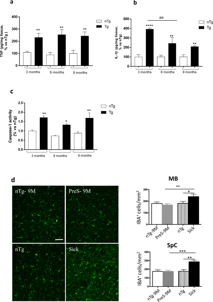
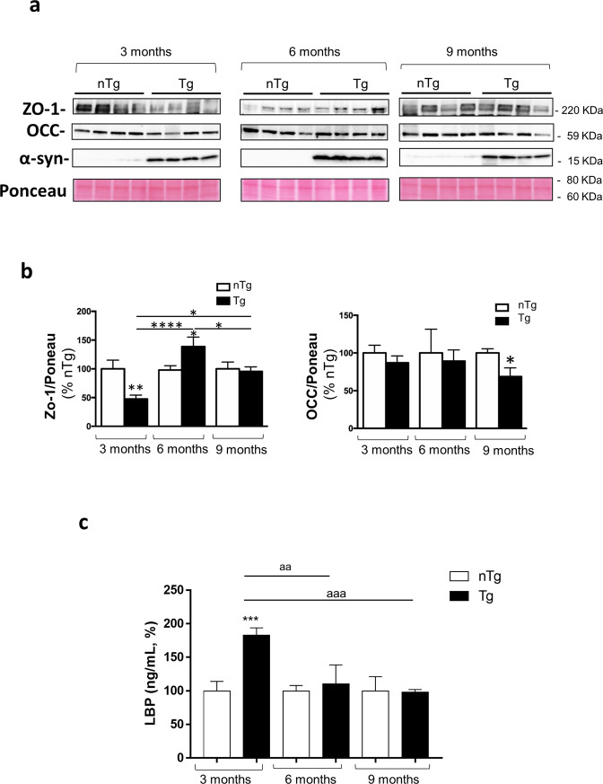
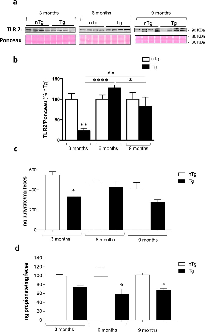
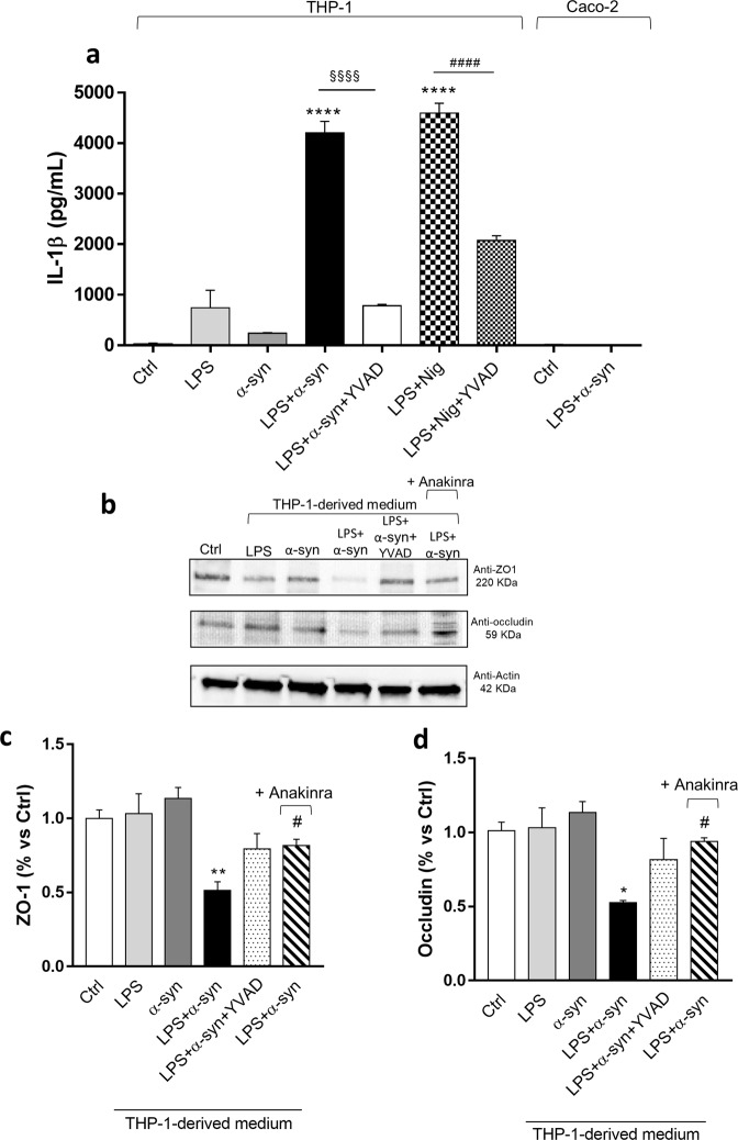
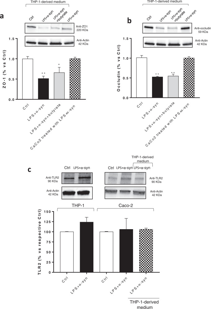
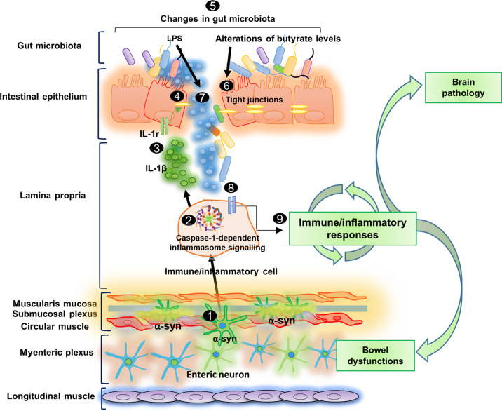
Bowel inflammation, impaired intestinal epithelial barrier (IEB), and gut dysbiosis could represent early events in Parkinson's disease (PD). This study examined, in a descriptive manner, the correlation among enteric α-synuclein, bowel inflammation, impairments of IEB and alterations of enteric bacteria in a transgenic (Tg) model of PD before brain pathology. Human A53T α-synuclein Tg mice were sacrificed at 3, 6, and 9 months of age to evaluate concomitance of enteric inflammation, IEB impairments, and enteric bacterial metabolite alterations during the early phases of α-synucleinopathy. The molecular mechanisms underlying the interplay between α-synuclein, activation of immune/inflammatory responses and IEB alterations were investigated with in vitro experiments in cell cultures. Tg mice displayed an increase in colonic levels of IL-1β, TNF, caspase-1 activity and enteric glia activation since 3 months of age. Colonic TLR-2 and zonulin-1 expression were altered in Tg mice as compared with controls. Lipopolysaccharide levels were increased in Tg animals at 3 months, while fecal butyrate and propionate levels were decreased. Co-treatment with lipopolysaccharide and α-synuclein promoted IL-1β release in the supernatant of THP-1 cells. When applied to Caco-2 cells, the THP-1-derived supernatant decreased zonulin-1 and occludin expression. Such an effect was abrogated when THP-1 cells were incubated with YVAD (caspase-1 inhibitor) or when Caco-2 were incubated with anakinra, while butyrate incubation did not prevent such decrease. Taken together, early enteric α-synuclein accumulation contributes to compromise IEB through the direct activation of canonical caspase-1-dependent inflammasome signaling. These changes could contribute both to bowel symptoms as well as central pathology.


Conflict of interest statement

The authors declare no competing interests.

Figures

Fig. 1
Fig. 1. Enteric inflammatory responses in pre-symptomatic Tg mice precede brain inflammation.
a TNF levels in colonic tissues from nTg and Tg mice at 3, 6, and 9 months of age. Values represent the means ± S.E.M. One-way ANOVA followed by Tukey post hoc test results: **P < 0.01, significant difference versus nTg age-matched controls; b IL-1β levels in colonic tissues from nTg and Tg mice at 3, 6, and 9 months of age. Values represent the means ± SEM. One-way ANOVA followed by Tukey post hoc test results: **P < 0.01, ****P < 0.001 significant difference versus nTg age-matched controls, and aaP < 0.01 significant difference; c Caspase-1 activity in colonic tissues from nTg and Tg mice at 3, 6, and 9 months of age. Values represent the means ± SEM. One-way ANOVA followed by Tukey post hoc test results: *P < 0.05 and **P < 0.01, significant difference versus nTg age-matched controls); d Confocal analysis of IBA-1 positive cells in MB and SpC of presymptomatic (9 months), sick and nTg mice. Images were acquired using a 40× objective of Leica TCS SP confocal laser-scanning microscope and cell density analysis was obtained using ImageJ. Values represent the means ± SEM. One-way ANOVA followed by Fisher post hoc test results: *P < 0.05; **P < 0.01; ***P < 0.001 significant difference. Scale bar, 100 nm. n = 3–5/group. IL-1β interleukin-1beta, MB midbrain, SpC spinal cord, nTg non-transgenic, Tg transgenic, TNF tumor necrosis factor.
Fig. 2
Fig. 2. Pre-symptomatic Tg mice display impairments of IEB integrity and permeability.
a Representative blots and (b) densitometric analysis of ZO-1 and Occ expression assessed by Western blot assay in colonic tissues from nTg and Tg mice at 3, 6, and 9 months of age. In the quantitative analysis protein levels were normalized using Ponceau staining and expressed as percentage of the mean of nTg age-matched controls. All values represent the means ± SEM. *P < 0.05, **P < 0.01, ****P < 0.0001, significant difference. One-way ANOVA followed by Tukey post hoc test. c Circulating LBP in nTg and Tg mice at 3, 6, and 9 months of age. All values represent the means ± SEM. ***P < 0.001, significant difference versus nTg age-matched controls), aaaP < 0.001 and aaP < 0.01. One-way ANOVA followed by Tukey post hoc test. n = 3–5/group. α-S α-synuclein, LBP lipopolysaccharide-binding protein, nTg non-transgenic, Tg transgenic, Occ occludin, ZO-1 zonulin-1.
Fig. 3
Fig. 3. Pre-symptomatic Tg mice display an altered TLR-2 expression in colonic tissues and a decrease in fecal butyrate and propionate levels since 3 months of age.
a Representative blots and (b) densitometric analysis of TLR-2 expression assessed by Western blot assay in colonic tissues from nTg and Tg mice at 3, 6, and 9 months of age. In the quantitative analysis protein levels were normalized using Ponceau staining and expressed as percentage of the mean of nTg age-matched controls. All values represent the mean ± SEM. *P < 0.05, **P < 0.01, ****P < 0.0001, significant difference. One-way ANOVA followed by Tukey post hoc test. c Butyrate and (d) propionate levels in the feces from nTg and Tg mice at 3, 6, and 9 months of age. All values represent the means ± SEM. Two-way ANOVA followed by Tukey post hoc test results: *P < 0.05, significant difference versus nTg age-matched controls). n = 3–5/group. nTg non-transgenic, SCFAs fermentation products of enteric bacteria, Tg transgenic, TLR-2 toll like receptor-2.
Fig. 4
Fig. 4. Enteric accumulation of α-syn triggers the release of IL-1β from activated macrophage, which, in turn, induces an increase in intestinal epithelial tight junction permeability.
a IL-1β levels in the supernatants of THP-1 cells treated with LPS alone or in combination with α-syn or Nig, in the presence or in the absence of YVAD. IL-1β levels in the supernatants of Caco-2 cells untreated or treated with LPS plus α-syn. All values represent the mean ± SEM. One-way ANOVA followed by Tukey post hoc test results: ****P < 0.0001 significant difference versus LPS-primed THP-1 (Ctrl); §§§§P < 0.0001, significant difference versus LPS-primed THP-1 treated with α-syn plus LPS; ####P < 0.0001, significant difference versus LPS-primed THP-1 treated with LPS plus Nig; b Representative blots and densitometric analysis of c ZO-1 and (d) occludin expression assessed by Western blot assay in cultured Caco-2 cells treated with conditioned medium derived from THP-1 cells. All values represent the mean ± SEM. One-way ANOVA followed by Tukey post hoc test results: *P < 0.05, **P < 0.01 significant difference versus Caco-2 cells (Ctrl); #P < 0.05 significant difference versus Caco-2 cells treated with α-syn plus LPS. n = 5 independent experiments. α-syn α-synuclein, IL-1β interleukin-1beta, LPS lipopolysaccharide, Nig nigericin, ZO-1 zonulin-1.
Fig. 5
Fig. 5. Incubation of α-syn plus LPS directly in the medium of Caco-2 cells does not alter the expression ZO-1 and occludin.
a, b Representative blots and densitometric analysis of ZO-1 and occludin expression assessed by Western blot assay in cultured Caco-2 cells treated with conditioned medium derived from THP-1 cells treated with α-syn plus LPS, in the absence or in the presence of butyrate, or with α-syn plus LPS directly in the medium. All values represent the mean± SEM. One-way ANOVA followed by Tukey’s post hoc test results: *P < 0.05, **P < 0.01 significant difference versus Caco-2 cells (Ctrl). c Representative blots and densitometric analysis of TLR-2 expression assessed by Western blot assay in cultured THP-1 and Caco-2 cells treated with LPS and α-syn and Caco-2 incubated with conditioned medium derived from THP-1 cells treated with α-syn and LPS. All values represent the mean ± SEM. Student’s t test and one-way ANOVA followed by Tukey’s post hoc test results: no significant difference versus respective Ctrl. n = 5 independent experiments. α-syn α-synuclein, LPS lipopolysaccharide, TLR-2 toll like receptor-2, ZO-1 zonulin-1.
Fig. 6
Fig. 6. Schematic representation of pathophysiological intestinal paths in presymptomatic A53T PD mice.
In the very early stages of PD before CNS pathology, the enteric α-syn accumulation 1 can promote the activation of immune/inflammatory signaling, including canonical caspase-1-dependent inflammasome pathways 2, with consequent massive release of IL-1β 3, which, in turn, impairs IEB 4, through the activation of IL-1 receptors on intestinal epithelial cells. In this setting, bowel inflammation and the impaired IEB can induce changes in SCFA levels 5, characterized by alterations of butyrate levels, that could contribute to IEB impairment 6, and, an increase in LPS concentration, which, translocating into the intestinal mucosa 7 could further contribute to the activation of immune/inflammatory pathways 8, thus generating a vicious circle that might lead to the chronicization of inflammatory processes and contribute both to intestinal symptoms and brain pathology 9.

References

    1. Fasano A, Visanji NP, Liu LW, Lang AE, Pfeiffer RF. Gastrointestinal dysfunction in Parkinson’s disease. Lancet Neurol. 2015;14:625–639. doi: 10.1016/S1474-4422(15)00007-1. - DOI - PubMed
    1. Pellegrini C, et al. Intestinal dysfunction in Parkinson’s disease: lessons learned from translational studies and experimental models. Neurogastroenterol. Motil.: Off. J. Eur. Gastrointest. Motil. Soc. 2016;28:1781–1791. doi: 10.1111/nmo.12933. - DOI - PubMed
    1. Pellegrini C, et al. Gastric motor dysfunctions in Parkinson’s disease: current pre-clinical evidence. Parkinsonism Relat. Disord. 2015;21:1407–1414. doi: 10.1016/j.parkreldis.2015.10.011. - DOI - PubMed
    1. Pellegrini C, Antonioli L, Colucci R, Blandizzi C, Fornai M. Interplay among gut microbiota, intestinal mucosal barrier and enteric neuro-immune system: a common path to neurodegenerative diseases? Acta Neuropathologica. 2018;136:345–361. doi: 10.1007/s00401-018-1856-5. - DOI - PubMed
    1. Keshavarzian A, et al. Colonic bacterial composition in Parkinson’s disease. Mov. Disord.: Off. J. Mov. Disord. Soc. 2015;30:1351–1360. doi: 10.1002/mds.26307. - DOI - PubMed