

Feasibility and clinical utility of handheld fundus cameras for retinal imaging
- PMID: 35022568
- PMCID: PMC9873676
- DOI: 10.1038/s41433-021-01926-y
Feasibility and clinical utility of handheld fundus cameras for retinal imaging
Erratum in
-
Correction: Feasibility and clinical utility of handheld fundus cameras for retinal imaging.Eye (Lond). 2023 Feb;37(2):380-381. doi: 10.1038/s41433-022-02041-2. Eye (Lond). 2023. PMID: 35614344 Free PMC article. No abstract available.
Abstract
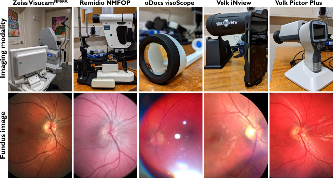
Background/objectives: Handheld fundus cameras are portable and cheaper alternatives to table-top counterparts. To date there have been no studies comparing feasibility and clinical utility of handheld fundus cameras to table-top devices. We compare the feasibility and clinical utility of four handheld fundus cameras/retinal imaging devices (Remidio NMFOP, Volk Pictor Plus, Volk iNview, oDocs visoScope) to a table-top camera (Zeiss VisucamNM/FA).
Subjects/methods: Healthy participants (n = 10, mean age ± SD = 21.0 ± 0.9 years) underwent fundus photography with five devices to assess success/failure rates of image acquisition. Participants with optic disc abnormalities (n = 8, mean age ± SD = 26.8 ± 15.9) and macular abnormalities (n = 10, mean age ± SD = 71.6 ± 15.4) underwent imaging with the top three scoring fundus cameras. Images were randomised and subsequently validated by ophthalmologists masked to the diagnoses and devices used.
Results: Image acquisition success rates (100%) were achieved in non-mydriatic and mydriatic settings for Zeiss, Remidio and Pictor, compared with lower success rates for iNview and oDocs. Image quality and gradeability were significantly higher for Zeiss, Remidio and Pictor (p < 0.0001) compared to iNview and oDocs. For cup:disc ratio estimates, similar levels of bias were seen for Zeiss (-0.09 ± SD:0.15), Remidio (-0.07 ± SD:0.14) and Pictor (-0.05 ± SD:0.16). Diagnostic sensitivities were highest for Zeiss (84.9%; 95% CI, 78.2-91.5%) followed by Pictor (78.1%; 95% CI, 66.6-89.5%) and Remidio (77.5%; 95% CI, 65.9-89.0%).
Conclusions: Remidio and Pictor achieve comparable results to the Zeiss table-top camera. Both devices achieved similar scores in feasibility, image quality, image gradeability and diagnostic sensitivity. This suggests that these devices potentially offer a more cost-effective alternative in certain clinical scenarios.
© 2022. The Author(s).
Conflict of interest statement
The authors declare no competing interests.
Figures


References
-
- Li B, Chen H, Zhang B, Yuan M, Jin X, Lei B, et al. Development and evaluation of a deep learning model for the detection of multiple fundus diseases based on colour fundus photography. Br J Ophthalmol. 2021. 10.1136/bjophthalmol-2020-316290. - PubMed
-
- Milea D, Najjar RP, Jiang Z, Ting D, Vasseneix C, Xu X, et al. Artificial intelligence to detect papilledema from ocular fundus photographs. N. Engl J Med. 2020;382:1687–95. 10.1056/NEJMoa1917130. - PubMed
MeSH terms
Grants and funding
LinkOut - more resources
Full Text Sources
