Review
. 2022 Mar;47(3):1032-1052.
doi: 10.1007/s00261-021-03396-y. Epub 2022 Jan 12.

Liver fibrosis quantification

Affiliations
Review

Liver fibrosis quantification

Sudhakar K Venkatesh et al. Abdom Radiol (NY). 2022 Mar.

Abstract

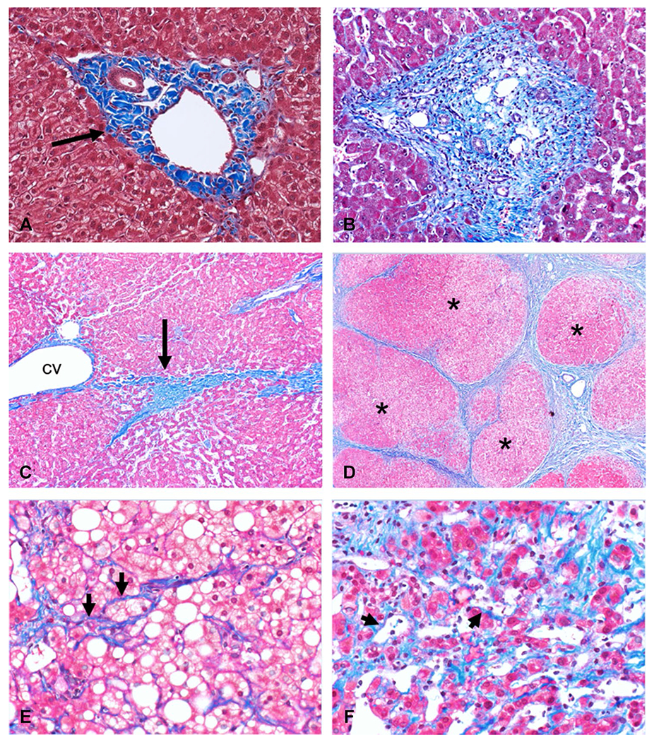
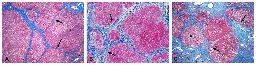
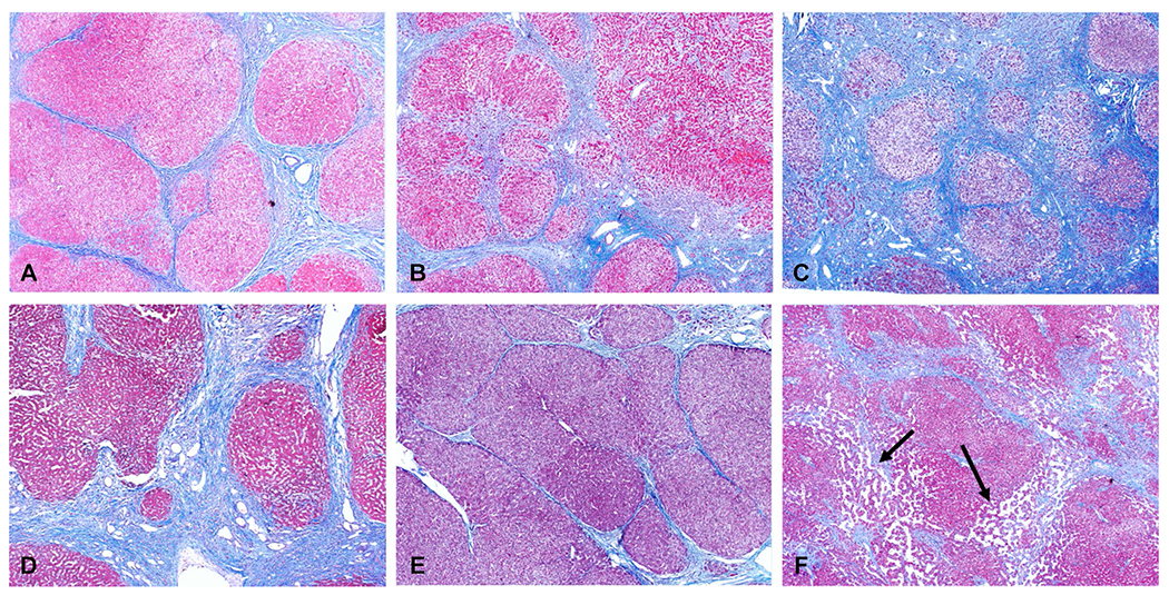
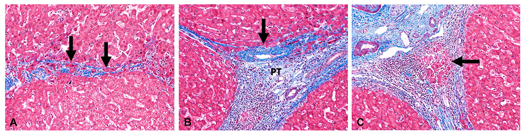
Liver fibrosis (LF) is the wound healing response to chronic liver injury. LF is the endpoint of chronic liver disease (CLD) regardless of etiology and the single most important determinant of long-term liver-related clinical outcomes. Quantification of LF is important for staging, to evaluate response to treatment and to predict outcomes. LF is traditionally staged by liver biopsy. However, liver biopsy is invasive and suffers from sampling errors when biopsy size is inadequate; therefore, non-invasive tests (NITs) have found important roles in clinical care. NITs include simple laboratory-based serum tests, panels of serum tests, and imaging biomarkers. NITs are validated against the liver biopsy and will be used in the future for evaluation of nearly all CLDs with invasive liver biopsy reserved for some cases. Both serum tests and some imaging biomarkers such as elastography are currently used clinically as surrogate markers for LF. Several other imaging biomarkers are still considered research and awaiting clinical application in the future. As the evaluation of imaging biomarkers will likely become the norm in the future, understanding pathogenesis of LF is important. Knowledge of properties measured by imaging biomarkers and its correlation with LF is important to understand the application of NITs by abdominal radiologists. In this review, we present a brief overview of pathogenesis of LF, spatiotemporal evolution of LF in different CLD, and severity assessment with liver biopsy. This will be followed by a brief discussion on properties measured by imaging biomarkers and their relationship to the LF.

Keywords: Diffusion; Elastography; Fibrosis burden; Hepatic fibrosis; Hepatobiliary uptake; Imaging biomarkers; Non-invasive tests; Surface nodularity; Susceptibility; T1-mapping; Volumetry.


Conflict of interest statement

Conflict of interest Financial interests: The author declares no financial interests. Non-financial interests: The author declares no non-financial interests.

Figures

Fig. 1
Fig. 1
Histological images (Masson-Trichrome stain) showing a normal portal tract with normal amount of collagen (black arrow, A) within the portal tract. In portal fibrosis or periportal fibrosis (B) there is increased collagen deposition that expands the portal tract. The next stage of bridging fibrosis (C) is characterized by fibrotic bands (black arrow, C) that connect portal tract to portal tract or portal tract to central vein (cv). At the cirrhosis stage (D), fibrosis bands surround regenerative nodules (asterisk). Pericellular fibrosis where collagen fibrils surround hepatocytes (arrowheads, E and F) is typically seen in steatohepatitis (E) and chronic venous outflow obstruction (F), respectively
Fig. 2
Fig. 2
Three examples of hepatitis C cirrhosis showing different degrees of fibrosis deposition. The regenerative nodules (asterisk) are larger in examples (A) and (B) compared to (C). The fibrous bands (black arrows) in (B) and (C) are thicker than that in (A)
Fig. 3
Fig. 3
Examples of cirrhosis from chronic hepatitis C (A), NASH-non-alcoholic steatohepatitis (B), ASH-alcoholic steatohepatitis (C), primary sclerosing cholangitis (D), auto immune hepatitis (E) and cardiac cirrhosis (F). Note the different degrees of fibrosis deposition. No hepatic steatosis is seen in both NASH and ASH examples which is common at this stage of chronic liver disease. Dilated sinusoidal spaces (arrows, F) are seen secondary to chronic venous outflow obstruction in this case of cardiac cirrhosis
Fig. 4
Fig. 4
Histologic changes associated with the regression of liver fibrosis. The specimens are Masson-Trichrome stained, with collagen fibers staining blue and the hepatocytes staining red. (A) Resorbing bridging fibrosis (arrows). (B) Adhered central vein (arrow) to the portal tract (PT). (C) Regression of cirrhosis with an early bud of hepatocytes (arrow) in a fibrous band
Fig. 5
Fig. 5
Contrast enhanced CT images from different patients illustrating the morphological changes in liver in chronic liver disease. Nodular outline of the liver (short arrows in A, B, and D), atrophic right lobe with enlarged caudate lobe (black arrow in B), enlarged left lobe (white arrow in B and C), increased abdominal fat or creeping fat sign (short black arrow, B), increased periportal space (double headed arrow, C), enlarged gall bladder fossa sign (white arrow, D), posterior hepatic notch sign (broken white arrow, B and E). Note splenomegaly (asterisk, B and D) consistent with portal hypertension. Compare with normal appearance of liver in a chronic hepatitis B patient (F) with biopsy proven stage 2 fibrosis. Morphological changes are usually seen in advanced fibrosis or cirrhosis and mostly absent in early fibrosis stages. GB = gall bladder
Fig. 6
Fig. 6
Examples of vibration controlled transient elastography (VCTE) (A, B), point shear wave elastography (pSWE) (C, D) and two-dimensional shear wave elastography (2D SWE) (E, F). A VCTE image in a 70-year-old woman with primary biliary cholangitis. The bulk modulus (E) is 5.1 kPa and within normal range. B VCTE image in a 49-year-old male with chronic hepatitis C with E value of 46.4 kPa consistent with cirrhosis. C pSWE image in a 31-year-old female with congestive hepatopathy with measured median velocity of 2.1 m/s corresponding to calculated value of 13.1 kPa consistent with advanced fibrosis and confirmed with biopsy. D pSWE image in a 36-year-old female with post Fontan congestive hepatopathy showing a median velocity of 4.25 m/s corresponding with 54 kPa consistent with biopsy confirmed cirrhosis. E 2D SWE image in a 50-year-old male with known hepatitis C. The median liver stiffness is 5.25 kPa which is within normal limits. F 2D SWE image in 64-year-old female with chronic liver disease of unknown etiology. The median stiffness is 16.7 kPa consistent with advanced fibrosis
Fig. 7
Fig. 7
Examples of magnetic resonance elastography (MRE). Axial T2W images (A, D, G), with corresponding level color wave images (D, E, H) and color stiffness maps with confidence map overlay (C, F, I). Regions of interest (solid lines) drawn within the confidence map which provides mean stiffness of liver parenchyma at that slice level. Top row: A 18-year-old male with primary sclerosing cholangitis (PSC) with mean liver stiffness of 1.8 kPa which is within normal limits and excluding significant fibrosis. Middle row: A 62-year-old male with PSC and mean liver stiffness of 3.3 kPa suggestive of mild fibrosis. Bottom row: A 70-year-old female with primary biliary cholangitis and non-alcoholic steatohepatitis overlap with a mean liver stiffness of 7.7 kPa consistent with stage 4 fibrosis or cirrhosis
Fig. 8
Fig. 8
Examples of T1-maps of liver. A A 50-year-old female with obesity and mean liver T1 of 740 ms. Liver biopsy confirmed a normal liver. B A 65-year-old female with obesity and mean liver T1 of 901 ms. Liver biopsy showed grade 1 steatosis and fatty liver, mild lobular inflammation, ballooning of hepatocytes, and stage 4 fibrosis consistent with non-alcoholic steatohepatitis. (Image courtesy, Drs. Jiahui Li and Meng Yin, Radiology, Mayo clinic, Rochester, MN, USA)
Fig. 9
Fig. 9
Examples of liver surface nodularity (LSN) scores derived from routine abdominal CT images. (A, B wide FOV-top row images; small FOV-bottom images) A A 46-year-old male with abdominal pain undergoes abdominal CT. The liver has normal morphological appearance, and the LSN score was normal at 1.59. B A 55-year-old male with abdominal distension undergoes nonenhanced abdominal CT. The patient had a history of hepatitis C chronic liver disease. The liver has liver surface nodularity and the LSN score was abnormal at 2.98 indicating cirrhosis (Image courtesy: Dr. Andrew Smith, University of Alabama Medical Center, UABC, Birmingham, AL)

References

    1. Anthony PP, Ishak KG, Nayak NC, Poulsen HE, Scheuer PJ, Sobin LH (1977) The morphology of cirrhosis: definition, nomenclature, and classification. Bull World Health Organ 55 (4):521–540 - PMC - PubMed
    1. Hernandez-Gea V, Friedman SL (2011) Pathogenesis of liver fibrosis. Annu Rev Pathol 6:425–456. 10.1146/annurev-pathol-011110-130246 - DOI - PubMed
    1. Rosenberg WM (2003) Rating fibrosis progression in chronic liver diseases. J Hepatol 38 (3):357–360. 10.1016/s0168-8278(03)00010-2 - DOI - PubMed
    1. Angulo P, Kleiner DE, Dam-Larsen S, Adams LA, Bjornsson ES, Charatcharoenwitthaya P, Mills PR, Keach JC, Lafferty HD, Stahler A, Haflidadottir S, Bendtsen F (2015) Liver Fibrosis, but No Other Histologic Features, Is Associated With Long-term Outcomes of Patients With Nonalcoholic Fatty Liver Disease. Gastroenterology 149 (2):389–397.e310. 10.1053/j.gastro.2015.04.043 - DOI - PMC - PubMed
    1. Horowitz JM, Venkatesh SK, Ehman RL, Jhaveri K, Kamath P, Ohliger MA, Samir AE, Silva AC, Taouli B, Torbenson MS, Wells ML, Yeh B, Miller FH (2017) Evaluation of hepatic fibrosis: a review from the society of abdominal radiology disease focus panel. Abdom Radiol (NY) 42 (8):2037–2053. 10.1007/s00261-017-1211-7 - DOI - PMC - PubMed

Publication types