. 2021 Sep 8:34:1-12.
doi: 10.1016/j.jare.2021.09.002. eCollection 2021 Dec.

Oxyphylla A ameliorates cognitive deficits and alleviates neuropathology via the Akt-GSK3β and Nrf2-Keap1-HO-1 pathways in vitro and in vivo murine models of Alzheimer's disease

Affiliations

Oxyphylla A ameliorates cognitive deficits and alleviates neuropathology via the Akt-GSK3β and Nrf2-Keap1-HO-1 pathways in vitro and in vivo murine models of Alzheimer's disease

Yaqi Bian et al. J Adv Res. .

Abstract

Introduction: Alzheimer's disease (AD) is a progressive brain disorder, and one of the most common causes of dementia and amnesia. Due to the complex pathogenesis of AD, the underlying mechanisms remain unclear. Although scientists have made increasing efforts to develop drugs for AD, no effective therapeutic agents have been found.

Objectives: Natural products and their constituents have shown promise for treating neurodegenerative diseases, including AD. Thus, in-depth study of medical plants, and the main active ingredients thereof against AD, is necessary to devise therapeutic agents.

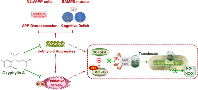
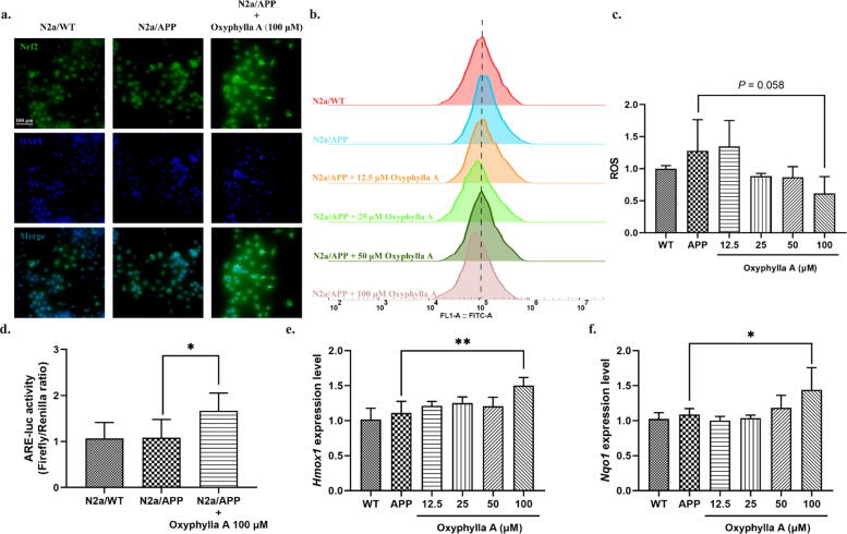
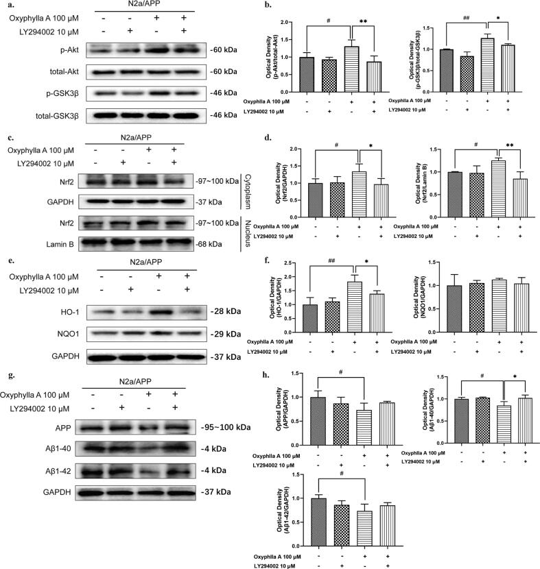
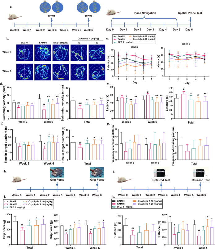
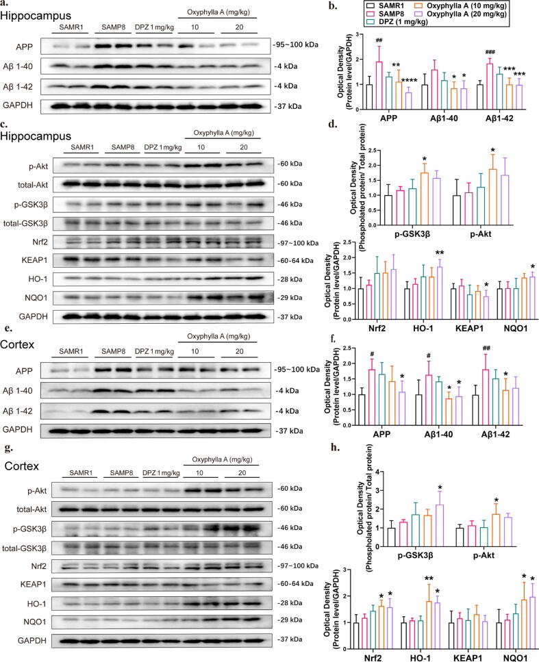
Methods: In this study, N2a/APP cells and SAMP8 mice were employed as in vitro and in vivo models of AD. Multiple molecular biological methods were used to investigate the potential therapeutic actions of oxyphylla A, and the underlying mechanisms.

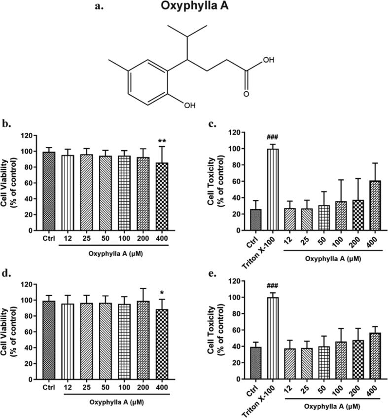
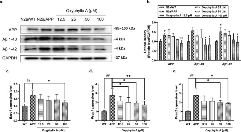
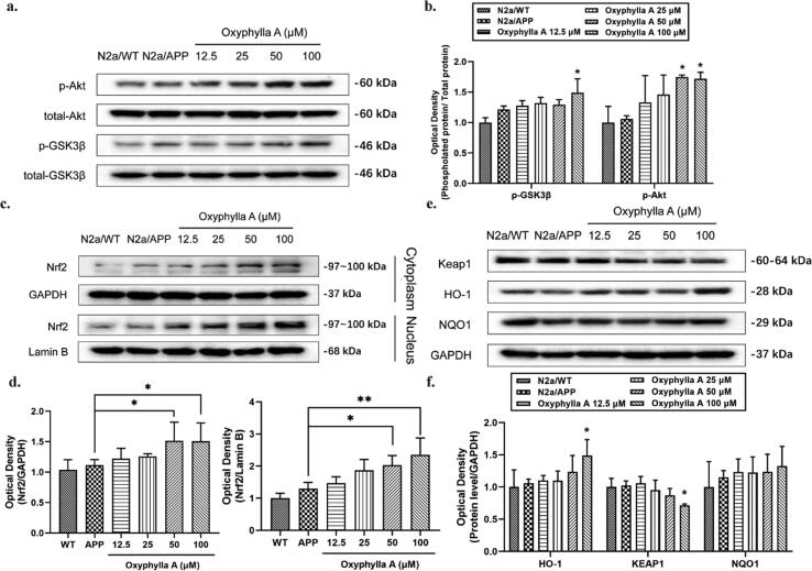
Results: Results showed that oxyphylla A, a novel compound extracted from Alpinia oxyphylla, could reduce the expression levels of amyloid precursor protein (APP) and amyloid beta (Aβ) proteins, and attenuate cognitive decline in SAMP8 mice. Further investigation of the underlying mechanisms showed that oxyphylla A exerted an antioxidative effect through the Akt-GSK3β and Nrf2-Keap1-HO-1 pathways.Conclusions.Taken together, our results suggest a new horizon for the discovery of therapeutic agents for AD.

Keywords: AD, Alzheimer’s disease; AOE, ethanolic extract of Alpinia oxyphylla; APP, amyloid precursor protein; ARE, antioxidant response element; ARE, antioxidant responsive element; Alzheimer’s disease; Amyloid beta proteins; Aβ, amyloid beta; GSK3, glycogen synthase kinase 3; HO-1, heme oxygenase-1; Keap1, Keleh-like ECH-associated protein; MWM, Morris Water Maze; NFTs, neurofibrillary tangles; NQO1, NAD(P)H:quinone oxidoreductase1; Nrf2, erythroid-derived 2-related factor 2; Oxidative stress; PD, Parkinson’s disease; PHF, paired helical filaments; RLU, relative luciferase units; ROS, reactive oxygen species; SAMP8; SAMP8 mice, senescence-accelerated mouse prone 8; oxyphylla A; pRL-TK, Renilla luciferase reporter plasmid.


Conflict of interest statement

The authors declare that they have no known competing financial interests or personal relationships that could have appeared to influence the work reported in this paper.

Figures

None
Graphical abstract
Fig. 1
Fig. 1
Cell viability and cell toxicity of oxyphylla A on N2a/WT and N2a/APP cells. Cells in different groups were incubated with various concentrations of oxyphylla A (12–400 μM) for 24 h. MTT and LDH were employed to assess cell viability and toxicity. (a) The chemical structure of oxyphylla A. (b) and (c) Statistical analysis of the cell viability and toxicity of oxyphylla A on N2a/WT cells. (d) and (e) Statistical analysis of the cell viability and toxicity of oxyphylla A on N2a/APP cells. The data are expressed as the mean ± SD of three independent experiments. *P < 0.05, **P < 0.01, compared to the control group; ###P < 0.001, compared to the Triton X-100 group.
Fig. 2
Fig. 2
Effects of oxyphylla A on the expression levels of APP, Aβ1-40 and Aβ1-42. (a) N2a/APP and N2a/WT cells were incubated with oxyphylla A (12.5–100 μM) and DMSO, respectively, for 24 h. Western blot was used to detect the expression levels of APP, Aβ1-40 and Aβ1-42. (b) Statistical analysis of the expression levels of APP, Aβ1-40 and Aβ1-42. (c)–(e) Relative mRNA levels of Bace1, Psen1 and Psen2. The data are expressed as the mean ± SD of three independent experiments. *P < 0.05, **P < 0.01, compared to the N2a/APP group; #P < 0.05, ##P < 0.01, compared to the N2a/WT group.
Fig. 3
Fig. 3
Effects of oxyphylla A on the activity levels of the Akt/GSK3β and Nrf2/Keap1/HO-1 pathways in N2a/APP cells. N2a/APP cells were incubated with various concentrations of oxyphylla A, while N2a/WT cells were incubated with DMSO for 24 h. Western blot was applied to detect the expression levels of total and phosphorylated proteins. (a)–(d) Western blot results and statistical analysis of the expression levels of p- GSK3β (ser9), p-Akt (ser473), Nrf2, Keap1, HO-1 and NQO1. The data are expressed as the mean ± SD of three independent experiments. *P < 0.05, **P < 0.01, compared to the N2a/APP group.
Fig. 4
Fig. 4
Effects of oxyphylla A on the activity of the Akt/GSK3β and Nrf2/Keap1/HO-1 pathways in N2a/APP cells. N2a/APP cells were incubated with various concentrations of oxyphylla A, while N2a/WT cells were incubated with DMSO for 24 h. (a) Representative immunofluorescence images of the translocation of Nrf2 from cytoplasm to nucleus. (b) and (c) Effects of oxyphylla A on ROS levels. (d) Firefly luciferase reporter plasmids carrying pARE-luc promoters were transfected into N2a/APP cells before oxyphylla A treatment. The Renilla luciferase reporter plasmid (pRL-TK) was used to normalize the transfection efficiency. Relative luciferase units (RLU) were statistically analyzed. (e) and (f) Relative mRNA levels of Hmox1 and Nqo1. The data are expressed as the mean ± SD of three independent experiments. *P < 0.05, **P < 0.01, compared to the N2a/APP group.
Fig. 5
Fig. 5
The role of Akt in the antioxidative effects of oxyphylla A on N2a/APP cells. N2a/APP cells were incubated with oxyphylla A for 24 h, with or without pretreatment with the Akt pathway inhibitor LY294002 for 2 h. Western blot was applied to assess the expression levels of total and phosphorylated proteins. (a)–(h) Western blot results and statistical analysis of the expression levels of p-Akt, p- GSK3β, Nrf2, HO-1, NQO1, APP, Aβ1-40 and Aβ1-42 in N2a/APP cells. The data are expressed as the mean ± SD of three independent experiments. *P < 0.05, **P < 0.01, compared to the N2a/APP group with treatment of LY294002 and oxyphylla A; #P < 0.05, ##P < 0.01, compared to the N2a/APP group with treatment of 0.1% (v/v) DMSO.
Fig. 6
Fig. 6
Behavioral analysis of the effects of oxyphylla A on SAMP8 mice in the MWM, grip force and rota-rod tests. (a) Experimental paradigm of the MWM. Five groups were devised in this study: the SAMR1, SAMP8, Donepezil-treated (1 mg/kg) and oxyphylla A-treated (10 or 20 mg/kg) groups. The animals were treated with different drugs by oral gavage, once a day for 6 weeks. The behavioral tests were performed in weeks 3 and 6. (b) Representative swimming patterns of all groups. (c) Latencies to find the target platform on different days in the navigation test. (d) Average swimming velocity. (e) Statistical analysis of latencies in the navigation test. (f) Frequencies of crossing the platform in the spatial probe test. (g) Time spent in the target quadrant during the spatial probe tests. (h) Experimental paradigm of the grip force test. (i) Statistical analysis of muscle strength in all groups. (j) Experimental paradigm of the Rota-rod test. (k) Statistical analysis of muscle endurance in all groups. The data are expressed as mean ± SD, n = 6–8 per group. *P < 0.05, **P < 0.01, compared to the SAMP8 group; #P < 0.05, ##P < 0.01, compared to the SAMR1 group.
Fig. 7
Fig. 7
Effects of oxyphylla A on the expression levels of amyloid proteins in SAMP8 mice. The animals in all groups were treated with various drugs by oral gavage, once a day for 6 weeks. When the experiment ended, all animals were sacrificed and different brain regions (hippocampus and cortex) were collected for western blot. (a–(d) Western blot results and statistical analysis of APP, Aβ1-40 and Aβ1-42 expression levels in the hippocampus and cortex of SAMP8 mice. (e)– (h) Western blot results and statistical analysis of p-Akt, p- GSK3β, Nrf2, Keap1, HO-1 and NQO1 expression levels in the hippocampus and cortex of SAMP8 mice. The data are expressed as the mean ± SD, n = 6 per group. *P < 0.05, **P < 0.01, ***P < 0.001, ****P < 0.0001, compared to the SAMP8 group; #P < 0.05, ##P < 0.01, ###P < 0.001, compared to the SAMR1 group.

References

    1. Lane C.A., Hardy J., Schott J.M. Alzheimer's disease. Eur J Neurol. 2018;25(1):59–70. - PubMed
    1. Wirths O., Bayer T.A. Motor impairment in Alzheimer's disease and transgenic Alzheimer's disease mouse models. Genes Brain Behav. 2008;7(Suppl 1):1–5. - PubMed
    1. Schirinzi T., Di Lorenzo F., Sancesario G.M., Di Lazzaro G., Ponzo V., Pisani A., et al. Amyloid-mediated cholinergic dysfunction in motor impairment related to Alzheimer's disease. J Alzheimers Dis. 2018;64(2):525–532. - PubMed
    1. Shin R.W., Ogomori K., Kitamoto T., Tateishi J. Increased tau accumulation in senile plaques as a hallmark in Alzheimer's disease. Am J Pathol. 1989;134(6):1365–1371. - PMC - PubMed
    1. Behrouz N., Defossez A., Delacourte A., Mazzuca M. The immunohistochemical evidence of amyloid diffuse deposits as a pathological hallmark in Alzheimer's disease. J Gerontol. 1991;46(6):B209–B212. - PubMed

Publication types