. 2021 Oct 1;11(4):e2021115.
doi: 10.5826/dpc.1104a115. eCollection 2021 Oct.

Prurigo Pigmentosa: Dermoscopic Evaluation

Affiliations

Prurigo Pigmentosa: Dermoscopic Evaluation

Hitaishi Mehta et al. Dermatol Pract Concept. .
No abstract available

Keywords: dermatoscopy; dermoscopy; keto rash; prurigo pigmentosa.


Conflict of interest statement

Competing interests:: None.

Figures
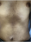
Figure 1
Figure 1
Clinical image of anterior trunk showing multiple papules surmounted on with macular pigmentation.
Figure 2
Figure 2
(A) Dermoscopic image of a fully developed lesion showing whitish scales (arrows), brown-red structureless areas with poorly defined borders (red stars), multiple irregularly distributed brownish blue-gray dots and globules (yellow stars), and blue-white veil like structures (white ovals) over a background of erythema (DermLite II, hybrid M, ×10, polarized).(B) Resolving lesions demonstrated brownish background reticular pigmentation surmounted by volcano-like islands composed of multiple irregular brown-gray dots and prominent linear blood vessels (white arrows, DermLite II, hybrid M, ×10, polarized). (C) Histopathology revealed mild hyperkeratosis, acanthosis, and superficial dermis showing perivascular inflammation on low power view. (D) Histopathology revealed basal cell vacuolization, pigment incontinence, apoptotic keratinocytes (arrow in inset), neutrophilic exocytosis, and interface dermatitis evident on high power magnification.

References

    1. Bolewska A, Słowińska M, Bożek P, Czuwara J, Paluchowska E, Owczarek W. Prurigo pigmentosa: not that uncommon? First case in Central and Eastern Europe. Dermoscopy. Postepy Dermatol Alergol. 2019;36(4):498–500. doi: 10.5114/ada.2019.87455. - DOI - PMC - PubMed
    1. Vinay K, Bishnoi A, Parsad D, Saikia UN, Sendhil Kumaran M. Dermatoscopic evaluation and histopathological correlation of acquired dermal macular hyperpigmentation. Int J Dermatol. 2017;56(12):1395–9. doi: 10.1111/ijd.13782. - DOI - PubMed
    1. Errichetti E, Stinco G, Lacarrubba F, Micali G. Dermoscopy of Darier’s disease. J Eur Acad Dermatol Venereol. 2016;30(8):1392–4. doi: 10.1111/jdv.13238. - DOI - PubMed
    1. Piccolo V, Corneli P, Russo T, Danielsson M, Zalaudek I, Argenziano G. Classic dowling degos disease: a rare genodermatosis. G Ital Dermatol Venereol. 2019 doi: 10.23736/S0392-0488.19.06386-7. - DOI - PubMed

LinkOut - more resources