. 2021 Dec;11(12):4045-4054.
doi: 10.1016/j.apsb.2021.03.036. Epub 2021 Mar 27.

Baicalin induces ferroptosis in bladder cancer cells by downregulating FTH1

Affiliations

Baicalin induces ferroptosis in bladder cancer cells by downregulating FTH1

Na Kong et al. Acta Pharm Sin B. 2021 Dec.

Abstract

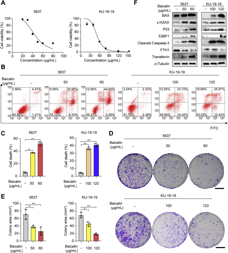
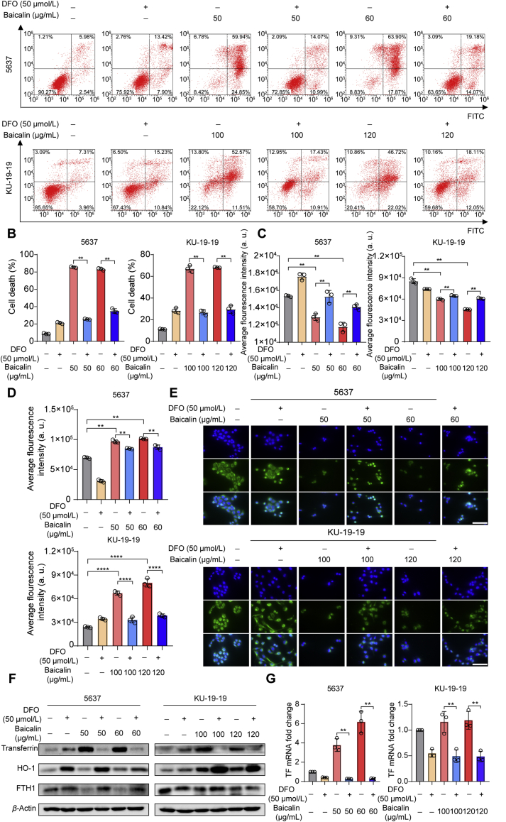
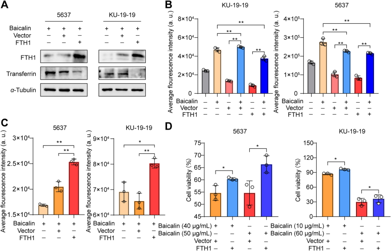
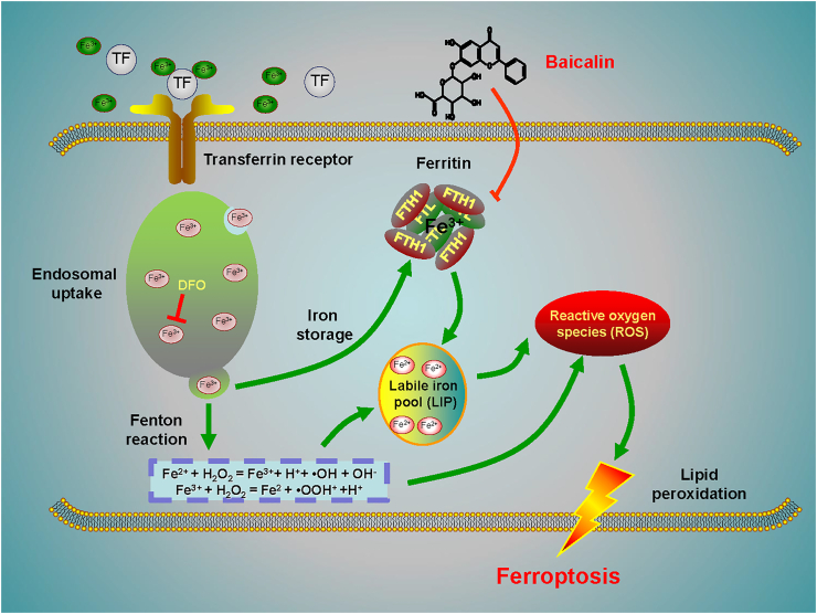
Ferroptosis is a non-apoptotic regulated cell death caused by iron accumulation and subsequent lipid peroxidation. Currently, the therapeutic role of ferroptosis on cancer is gaining increasing interest. Baicalin an active component in Scutellaria baicalensis Georgi with anticancer potential various cancer types; however, the effects of baicalein on bladder cancer and the underlying molecular mechanisms remain largely unknown. In the study, we investigated the effect of baicalin on bladder cancer cells 5637 and KU-19-19. As a result, we show baicalin exerted its anticancer activity by inducing apoptosis and cell death in bladder cancer cells. Subsequently, we for the first time demonstrate baicalin-induced ferroptotic cell death in vitro and in vivo, accompanied by reactive oxygen species (ROS) accumulation and intracellular chelate iron enrichment. The ferroptosis inhibitor deferoxamine but not necrostatin-1, chloroquine (CQ), N-acetyl-l-cysteine, l-glutathione reduced, or carbobenzoxy-valyl-alanyl-aspartyl-[O-methyl]-fluoromethylketone (Z-VAD-FMK) rescued baicalin-induced cell death, indicating ferroptosis contributed to baicalin-induced cell death. Mechanistically, we show that ferritin heavy chain 1 (FTH1) was a key determinant for baicalin-induced ferroptosis. Overexpression of FTH1 abrogated the anticancer effects of baicalin in both 5637 and KU19-19 cells. Taken together, our data for the first time suggest that the natural product baicalin exerts its anticancer activity by inducing FTH1-dependent ferroptosis, which will hopefully provide a prospective compound for bladder cancer treatment.

Keywords: Baicalin; Bladder cancer; Deferoxamine; FTH1; Ferroptosis.


Figures

Image 1
Graphical abstract
Figure 1
Figure 1
The cell viability of 5637 and KU-19-19 cells was detected after the treatment with baicalin. (A) The cell viability of 5637 and KU-19-19 was examined by CCK-8 assay after the treatment with various concentrations of baicalin for 24 h. (B) and (C) Representative results of annexin V-FITC/PI staining and quantitative analysis after the treatment with baicalin for 24 h, mean ± SD, n = 3; ∗P < 0.05, ∗∗P < 0.01. (D) and (E) Representative results of the colony formation and quantitative analysis, mean ± SD, n = 3; ∗P < 0.05, ∗∗P < 0.01. Scale: 30 mm. (F) The expression of BCl-2-associated X (BAX), P53 binding protein 1 (53BP1), phosphorylated histone H2AX (γ-H2AX), P53, ferritin heavy chain 1 (FTH1), transferrin, and cleaved caspase-3 was determined by Western blotting.
Figure 2
Figure 2
The effect of baicalin alone or in combination with other cell death inhibitors on the inhibition of growth of bladder cancer cells. (A) 5637 and KU-19-19 were treated with baicalin accompanied with or without Nec-1 for 24 h, then cell viability was tested. (B) 5637 and KU-19-19 were treated with baicalin along with or without chloroquine (CQ) for 24 h, then cell viability was tested. (C) Pretreatment with or without carbobenzoxy-valyl-alanyl-aspartyl-[O-methyl]-fluoromethylketone (Z-VAD-FMK), 5637 and KU-19-19 cells were treated with baicalin for 24 h, then cell viability was assayed. (D) 5637 and KU-19-19 were treated with baicalin with or without deferoxamine (DFO) for 24 h and cell viability was analyzed, ∗∗P < 0.01. (E) 5637 and KU-19-19 were treated with baicalin with or without glutathione (GSH) for 24 h, then cell viability was analyzed, mean ± SD, n = 3; ∗P < 0.05, ∗∗P < 0.01. (F) 5637 and KU-19-19 were treated with baicalin along with or without N-acetylcysteine (NAC) for 24 h and the cell viability was assayed, mean ± SD, n = 3; ∗∗P < 0.01.
Figure 3
Figure 3
DFO could trigger significantly the treatment of baicalin in bladder cancer cells. (A) and (B) The cell death was observed by flow cytometer with the treatment of baicalin with or without DFO, mean ± SD, n = 3, ∗∗P < 0.01. (C) 5637 and KU-19-19 were treated with baicalin along with or without DFO for 24 h, then intracellular chelate iron was analyzed, mean ± SD, n = 3; ∗P < 0.05, ∗∗P < 0.01. (D) The reactive oxygen species (ROS) level was analyzed by a flow cytometer, mean ± SD, n = 3; ∗∗P < 0.01. (E) Mito-Tracker Green staining was made to detect mitochondria damage. Scale: 100 μm. (F) The expression of FTH1, HO-1, transferrin in bladder cancer cells was detected after the treatment with baicalin with or without DFO for 24 h by Western blotting. (G) qRT-PCR was performed to detect the mRNA expression of transferrin, mean ± SD, n = 3; ∗∗P < 0.01.
Figure 4
Figure 4
FTH1 was a key determinant for baicalin-induced ferroptosis. (A) The expression of FTH1 and transferrin was detected by Western blotting. (B) Untransfected cells and FTH1-transfected cells were treated with or without baicalin for 24 h and then ROS level was detected, mean ± SD, n = 3; ∗∗P < 0.01. (C) FTH1-transfected 5637 and KU-19-19 cells were treated with baicalin for 24 h and then intracellular chelate iron was analyzed, mean ± SD, n = 3; ∗P < 0.05, ∗∗P < 0.01. (D) FTH1-transfected 5637 and KU-19-19 cells were treated with baicalin for 24 h and then the cell viability was detected by CCK-8, mean ± SD, n = 3; ∗P < 0.05.
Figure 5
Figure 5
Baicalin triggered ferroptosis in bladder cancer xenograft model. (A) An image of tumor samples in each group. (B) Tumor volume in each group was analyzed. Data are expressed as the mean ± SD, n = 3; ∗∗P < 0.01. (C) Mice weight in each group was analyzed. (D) and (E) Representative results of free iron deposition and quantitative analysis, mean ± SD, n = 3; ∗∗P < 0.01. Scale: 50 μm and 2.5 mm. (F) The expression of FTH1 was determined by immunohistochemical staining. Scale: 120 μm.
Figure 6
Figure 6
A schematic diagram about the central role of baicalin in ferroptosis induction in bladder cancer. Baicalin exerts its anticancer activity in bladder cancer by inducing FTH1-dependent ferroptosis, which will hopefully provide great therapeutic potential for bladder cancer treatment.

References

    1. Yu B., Choi B., Li W., Kim D.H. Magnetic field boosted ferroptosis-like cell death and responsive MRI using hybrid vesicles for cancer immunotherapy. Nat Commun. 2020;11:3637. - PMC - PubMed
    1. Wang J., Yin X., He W., Xue W., Zhang J., Huang Y. SUV39H1 deficiency suppresses clear cell renal cell carcinoma growth by inducing ferroptosis. Acta Pharm Sin B. 2021;11:406–419. - PMC - PubMed
    1. Alu A., Han X., Ma X., Wu M., Wei Y., Wei X. The role of lysosome in regulated necrosis. Acta Pharm Sin B. 2020;10:1880–1903. - PMC - PubMed
    1. Qiu Y., Cao Y., Cao W., Jia Y., Lu N. The application of ferroptosis in diseases. Pharmacol Res. 2020;159:104919. - PubMed
    1. Stockwell B.R., Jiang X., Gu W. Emerging mechanisms and disease relevance of ferroptosis. Trends Cell Biol. 2020;30:478–490. - PMC - PubMed