. 2021 Dec 24;8(1):e08673.
doi: 10.1016/j.heliyon.2021.e08673. eCollection 2022 Jan.

Human telomerase reverse transcriptase promotes the epithelial to mesenchymal transition in lung cancer cells by enhancing c-MET upregulation

Affiliations

Human telomerase reverse transcriptase promotes the epithelial to mesenchymal transition in lung cancer cells by enhancing c-MET upregulation

Ram Raj Prasad et al. Heliyon. .

Abstract

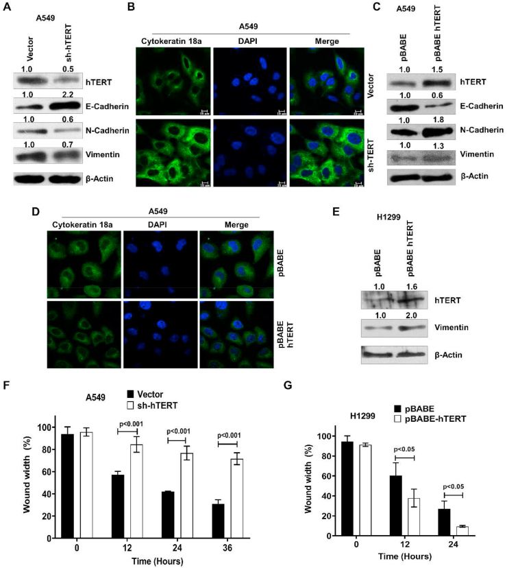
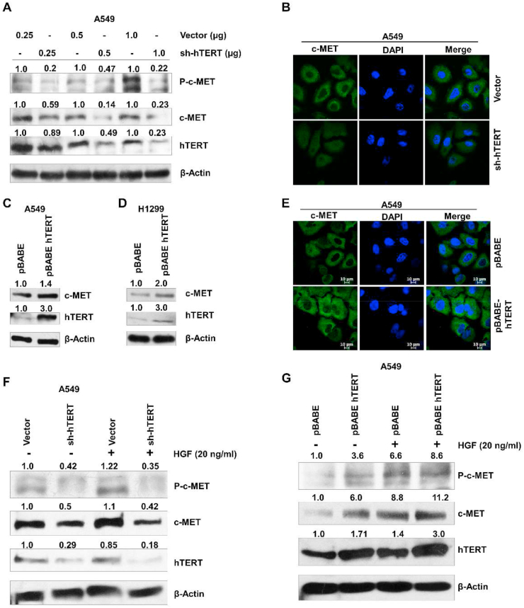
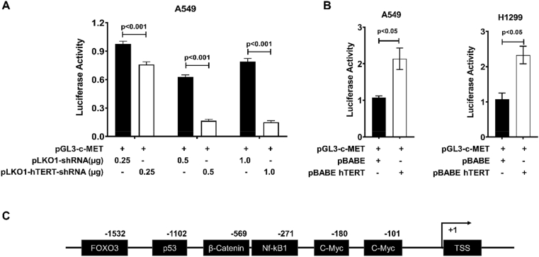
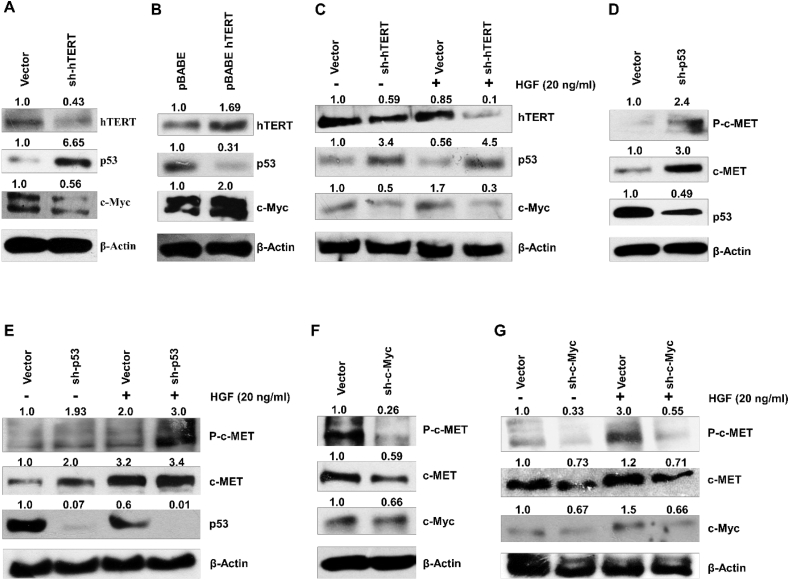
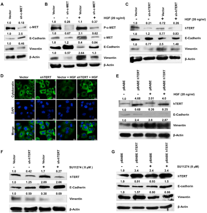
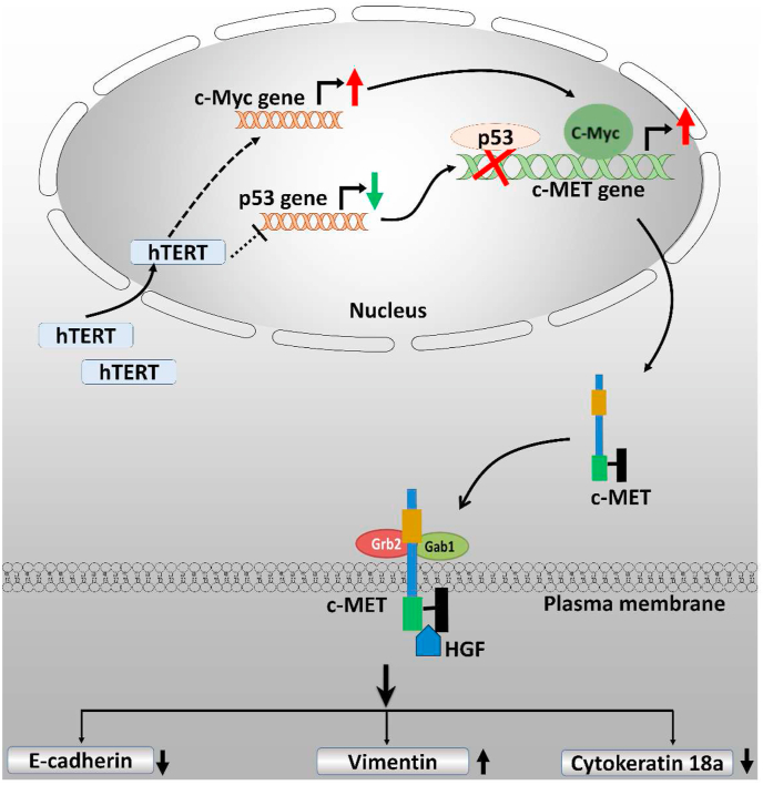
Human telomerase reverse transcriptase (hTERT), the essential catalytic subunit of telomerase, is associated with telomere homeostasis to prevent replicative senescence and cellular aging. However, hTERT reactivation also has been linked to the acquisition of several hallmarks of cancer, although the underlying mechanism beyond telomere extension remains elusive. This study demonstrated that hTERT overexpression promotes, whereas its inhibition by shRNA suppresses, epithelial-mesenchymal transition (EMT) in lung cancer cells (A549 and H1299). We found that hTERT modulates the expression of EMT markers E-cadherin, vimentin, and cytokeratin-18a through upregulation of the c-MET. Ectopic expression of hTERT induces expression of c-MET, while hTERT-shRNA treatment significantly decreases the c-MET level in A549 and H1299 through differential expression of p53 and c-Myc. Reporter assay suggests the regulation of c-MET expression by hTERT to be at the promoter level. An increase in c-MET level significantly promotes the expression of mesenchymal markers, including vimentin and N-cadherin, while a notable increase in epithelial markers E-cadherin and cytokeratin-18a is observed after the c-MET knockdown in A549.

Keywords: EMT; c-MET; c-Myc; hTERT; p53.


Conflict of interest statement

The authors declare no conflict of interest.

Figures

Figure 1
Figure 1
hTERT promotes the epithelial to mesenchymal transition and cell migration in A549 and H1299. A) Immunoblot analysis of E-cadherin, N-cadherin, and vimentin on hTERT knockdown B) Immunofluorescence assay showing knockdown of hTERT in A549 cells increased the expression level of cytokeratin-18a. C, D) hTERT overexpression in A549 leads to an increased expression of mesenchymal marker (N-cadherin and vimentin) and decreased epithelial markers (E-cadherin and cytokeratin-18a). E) hTERT expression plasmid was also transfected in H1299 cells, showing increased mesenchymal protein (vimentin) on hTERT overexpression. F, G) Migration assay in hTERT knockdown and overexpression in lung cancer cells (A549 and H1299), hTERT down-regulation reduced, while overexpression promoted the cell migration (Mean ± SE, n = 3). Full and non-adjusted images of immunoblots were shown in the supplementary material (Figure SM 1).
Figure 2
Figure 2
c-MET expression is regulated by hTERT. A) Immunoblot analysis of phospho-c-MET, c-MET proteins after treatment with different doses of hTERT shRNA in A549. hTERT shRNA reduced the c-MET expression in a dose-dependent manner. B) Immunofluorescence of c-MET in hTERT knockdown A549 also shows the positive association between hTERT and c-MET expression. C, D) hTERT overexpression in A549 (left panel) and H1299 (right panel) shows the increased level of c-MET expression. E) Immunofluorescence of the c-MET protein in hTERT overexpressing A549 cells. F) hTERT knockdown A549 cells were starved for 12 h then stimulated by HGF (20 ng/ml) for 24 h. Expression of phospho and total c-MET were analyzed by western blotting. HGF treatment showed no significant promoting effect on the c-MET expression in cells knocked down for hTERT expression. G) Similar to the previous experiment, for pBABE-hTERT transfection, A549 cells were starved and treated with HGF. Western blots showed that hTERT overexpression has a synergistic stimulatory effect with HGF on the expression of c-MET. Full and non-adjusted images of immunoblots were shown in the supplementary material (Figure SM 2)
Figure 3
Figure 3
hTERT expression induced c-MET promoter activity. A) A549 cells were co-transfected with vector and hTERT shRNA plasmid with pGL3-c-MET promoter construct. c-MET promoter activity was assayed using a Luciferase reporter kit. c-MET promoter activity decreased with hTERT downregulation in a dose-dependent manner (Error bars represent mean ± SE, n = 3). B) The luciferase promoter-reporter assay in cells overexpressing hTERT, pBABE-hTERT and control plasmids were co-transfected with pGL3-c-MET promoter construct in A549 and H1299 cells. Results show an increase in c-MET promoter activity on hTERT overexpression in A549 and H1299 lung cancer cells (Mean ± SE, n = 3). Luciferase assay in A549 and H1299 was done independently, and the values of promoter activity were normalized for total cell protein and are shown relative to activity in control cells. C) Bioinformatics analysis of the c-MET promoter region showed many putative transcription factor-binding sites for FOXO3, p53, beta-catenin, NF-kB1, and c-Myc.
Figure 4
Figure 4
p53 and c-Myc play a critical role in hTERT mediated regulation of c-MET expression. Immunoblot analysis is showing the p53 and c-Myc expression as influenced by (A) hTERT downregulation and (B) over-expression in A549. (C) p53 and c-Myc expression were observed in A549 cells after hTERT knockdown and treatment with HGF. (D) Immunoblot analysis of c-MET, phospho c-MET expression in p53 knockdown A549 cells. (E) Western blot analysis of c-MET and Phospho-c-MET in p53-shRNA transfected A549 cells followed by HGF treatment showed that p53 knockdown increased the c-MET level, and HGF treatment further elevated the expression and phosphorylation of c-MET. (F) Western blot analysis of Phospho-c-MET and c-MET in c-Myc knockdown A549. (G) Western blot analysis of c-MET and Phospho-c-MET in A549 cells knocked down for c-Myc and treated with HGF. c-Myc downregulation reduced the c-MET level, and further HGF treatment in c-Myc knockdown A549 cells does not significantly restore c-MET expression. Full and non-adjusted images of immunoblots were shown in the supplementary material (Figure SM 3)
Figure 5
Figure 5
c-MET induced EMT is positively associated with hTERT expression. (A) Western blot showing increased E-cadherin expression and decreased vimentin expression following c-MET knockdown A549 cells. (B) c-MET knocked down A549 cells were starved for 12 h and treated with HGF (20 ng/ml) for 24 h. Western blots show inverse relationship between expression of c-MET and epithelial marker E-cadherin and a direct relationship with mesenchymal marker vimentin. C) hTERT knockdown A549 cells were treated with HGF and analyzed for expression of E-cadherin and vimentin. shRNA targeting hTERT. Western blots show that knocking down hTERT expression promoted the expression of epithelial marker E-cadherin and decreased the mesenchymal protein vimentin in cells. D) Immunofluorescence staining of cytokeratin-18a was done in hTERT knockdown A549 cells treated with HGF. HGF treatment reduced the cytokeratin level, while hTERT-shRNA treatment restored the cytokeratin expression. E) Stably hTERT over-expressing A549 cells were starved and then stimulated by HGF (20 ng/ml), leading to increased expression of vimentin and suppression of E-cadherin synergistically. F) hTERT downregulated A549 cells were treated with the 8 μM SU11274 (c-MET inhibitor), and expression of E-cadherin and vimentin were detected by western blotting. G) Immunoblot analysis of epithelial marker E-cadherin and vimentin following SU11274 (8 μM) treatment in hTERT over-expressing A549 cells showed lower expression of this mesenchymal marker. Full and non-adjusted images of immunoblots were shown in the supplementary material (Figure SM 4)
Figure 6
Figure 6
A model is showing the role of hTERT in epithelial to mesenchymal transition by enhancing the c-MET upregulation.

References

    1. Kim N.W., Piatyszek M.A., Prowse K.R., Harley C.B., West M.D., Ho P.L., Coviello G.M., Wright W.E., Weinrich S.L., Shay J.W. Specific association of human telomerase activity with immortal cells and cancer. Science. 1994;266:2011–2015. - PubMed
    1. Hayflick L. The cell biology of aging. Clin. Geriatr. Med. 1985;1:15–27. - PubMed
    1. Stewart S.A., Hahn W.C., O’Connor B.F., Banner E.N., Lundberg A.S., Modha P., Mizuno H., Brooks M.W., Fleming M., Zimonjic D.B., Popescu N.C., Weinberg R.A. Telomerase contributes to tumorigenesis by a telomere length-independent mechanism. Proc. Natl. Acad. Sci. U. S. A. 2002;99:12606–12611. - PMC - PubMed
    1. Martínez P., Blasco M.A. Telomeric and extra-telomeric roles for telomerase and the telomere-binding proteins. Nat. Rev. Cancer. 2011;11:161–176. - PubMed
    1. Guo W., Lu J., Dai M., Wu T., Yu Z., Wang J., Chen W., Shi D., Yu W., Xiao Y., Yi C., Tang Z., Xu T., Xiao X., Yuan Y., Liu Q., Du G., Deng W. Transcriptional coactivator CBP upregulates hTERT expression and tumor growth and predicts poor prognosis in human lung cancers. Oncotarget. 2014;5:9349–9361. - PMC - PubMed