

Anatomical and topographical variations in the distribution of brain metastases based on primary cancer origin and molecular subtypes: a systematic review
- PMID: 35024611
- PMCID: PMC8739649
- DOI: 10.1093/noajnl/vdab170
Anatomical and topographical variations in the distribution of brain metastases based on primary cancer origin and molecular subtypes: a systematic review
Abstract
Background: While it has been suspected that different primary cancers have varying predilections for metastasis in certain brain regions, recent advances in neuroimaging and spatial modeling analytics have facilitated further exploration into this field.
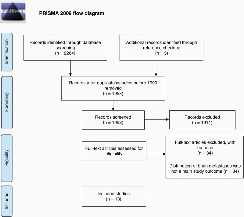
Methods: A systematic electronic database search for studies analyzing the distribution of brain metastases (BMs) from any primary systematic cancer published between January 1990 and July 2020 was conducted using PRISMA guidelines.
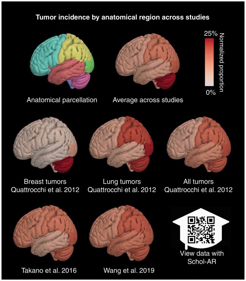
Results: Two authors independently reviewed 1957 abstracts, 46 of which underwent full-text analysis. A third author arbitrated both lists; 13 studies met inclusion/exclusion criteria. All were retrospective single- or multi-institution database reviews analyzing over 8227 BMs from 2599 patients with breast (8 studies), lung (7 studies), melanoma (5 studies), gastrointestinal (4 studies), renal (3 studies), and prostate (1 study) cancers. Breast, lung, and colorectal cancers tended to metastasize to more posterior/caudal topographic and vascular neuroanatomical regions, particularly the cerebellum, with notable differences based on subtype and receptor expression. HER-2-positive breast cancers were less likely to arise in the frontal lobes or subcortical region, while ER-positive and PR-positive breast metastases were less likely to arise in the occipital lobe or cerebellum. BM from lung adenocarcinoma tended to arise in the frontal lobes and squamous cell carcinoma in the cerebellum. Melanoma metastasized more to the frontal and temporal lobes.
Conclusion: The observed topographical distribution of BM likely develops based on primary cancer type, molecular subtype, and genetic profile. Further studies analyzing this association and relationships to vascular distribution are merited to potentially improve patient treatment and outcomes.
Keywords: brain metastases; distribution; magnetic resonance imaging; topographical variations.
© The Author(s) 2021. Published by Oxford University Press, the Society for Neuro-Oncology and the European Association of Neuro-Oncology.
Figures
References
-
- Nathoo N, Toms SA, Barnett GH. Metastases to the brain: current management perspectives. Expert Rev Neurother. 2004;4(4):633–640. - PubMed
Publication types
Grants and funding
LinkOut - more resources
Full Text Sources
Research Materials
Miscellaneous