Case Reports
. 2022 Feb;44(2):201-205.
doi: 10.1007/s00276-021-02881-9. Epub 2022 Jan 13.

Diagnosis of ulnar nerve entrapment anterior to the medial epicondyle by ultrasound elastography and diffusion tensor imaging with fiber tractography: a case report

Affiliations
Case Reports

Diagnosis of ulnar nerve entrapment anterior to the medial epicondyle by ultrasound elastography and diffusion tensor imaging with fiber tractography: a case report

Guillaume Jaques et al. Surg Radiol Anat. 2022 Feb.

Abstract

Ulnar/cubital tunnel syndrome is the second most common compressive neuropathy of the upper limb. Permanent location of the ulnar nerve anterior to the medial epicondyle is extremely rare, with only five cases reported in the literature. Using ultrasound elastography and diffusion tensor imaging with fiber tractography, we diagnosed a case in which ulnar nerve entrapment was associated with anterior nerve location. Surgical release confirmed the diagnosis and the patient was symptom free 3 months after surgery.

Keywords: Cubital tunnel syndrome; Diffusion tensor imaging; Elastography; Nerve surgery; Tractography; Ulnar nerve.


Conflict of interest statement

There is no potential conflicts of interest.

Figures

Fig. 1
Fig. 1
Conventional ultrasound and SWE, and DTI with fiber tractography. a Axial/transverse B-mode ultrasound image of the left ulnar nerve with CSA measurement at the compression site (0.08 cm2, white arrow). The ulnar nerve is located anterior to the medial epicondyle (asterisk). b SWE image and measurement of the left ulnar nerve in complete elbow flexion (mean, 79.1 kPa; SD, 6.6 kPa). c SWE image and measurement of the left ulnar nerve in complete elbow extension (mean, 107.2 kPa; SD, 4.5 kPa). DTI fiber tractography reconstructions of the right and left ulnar nerves. d Right elbow showing the normal pathway of the right ulnar nerve located posterior to the medial epicondyle within the cubital tunnel. e Abnormal “S-shape” pathway of the left ulnar nerve running anterior to the medial epicondyle outside the cubital tunnel
Fig. 2
Fig. 2
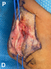
Intraoperative photograph of the medial aspect of the left elbow showing complete surgical release of the left ulnar nerve located anterior to the medial epicondyle (asterisk). P proximal; D distal

References

    1. Aziz MA. Anatomical defects in a case of trisomy 13 with a D/D translocation. Teratology. 1980;22:217–227. doi: 10.1002/tera.1420220211. - DOI - PubMed
    1. Bäumer P, Dombert T, Staub F, Kaestel T, Bartsch AJ, Heiland S, Bendszus M, Pham M. Ulnar neuropathy at the elbow: MR neurography–nerve T2 signal increase and caliber. Radiology. 2011;260:199–206. doi: 10.1148/radiol.11102357. - DOI - PubMed
    1. Cotten A, Haddad F, Hayek G, Lefebvre G, Dodré E, Budzik JF. Tractography: possible applications in musculoskeletal radiology. Semin Musculoskelet Radiol. 2015;19:387–395. doi: 10.1055/s-0035-1563736. - DOI - PubMed
    1. Davis GA. Ulnar nerve volar to medial epicondyle: an anatomical variation. Case illustration. J Neurosurg. 2006;104:625. doi: 10.3171/jns.2006.104.4.625. - DOI - PubMed
    1. Durand S, Raffoul W, Christen T, Pedrazzi N. Post-operative assessment of ulnar nerve tension using shear-wave elastography. Neurol Int. 2021;13:469–476. doi: 10.3390/neurolint13030046. - DOI - PMC - PubMed

Publication types

MeSH terms

LinkOut - more resources