. 2022 Jan 14;375(6577):221-225.
doi: 10.1126/science.abj8432. Epub 2022 Jan 13.

Bacterial gasdermins reveal an ancient mechanism of cell death

Affiliations

Bacterial gasdermins reveal an ancient mechanism of cell death

Alex G Johnson et al. Science. .

Abstract

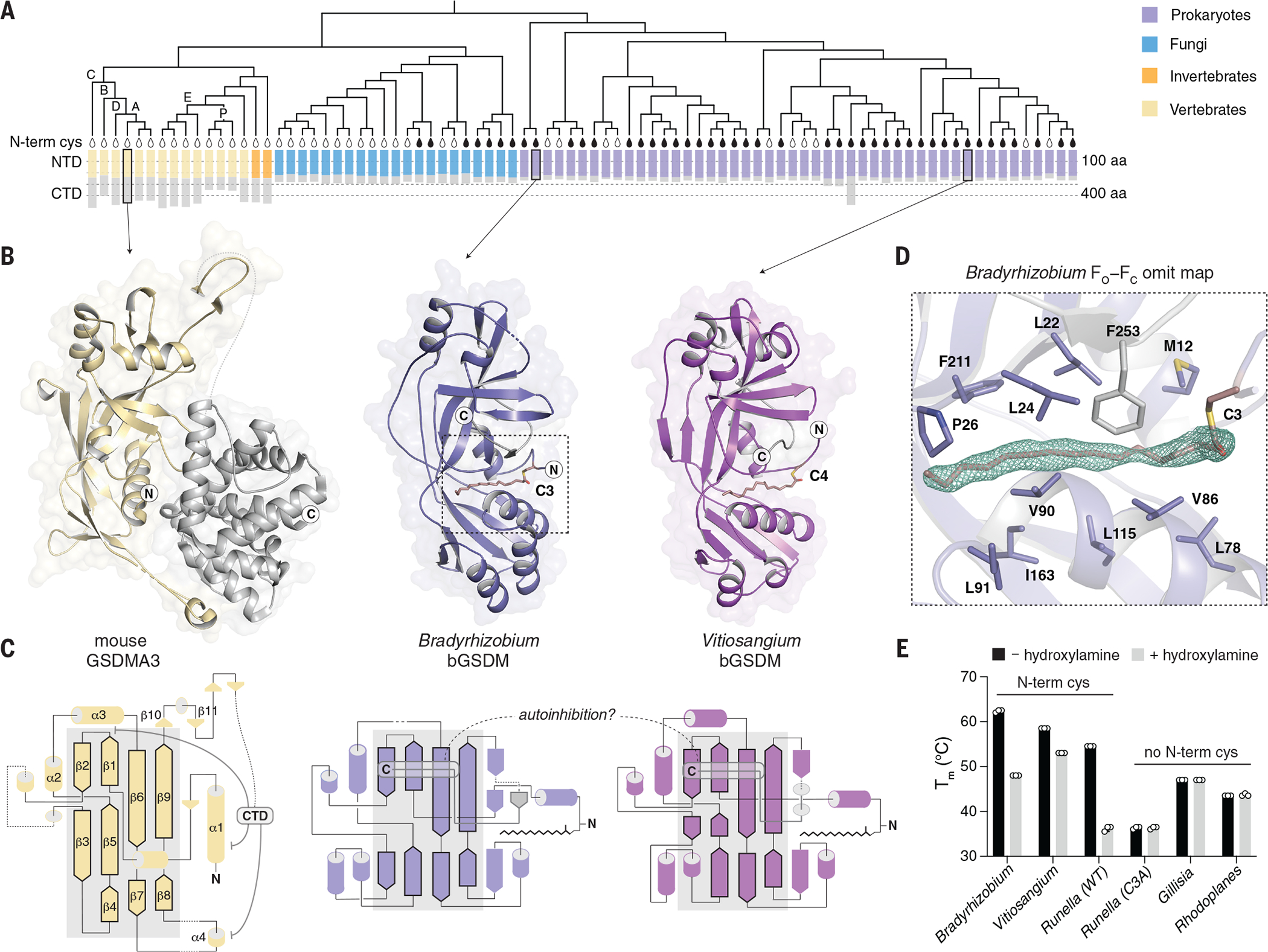
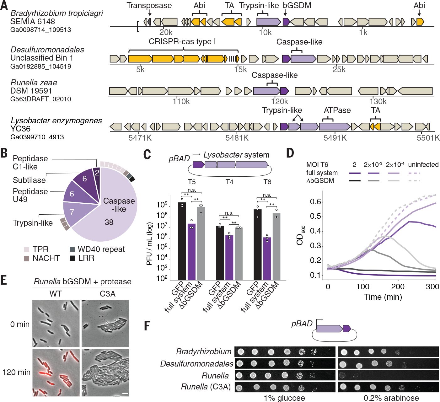
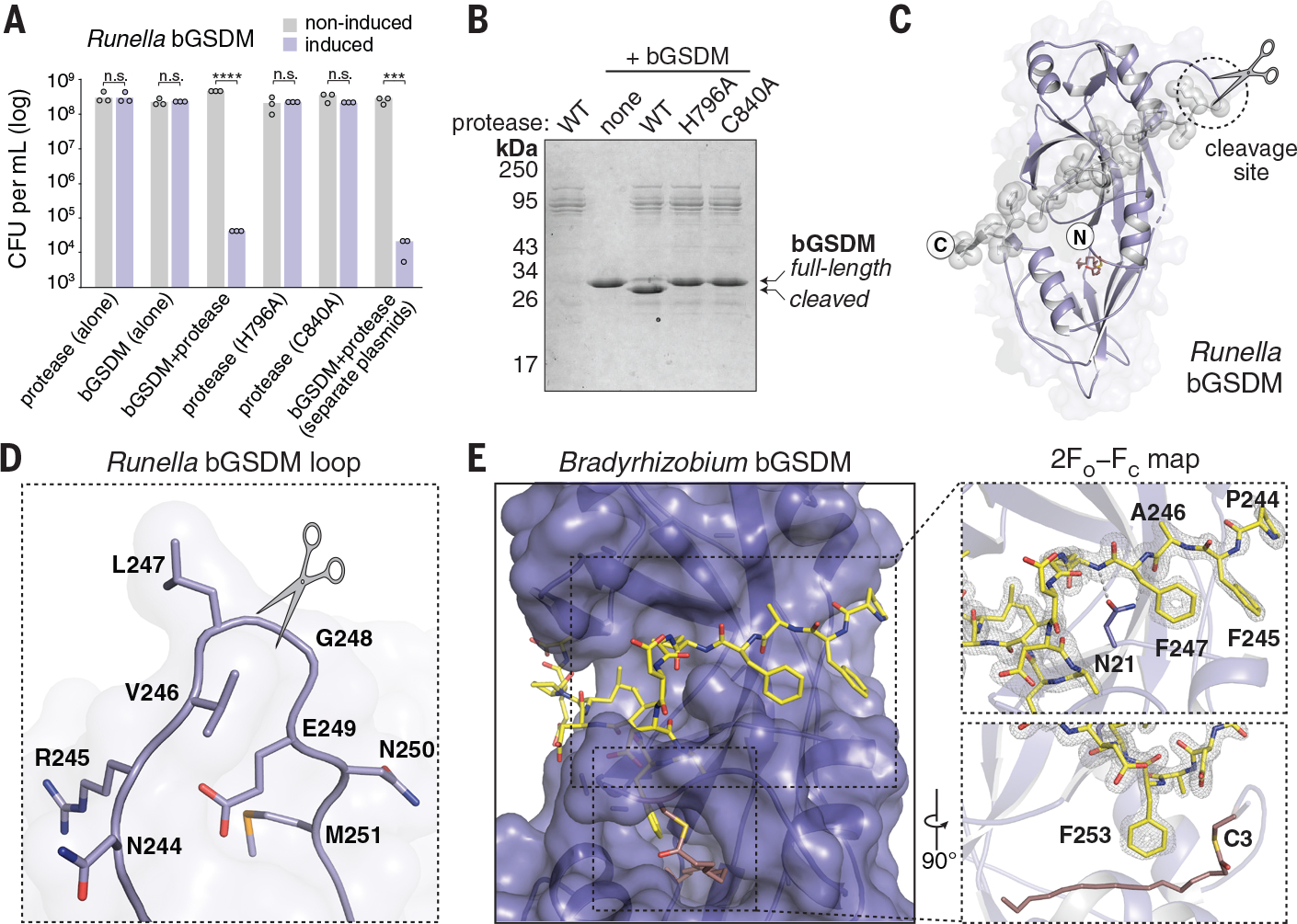
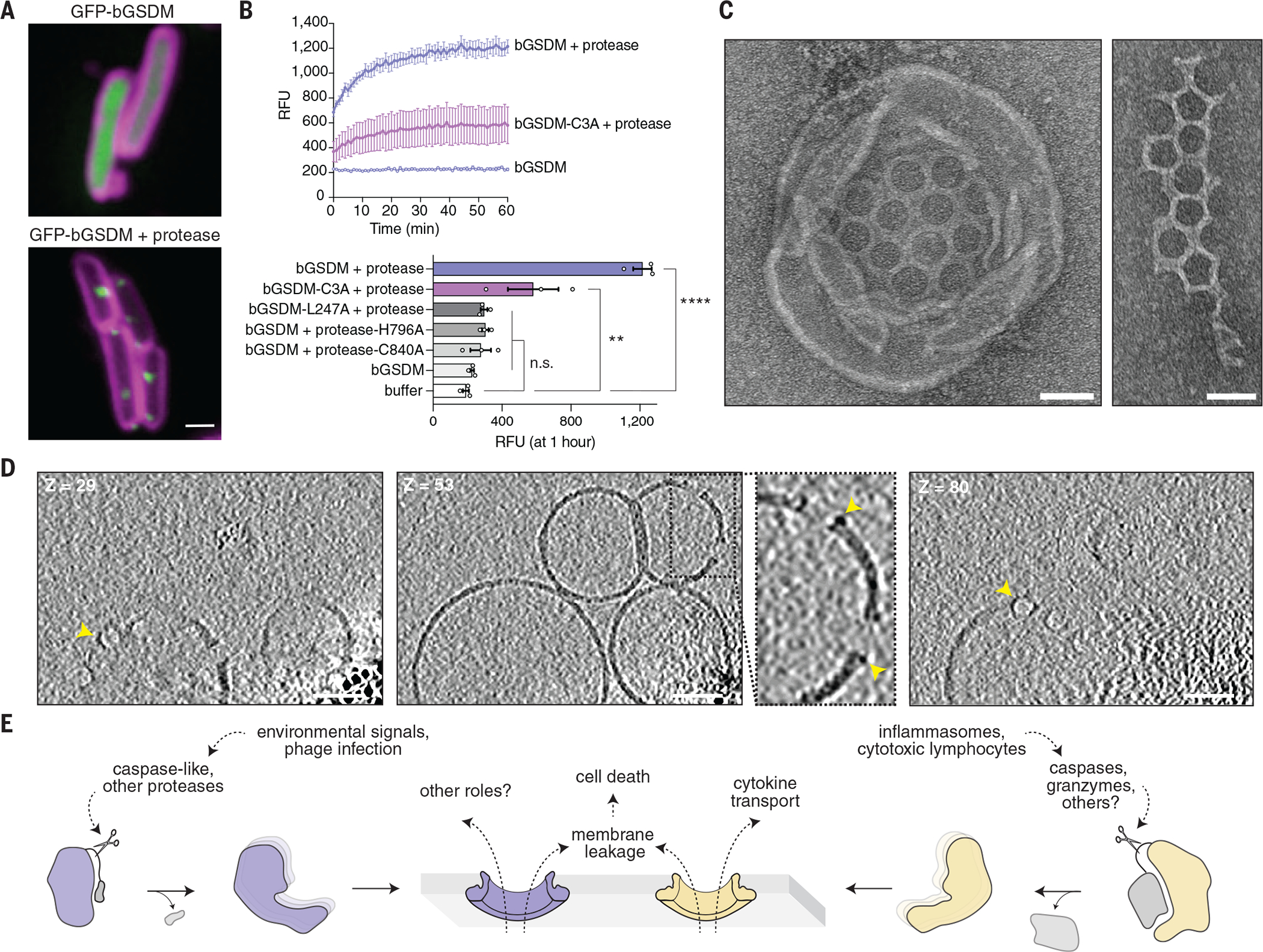
Gasdermin proteins form large membrane pores in human cells that release immune cytokines and induce lytic cell death. Gasdermin pore formation is triggered by caspase-mediated cleavage during inflammasome signaling and is critical for defense against pathogens and cancer. We discovered gasdermin homologs encoded in bacteria that defended against phages and executed cell death. Structures of bacterial gasdermins revealed a conserved pore-forming domain that was stabilized in the inactive state with a buried lipid modification. Bacterial gasdermins were activated by dedicated caspase-like proteases that catalyzed site-specific cleavage and the removal of an inhibitory C-terminal peptide. Release of autoinhibition induced the assembly of large and heterogeneous pores that disrupted membrane integrity. Thus, pyroptosis is an ancient form of regulated cell death shared between bacteria and animals.


Conflict of interest statement

Competing Interests: R.S. is a scientific cofounder and advisor of BiomX, Pantheon Bioscience, and Ecophage. The remaining authors have no competing financial interests to declare.

Figures

Fig. 1.
Fig. 1.. Structures of bGSDMs reveal homology with mammalian cell death effectors.
(A) Gasdermin phylogenetic tree. The sizes of the gasdermin NTDs and CTDs are depicted. Vertebrate gasdermins are labeled with single letters indicating human gasdermins GSDMA to GSDME (“A” to “E”), and “P” depicts pejvakin. The black teardrop indicates a conserved N-terminal cysteine(N-termin cys). A representative set of 20 fungal gasdermins are included in the tree. aa, amino acid. (B) Crystal structures of bGSDMs from species of the genera Bradyrhizobium and Vitiosangium. bGSDM structures reveal homology to the NTD of mammalian gasdermins in an inactive conformation including mouse GSDMA3 (Protein Data Bank ID 5B5R). (C) Gasdermin topology diagrams indicate a conserved central core of the bacterial and mammalian NTD. bGSDMs notably lack the CTD required for autoinhibition of mammalian gasdermins and instead encode a short C-terminal peptide. (D) Simulated annealing FO−FC omit map (contoured at 3.0 σ) from the Bradyrhizobium bGSDM fit with a palmitoyl modification at C3. Omit map is shown as green mesh and select residues forming a hydrophobic pocket around the palmitoyl group are indicated. (E) Melting temperatures (Tm) of bGSDMs with and without N-terminal cysteines, as determined with thermofluor assays. Data are the mean and standard deviation of three technical replicates and are representative of three independent experiments. WT, wild-type. Single-letter abbreviation for the amino acid residues are as follows: A, Ala; C, Cys; D, Asp; E, Glu; F, Phe; G, Gly; H, His; I, Ile; K, Lys; L, Leu; M, Met; N, Asn; P, Pro; Q, Gln; R, Arg; S, Ser; T, Thr; V, Val; W, Trp; and Y, Tyr.
Fig. 2.
Fig. 2.. bGSDMs are associated with proteases, defend from phages, and execute cell death.
(A) Representative instances of bGSDMs and associated proteases, in their genomic neighborhoods. Genes known to be involved in anti-phage defense are shown in yellow. TA, toxin-antitoxin; Abi, abortive infection; ATPase, adenosine triphosphatase. (B) Types of proteases found adjacent to bGSDMs (n = 59). Some bGSDMs appear with more than one adjacent protease. Caspase-like proteases include peptidase C14 (n = 15) and CHAT (n = 23). Cases in which the protease gene also encodes an additional domain are indicated. TPR, tetratricopeptide repeat; LRR, leucine-rich repeat. (C) A bGSDM-containing operon protects against phages. The efficiency of plating of phages on E. coli MG1655 cells expressing the Lysobacter bGSDM WT or mutated operon is shown. Data represent plaque-forming units (PFU) per milliliter and are the averages of three independent replicates, with individual data points overlaid. GFP represents a control strain. Statistical significance was determined by a one-way analysis of variance (ANOVA) and Tukey multiple comparison test. Not significant (n.s.) ≥ 0.05, **P = 0.001 to 0.01. (D) Growth of liquid cultures of E. coli expressing the WT and mutated Lysobacter bGSDM operons. Cells were infected with phage T6. For each experiment, data represent one out of three biological replicates (replicates are shown in Fig. S6). OD600, optical density at 600 nm. (E) The Runella bGSDM operon causes cell death. E. coli DH5α cells expressing the Runella protease and WT or C3A mutated bGSDM were examined by time lapse microscopy. Overlay images from PI (red) and phase contrast of cells captured at the start of the experiment and after 120 min of incubation are shown. Scale bar, 2 μm. (F) bGSDM operons are toxic. Cells encoding protease and WT or mutated bGDSM were plated in 10-fold serial dilution on LB-agar in conditions that repress operon expression (1% glucose) or induce expression (0.2% arabinose).
Fig. 3.
Fig. 3.. bGSDMs are activated by proteolytic cleavage.
(A) Toxicity of Runella bGSDM in vivo requires the associated protease. Bacteria expressing WT and mutated versions of the Runella bGSDM–protease operon were grown on LB-agar in conditions that repress or induce expression. Data represent colony-forming units (CFU) per milliliter, and bar graphs represent an average of three independent replicates, with individual data points overlaid. Asterisks indicate statistically significant differences compared with the respective noninduced control using two-sided t-test. n.s. ≥ 0.05; ***P = 0.0001–0.001; ****P < 0.0001. (B) Runella bGSDM cleavage by its associated protease is dependent on catalytic histidine and residues in vitro. 15% SDS-polyacrylamide gel electrophoresis (SDS-PAGE) were run after cleavage at room temperature for 18 h and visualized by Coomassie staining. (C) The Runella bGSDM crystal structure and protease cleavage site. The Runella bGSDM structure is shown in lavender with the last 21 amino acids highlighted as gray spheres. (D) Close-up view of the Runella bGSDM cleavage site wherein cleavage occurs after the P1 L247 residue. (E) Structural overview of the Bradyrhizobium CTD and autoinhibitory interactions. The bGSDM is colored purple except for its last 16 residues, which are colored yellow. Insets show interactions of F245 and F247 adjacent to D21 of the N-terminal β sheet (top) and F253 and the palmitoyl modification at C3 (bottom). The 2FO−FC (contoured at 1.5 σ) map is shown as gray mesh fit to the last 16 residues.
Fig. 4.
Fig. 4.. Cleaved bGSDMs form membrane pores to elicit cell death.
(A) GFP was fused to the N-terminus of the Runella bGSDM. Cells expressing GFP-bGSDM alone (top) or with the caspase-like protease (bottom) are shown. GFP is colored green. Membrane dye (FM4-64) is in magenta. Scale bar, 1 μm. (B) Cleaved Runella gasdermin permeabilizes liposome membranes. Relative fluorescence units (RFU) were measured continuously from cleavage reactions of dioleoylphosphatidylcholine (DOPC) liposomes loaded with TbCl3 with an external solution containing 20 μM dipicolonic acid (DPA). The top plot represents an example of time-course liposome leakage, whereas the bottom bar chart shows values for each condition at 60 min. Error bars represent the SEM of three technical replicates and statistical significance was determined by one-way ANOVA and Tukey multiple comparison test. n.s. ≥ 0.05; **P = 0.001–0.01; ****P < 0.0001. (C) Negative stain electron microscopy of Runella gasdermin pores in DOPC liposomes (left) and in mesh-like arrays (right). Scale bars, 50 nm. (D) Slices from representative tomogram (1 of 10) of Runella gasdermin pores in DOPC liposomes, at three different depths (Z). Yellow arrowheads indicate pores inserted within the liposome membrane. Scale bars, 50 nm. (E) Model of pyroptosis for bGSDMs and mammalian gasdermins.

Comment in

References

    1. Kayagaki N, Stowe IB, Lee BL, O’Rourke K, Anderson K, Warming S, Cuellar T, Haley B, Roose-Girma M, Phung QT, Liu PS, Lill JR, Li H, Wu J, Kummerfeld S, Zhang J, Lee WP, Snipas SJ, Salvesen GS, Morris LX, Fitzgerald L, Zhang Y, Bertram EM, Goodnow CC, Dixit VM, Caspase-11 cleaves gasdermin D for non-canonical inflammasome signalling. Nature. 526, 666–671 (2015). - PubMed
    1. Shi J, Zhao Y, Wang K, Shi X, Wang Y, Huang H, Zhuang Y, Cai T, Wang F, Shao F, Cleavage of GSDMD by inflammatory caspases determines pyroptotic cell death. Nature. 526, 660–665 (2015). - PubMed
    1. He WT, Wan H, Hu L, Chen P, Wang X, Huang Z, Yang ZH, Zhong CQ, Han J, Gasdermin D is an executor of pyroptosis and required for interleukin-1β secretion. Cell Res. 25, 1285–1298 (2015). - PMC - PubMed
    1. Liu X, Zhang Z, Ruan J, Pan Y, Magupalli VG, Wu H, Lieberman J, Inflammasome-activated gasdermin D causes pyroptosis by forming membrane pores. Nature. 535, 153–158 (2016). - PMC - PubMed
    1. Ding J, Wang K, Liu W, She Y, Sun Q, Shi J, Sun H, Wang DC, Shao F, Pore-forming activity and structural autoinhibition of the gasdermin family. Nature. 535, 111–116 (2016). - PubMed

Publication types

MeSH terms

Supplementary concepts