. 2022 Jan 14;375(6577):167-172.
doi: 10.1126/science.abm7285. Epub 2022 Jan 13.

Cryo-EM structures of amyloid-β 42 filaments from human brains

Affiliations

Cryo-EM structures of amyloid-β 42 filaments from human brains

Yang Yang et al. Science. .

Abstract

Filament assembly of amyloid-β peptides ending at residue 42 (Aβ42) is a central event in Alzheimer’s disease. Here, we report the cryo–electron microscopy (cryo-EM) structures of Aβ42 filaments from human brains. Two structurally related S-shaped protofilament folds give rise to two types of filaments. Type I filaments were found mostly in the brains of individuals with sporadic Alzheimer’s disease, and type II filaments were found in individuals with familial Alzheimer’s disease and other conditions. The structures of Aβ42 filaments from the brain differ from those of filaments assembled in vitro. By contrast, in AppNL-F knock-in mice, Aβ42 deposits were made of type II filaments. Knowledge of Aβ42 filament structures from human brains may lead to the development of inhibitors of assembly and improved imaging agents.


Conflict of interest statement

Competing interests: The authors declare that they have no competing interests.

Figures

Figure 1
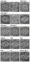
Figure 1. Cryo-EM maps of Type I, Type Ib and Type II Aβ42 filaments from brain.
Five cases of Alzheimer’s disease [three sporadic (sAD cases 1-3) and two familial (fAD case 1, mutation in APP encoding V717F; fAD case 2, mutation in PSEN1 encoding F105L)]; other human diseases [aging-related tau astrogliopathy (ARTAG), Parkinson’s disease dementia (PDD), dementia with Lewy bodies (DLB), frontotemporal dementia (FTD) caused by a GRN mutation and pathological aging (PA)]; and homozygous mice of the App NL-F knock-in line. For each map, a sum of the reconstructed densities for several XY-slices, approximating one β-rung, is shown. Filament types (Type I, Type Ib and Type II) are indicated on the top left; the percentages of a given filament type among Aβ42 filaments in the dataset are shown on the top right. The same scales apply to all panels.
Figure 2
Figure 2. Structures of Type I and Type II Aβ42 filaments from brain.
(A) Amino acid sequence of Aβ1-42. Type I filaments (in orange) extend from G9-A42; Type II filaments (in blue) from V12-A42. Thick connecting lines with arrowheads indicate β-strands (β1-β5 and β1-β4). (B,C) Cryo-EM density map (in transparent grey) and atomic models for Type I (B) and Type II (C) filaments. Each filament type is made of two identical protofilaments shown in orange (Type I) and blue (Type II). The density maps are displayed using the zone feature in ChimeraX at a distance of 2 Å. Associated solvent molecules are shown in white and putative metals in teal (B) and purple (C). (D,E) Schematics of Type I (D) and Type II (E) Aβ42 folds. They were produced using atom2svg.py (https://doi.org/10.5281/zenodo.4090924). Negatively charged residues are shown in red, positively charged residues in blue, polar residues in green, non-polar residues in white, sulfur-containing residues in yellow and glycines in pink. Thick connecting lines with arrowheads indicate β-strands.
Figure 3
Figure 3. Protofilament folds and putative metal ion-binding sites of Type I and Type II Aβ42 filaments.
(A) Superposition of the structures of F20-V24 arcs overlaid on the corresponding section of the 2.5 Å electron density map of Type I filaments. Putative metal ions are shown as teal and purple spheres. (B,C) Side views of putative metal ion-binding sites in Type I (teal) and Type II (purple) protofilaments, superimposed on the corresponding density maps. (D) Superposition of Type I (orange) and Type II (blue) protofilaments, based on the central layer of their S-shaped domains. (E,F) Side views of Type I (E) and Type II (F) protofilaments along the central β3 strand. The centre layer monomers in five successive rungs are shown in cartoon, with β-strands shown as arrows.
Figure 4
Figure 4. Comparison of protofilaments and filaments of brain Aβ42 with those of seeded recombinant Aβ40, recombinant Aβ42 and recombinant Aβ40ΔE22.
(A) Comparison of the cryo-EM structures of human brain Type I and Type II Aβ42 protofilaments with the cryo-EM structure of seeded recombinant Aβ40 protofilaments. Type I is in orange; Type II is in blue; seeded Aβ40 (PDB 6W0O) is in grey. (B) Comparison of cryo-EM structures of human brain Type I and Type II protofilaments with cryo-EM and NMR structures of recombinant protofilaments. PDBs and colour codes for recombinant Aβ42 filaments: 5OQV, wheat; 2NAO, dark grey; 2MXU, grey; 5KK3, light grey. (C) Comparison of cryo-EM structures of human brain Type I filaments and NMR structures of recombinant Aβ40ΔE22 filaments. Human brain Type I is in orange; recombinant Aβ40ΔE22 (PDB 2MVX) is in grey, with residues around ΔE22 shown in green. (D) Comparison of cryo-EM structures of Aβ42 filaments from the brains of mouse knock-in line App NL-F with human brain Type II filaments. Mouse brain filaments are in green; human brain Type II filaments are in blue.

Comment in

References

    1. Long JM, Holtzman DM. Alzheimer’s disease: An update on pathobiology and treatment strategies. Cell. 2019;179:312–339. - PMC - PubMed
    1. Hardy JA, Higgins GA. Alzheimer’s disease: The amyloid cascade hypothesis. Science. 1992;256:184–185. - PubMed
    1. Haass C, Kaether C, Thinakran G, Sisodia S. Trafficking and proteolytic processing ofAPP . Cold Spring Harb Perspect Med. 2012;2:a006270. - PMC - PubMed
    1. Suzuki N, Cheung TT, Cai XD, Odaka A, Otvos L, Eckman C, Golde TE, Younkin SG. An increased percentage of long amyloid beta secreted by familial amyloid beta protein precursor (beta APP 717) mutants. Science. 1994;264:1336–1340. - PubMed
    1. Scheuner D, Eckman C, Jensen M, Song X, Citron M, Suzuki N, Bird TD, Hardy J, Hutton M, Kukull W, Larson E, et al. Secreted amyloid beta-protein similar to that in the senile plaques of Alzheimer’s disease is increased in vivo by the presenilin 1 and 2 and APP mutations linked to familial Alzheimer’s disease. Nature Med. 1996;2:864–870. - PubMed

Publication types

MeSH terms