. 2022 Feb 15;132(4):e153397.
doi: 10.1172/JCI153397.

Androgen receptor signaling promotes Treg suppressive function during allergic airway inflammation

Affiliations

Androgen receptor signaling promotes Treg suppressive function during allergic airway inflammation

Vivek D Gandhi et al. J Clin Invest. .

Abstract

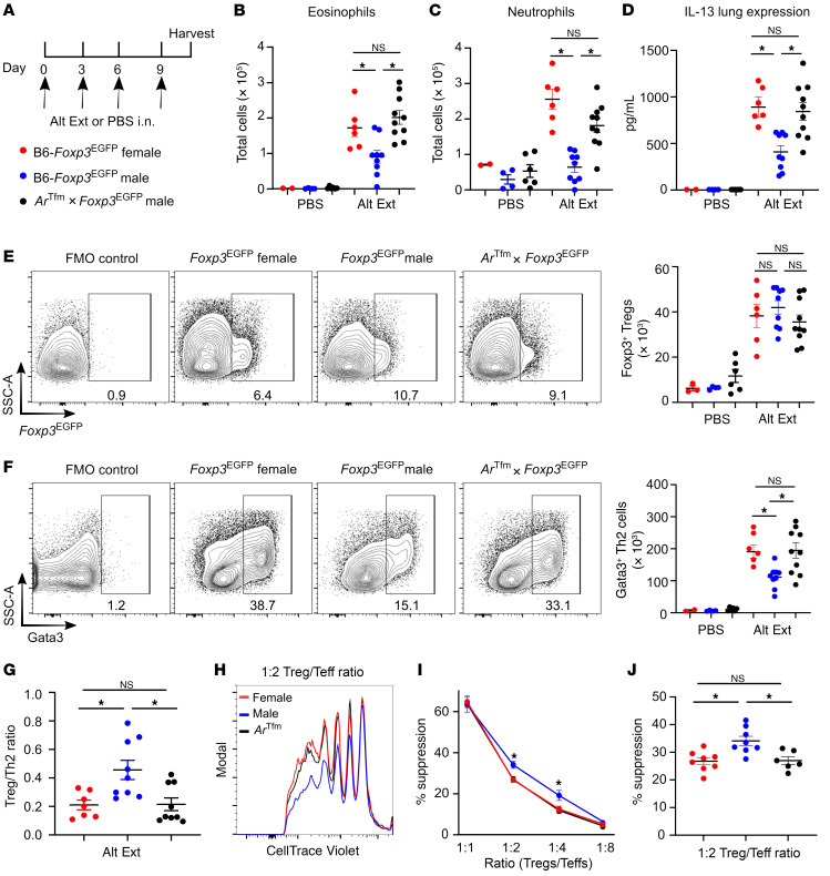
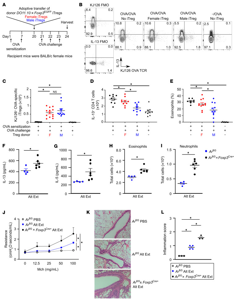
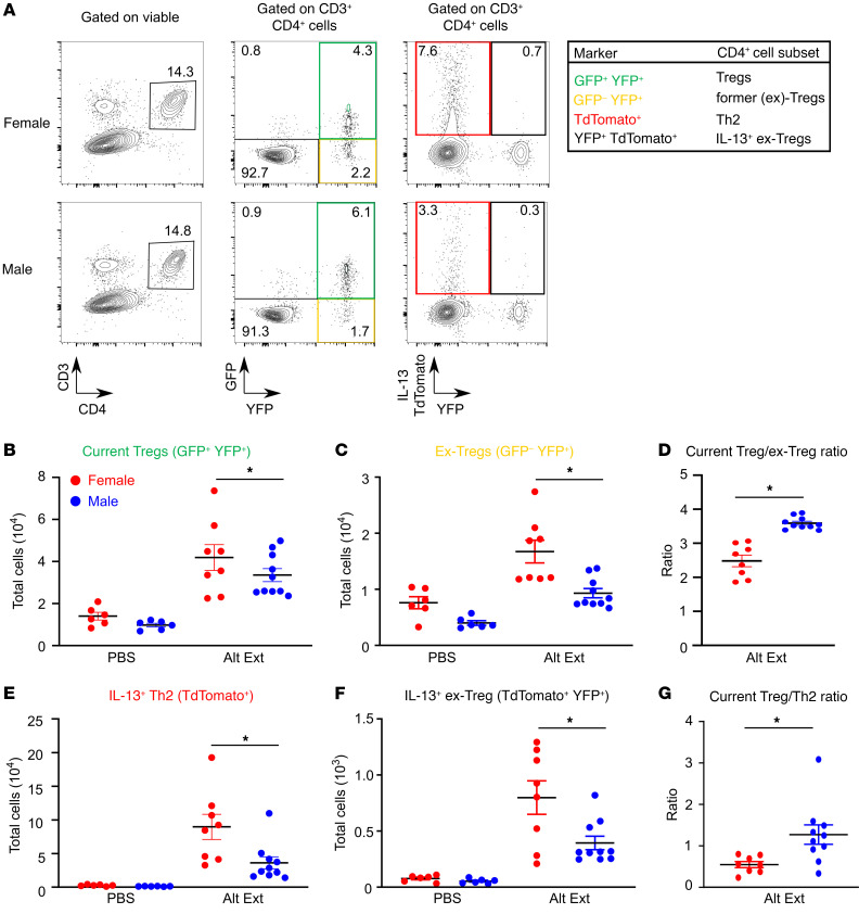
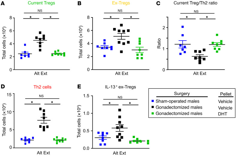
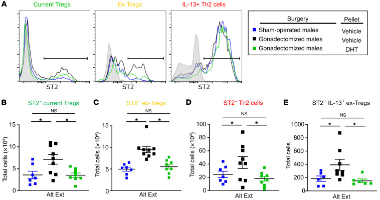
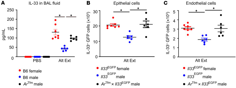
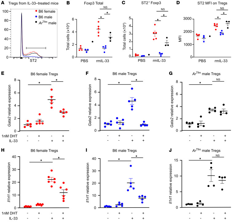
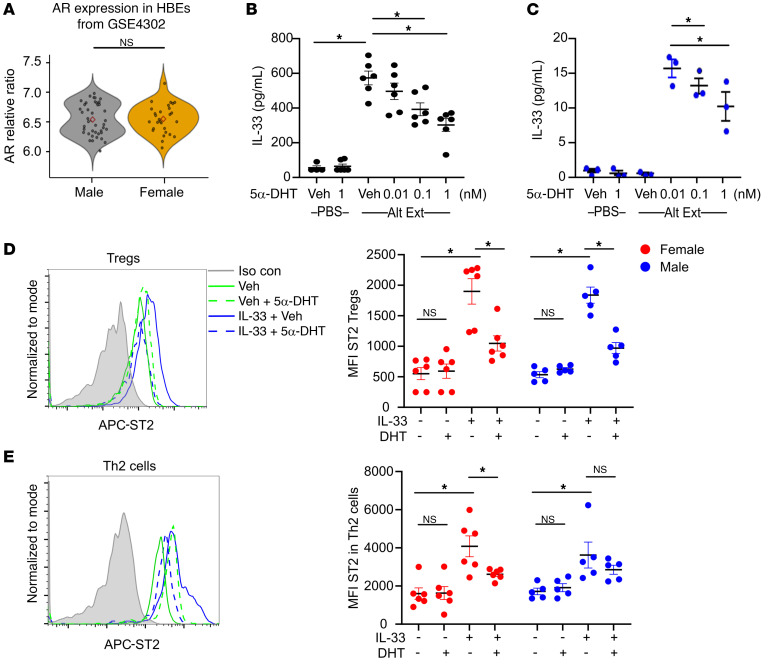
Women have higher prevalence of asthma compared with men. In asthma, allergic airway inflammation is initiated by IL-33 signaling through ST2, leading to increased IL-4, IL-5, and IL-13 production and eosinophil infiltration. Foxp3+ Tregs suppress and ST2+ Tregs promote allergic airway inflammation. Clinical studies showed that the androgen dehydroepiandrosterone (DHEA) reduced asthma symptoms in patients, and mouse studies showed that androgen receptor (AR) signaling decreased allergic airway inflammation. Yet the impact of AR signaling on lung Tregs remains unclear. Using AR-deficient and Foxp3 fate-mapping mice, we determined that AR signaling increased Treg suppression during Alternaria extract (Alt Ext; allergen) challenge by stabilizing Foxp3+ Tregs and limiting the number of ST2+ ex-Tregs and IL-13+ Th2 cells and ex-Tregs. AR signaling also decreased Alt Ext-induced ST2+ Tregs in mice by limiting expression of Gata2, a transcription factor for ST2, and by decreasing Alt Ext-induced IL-33 production from murine airway epithelial cells. We confirmed our findings in human cells where 5α-dihydrotestosterone (DHT), an androgen, decreased IL-33-induced ST2 expression in lung Tregs and decreased Alt Ext-induced IL-33 secretion in human bronchial epithelial cells. Our findings showed that AR signaling stabilized Treg suppressive function, providing a mechanism for the sex difference in asthma.

Keywords: Asthma; Inflammation; Pulmonology; Sex hormones; T cells.


Conflict of interest statement

Conflict of interest: The authors have declared that no conflict of interest exists.

Figures

Figure 1
Figure 1. AR signaling decreases allergic airway inflammation and increases Treg suppressive function.
(A) Experimental design of Alt Ext intranasal challenge model in B6-Foxp3EGFP female, B6-Foxp3EGFP male, and ArTfm Foxp3EGFP male mice. (B and C) Eosinophil and neutrophil counts in BAL fluid. (D) IL-13 protein expression in whole-lung homogenates. (E) Representative dot plots from Alt Ext–challenged mice displaying lung Tregs (Foxp3+CD4+ T cells) with quantitation to the right. (F) Representative dot plots from Alt Ext–challenged mice displaying lung Th2 cells (Gata3+CD4+ T cells) with quantitation to the right. In both E and F, cells were pre-gated on viable CD3+CD4+ T cells. FMO, fluorescence minus one. (G) Ratio of Tregs to Th2 cells in lungs of Alt Ext–challenged mice. (BG) Data are expressed as mean ± SEM; n = 2–10 from 2 experiments. *P < 0.05, ANOVA with Tukey’s post hoc analysis. (HJ) Treg suppression assay using Tregs from B6-Foxp3EGFP female, B6-Foxp3EGFP male, or ArTfm Foxp3EGFP male mice and T effector cells from B6-Foxp3EGFP female mice. (H) Representative samples of T effector cell (Teff) proliferation at 1:2 Treg/T effector ratio. (I and J) Representative experiment and percentage suppression of Tregs at various ratios. (HJ) Data are expressed as mean ± SEM; n = 6–8 from 3 experiments. *P < 0.05, ANOVA with Tukey’s post hoc analysis.
Figure 2
Figure 2. AR signaling increases the suppressive function of Tregs during ongoing allergic airway inflammation.
(A) Experimental model for OVA-induced allergic airway inflammation with adoptive transfer of OVA-specific iTregs from DO11.10 Foxp3EGFP male or female mice and T effectors from DO11.10 female mice. (B) Representative dot plots showing OVA-specific iTregs and IL-13 production in lungs of recipient mice. Cells were pre-gated on viable, CD3+CD4+ cells. (C and D) Quantification of OVA-specific iTregs and IL-13+CD4+ T cells in lungs of recipient mice. (E) Percentage of eosinophils in BAL fluid from recipient mice. (BE) Data are expressed as mean ± SEM; n = 9–11 from 2 separate experiments. *P < 0.05, ANOVA with Tukey’s post hoc analysis. (FI) Arfl/0 and Arfl/0 Foxp3Cre+ male mice were intranasally challenged with Alt Ext, and BAL and lungs were harvested 1 day after the last challenge. (F and G) Whole-lung IL-13 and IL-5 levels. (H and I) BAL eosinophil and neutrophil cell counts. Data are expressed as mean ± SEM; n = 4–6. *P < 0.05, Student’s t test. (JL) Arfl/0 and Arfl/0 Foxp3Cre+ male mice were intranasally challenged with Alt Ext or PBS, and lungs were harvested 2 days after the last Alt Ext or PBS challenge for airway physiology or histology. (J) Airway resistance to increasing concentrations of nebulized methacholine (Mch) was determined. (K and L) Airway inflammation. Whole lungs were stained with H&E, and airway inflammation was scored. Original magnification of images, ×10. (JL) Data are expressed as mean ± SEM; n = 3–4. *P < 0.05, ANOVA with repeated analysis and Tukey’s post hoc test for J and ANOVA with Tukey’s post hoc analysis for L.
Figure 3
Figure 3. Tregs from Alt Ext–challenged male mice are more stable than Tregs from female mice.
Foxp3GFP/YFPIl13TdTomato female and male mice underwent Alt Ext protocol. (A) Representative dot plots of lung CD4+ T cells in Alt Ext–challenged mice showing current Tregs, ex-Tregs, Th2 cells, and IL-13+ ex-Tregs. (B and C) Numbers of current Tregs and ex-Tregs in lungs of mice. (D) Ratio of current Tregs to ex-Tregs. (E and F) Numbers of IL-13+ Th2 cells and IL-13+ ex-Tregs in lungs of mice. (G) Ratio of current Tregs to Th2 cells. Data are mean ± SEM; n = 6–10 from 2 separate experiments. *P < 0.05, ANOVA with Tukey’s post hoc analysis (BF), Student’s t test (G).
Figure 4
Figure 4. AR signaling improves Treg stability during ongoing allergic airway inflammation.
Foxp3GFP/YFPIl13TdTomato male mice underwent gonadectomy or sham operation at 3–4 weeks of age. At 8 weeks old, 5α-DHT or vehicle slow-release pellets were subcutaneously placed into mice. At 11 weeks old, mice underwent Alt Ext protocol. (A and B) Numbers of current Tregs and ex-Tregs in lungs of mice. (C) Ratio of current Tregs to Th2 cells. (D) Numbers of IL-13+ Th2 cells in lungs of mice. (E) Numbers of IL-13+ ex-Tregs in lungs of mice. Data are mean ± SEM; n = 7–9 from 2 separate experiments. *P < 0.05, ANOVA with Tukey’s post hoc analysis.
Figure 5
Figure 5. AR signaling decreases ST2+ Tregs after allergen challenge.
Foxp3GFP/YFPIl13TdTomato male mice underwent gonadectomy or sham operation at 3–4 weeks of age. At 8 weeks old, 5α-DHT or vehicle slow-release pellets were subcutaneously placed into mice. After 3 weeks, mice underwent Alt Ext protocol. (AE) Histograms and quantification of ST2-expressing Tregs, ex-Tregs, Th2 cells, and IL-13+ ex-Tregs. Data are mean ± SEM; n = 7–9 from 2 separate experiments. *P < 0.05, ANOVA with Tukey’s post hoc analysis.
Figure 6
Figure 6. AR signaling decreases IL-33 production and secretion in mice.
(A) B6 female, B6 male, and ArTfm male mice were challenged with Alt Ext or PBS, and BAL fluid was collected 1 hour after the final Alt Ext challenge. IL-33 production was examined by ELISA. (B and C) Il33EGFP female, Il33EGFP male, and ArTfm Il33EGFP male mice underwent the Alt Ext protocol, and IL-33 (GFP)+ epithelial cells and endothelial cells were determined in the lung by flow cytometry. Data are mean ± SEM; n = 6 from 2 separate experiments. *P < 0.05, ANOVA with Tukey’s post hoc analysis.
Figure 7
Figure 7. AR signaling decreases IL-33–induced ST2 expression on Tregs by decreasing Gata2 expression.
B6 female, B6 male, and ArTfm male mice were given rmIL-33 (300 ng) or vehicle (“–”, PBS) every 3 days for 9 days total. On day 10, lungs were harvested for analysis. (A) ST2 expression on lung Tregs. (B and C) Quantification of Tregs and ST2+ Tregs in PBS and rmIL-33 groups of mice. (D) ST2 MFI on Tregs. (EJ) Splenic Tregs were isolated from mice and restimulated in the presence of 5α-DHT (1 nM) and/or rmIL-33 (100 ng/mL) for 3 days. Gata2 and Il1rl1 (ST2) relative expression to vehicle was determined based on expression of Gapdh. Data are mean ± SEM; n = 3–5. *P < 0.05, ANOVA with Tukey’s post hoc analysis.
Figure 8
Figure 8. 5α-DHT decreases IL-33 secretion on human airway epithelial cells and reduces ST2 expression on human lung Tregs.
(A) Ar expression in human bronchial epithelial cells (HBEs) from male and female subjects (control and asthma participants are combined) in the GEO GSE4302 study with statistical analysis by Student’s t test. (B and C) hBE33 cells or primary, differentiated HBEs were treated for 24 hours with vehicle (Veh) or 5α-DHT (0–1 nM) and stimulated with Alt Ext (30 μg/mL) for 1 hour. IL-33 was measured in supernatants by ELISA. hBE33 cells, n = 4 from 2 separate experiments; HBEs from male asthmatic individuals, n = 3 donors. Data are mean ± SEM. *P < 0.05, ANOVA with Tukey’s post hoc analysis. (D and E) CD45+ cells isolated from excised human lungs were stimulated with anti-CD3 and anti-CD28 in the presence of 5α-DHT (1 nM), IL-33 (40 ng/mL), and/or vehicle for 24 hours. Tregs (CD3+CD4+Foxp3+) and Th2 cells (CD3+CD4+Gata3+) were pre-gated, and ST2 MFI was determined for each group. Histogram of ST2 in Tregs or Th2 cells is shown. Data are mean ± SEM; n = 6 female and 5 male donors. *P < 0.05, ANOVA with Tukey’s post hoc analysis.

References

    1. DeBoer MD, et al. Effects of endogenous sex hormones on lung function and symptom control in adolescents with asthma. BMC Pulm Med. 2018;18(1):58–68. doi: 10.1186/s12890-018-0612-x. - DOI - PMC - PubMed
    1. Zein JG, Erzurum SC. Asthma is different in women. Curr Allergy Asthma Rep. 2015;15(6):28. doi: 10.1007/s11882-015-0528-y. - DOI - PMC - PubMed
    1. Zein JG, et al. Benefits of airway androgen receptor expression in human asthma. Am J Respir Cri Care Med. 2021;204(3):285–293. doi: 10.1164/rccm.202009-3720OC. - DOI - PMC - PubMed
    1. Marozkina N, et al. Dehydroepiandrosterone supplementation may benefit women with asthma who have low androgen levels: a pilot study. Pulm Ther. 2019;5(2):213–220. doi: 10.1007/s41030-019-00101-9. - DOI - PMC - PubMed
    1. Zein J, et al. HSD3B1 genotype identifies glucocorticoid responsiveness in severe asthma. Proc Natl Acad Sci U S A. 2020;117(4):2187–2193. doi: 10.1073/pnas.1918819117. - DOI - PMC - PubMed

Publication types

Substances