. 2022 Jan 13;17(1):e0259872.
doi: 10.1371/journal.pone.0259872. eCollection 2022.

Calmodulin binds the N-terminus of the functional amyloid Orb2A inhibiting fibril formation

Affiliations

Calmodulin binds the N-terminus of the functional amyloid Orb2A inhibiting fibril formation

Maria A Soria et al. PLoS One. .

Abstract

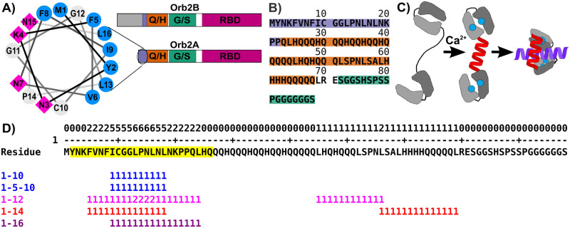
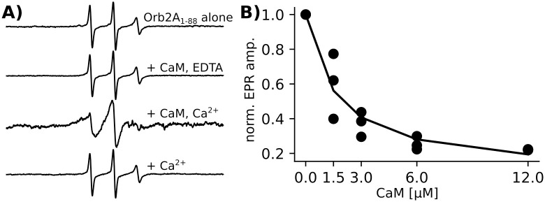
The cytoplasmic polyadenylation element-binding protein Orb2 is a key regulator of long-term memory (LTM) in Drosophila. The N-terminus of the Orb2 isoform A is required for LTM and forms cross-β fibrils on its own. However, this N-terminus is not part of the core found in ex vivo fibrils. We previously showed that besides forming cross-β fibrils, the N-terminus of Orb2A binds anionic lipid membranes as an amphipathic helix. Here, we show that the Orb2A N-terminus can similarly interact with calcium activated calmodulin (CaM) and that this interaction prevents fibril formation. Because CaM is a known regulator of LTM, this interaction could potentially explain the regulatory role of Orb2A in LTM.


Conflict of interest statement

The authors have declared that no competing interests exist.

Figures

Fig 1
Fig 1
A) Domain layout of Orb2A and Orb2B, including the amphipathic helix at the N-terminus of Orb2A. The extended N-terminus of Orb2B is highlighted in gray, the amphipathic region in purple, the Q/H-rich domain in orange, the G/S-rich domain in green, and the RBD in pink. B) Sequence of the N-terminal 88 amino acids of Orb2A used throughout this study. The colors correspond to the domains in A. C) Mechanism by which Ca2+/CaM binds to amphipathic helices. Blue circles represent Ca2+, the red helix is the linker domain of CaM, the purple helix is a hypothetical CaM binding partner. D) CaM binding site prediction using the calmodulin meta-analysis predictor. Overall per residue score that is equal to the number of times a residue is found in a canonical CaM-binding motif is shown in the first line followed by the Orb2A1-88 sequence with the identified potential binding motif highlighted in yellow. Matches to canonical CaM binding motifs are shown at the bottom.
Fig 2
Fig 2. Activated CaM binds Orb2A1-88.
A) EPR spectra of Orb2A1-88 labeled at position 10 (10R1) show no change in linewidth with the addition of CaM together with EDTA or Ca2+alone. However, the linewidth increased with the addition of CaM together with Ca2+. CaM was added at a 2:1 molar ratio to Orb2A1-88, Ca2+ and EDTA were added to a final concentration of 1 mM. EPR spectra intensity was normalized to their central linewidth. B) Change in EPR central line amplitude of 6 μM Orb2A1-88 10R1 with increasing concentrations of CaM. Ca2+ was present in excess (10 mM). Three biological replicates are shown together with a fit to a one-site binding hyperbolic function to determine the dissociation constant.
Fig 3
Fig 3. CaM binds the amphipathic N-terminus of Orb2A1-88.
A) EPR spectra in the absence of CaM (blue) and with the addition of activated CaM (i.e. with 10 mM Ca2+) at a 1:1 molar ratio (red). B) Relative EPR amplitude after the addition of CaM compared to without CaM for each site. The low relative amplitude of the EPR spectra for the amphipathic N-terminus (purple) relative to the Q/H-rich domain (orange), and the G/S-rich domain (green), confirms that CaM binds the N-terminus of Orb2A.
Fig 4
Fig 4. CaM binding prevents Orb2A1-88 aggregation.
Change in EPR spectral amplitude of Orb2A1-88 labeled at residues 10 and 34 with and without activated CaM (1:2 molar ratio, 10 mM Ca2+). Amplitudes were normalized to the first spectrum of each kinetic experiment. In addition, EPR spectra corresponding to the beginning and end of each curve are shown. The EPR amplitude in the absence of CaM decreases significantly for 10R1, i.e. in the center of the amphipathic region, and over time also for 34R1, inside the Q/H-rich domain. This decrease is compatible with aggregation of the protein into cross-β fibrils. No change in amplitude was observed for CaM bound Orb2A1-88 suggesting that this interaction prevented fibril formation.
Fig 5
Fig 5. CaM + Ca2+ prevents fibril formation of Orb2A1-88.
A) ThT fluorescence kinetics of Orb2A1-88 alone (red) and Orb2A1-88 with CaM + Ca2+at a 1:1 molar ratio (blue). Error bars representing the standard deviation of three biological replicates are shown in case they are larger than the marker. The fluorescence in the presence of CaM is high because CaM + Ca2+ induces Tht fluorescence on its own. B) EM images of samples as described in A, taken after 9 days of incubation.
Fig 6
Fig 6. Activated CaM prevents fibril formation of Orb2A1-88.
Addition of Ca2+activated CaM sequesters the N-terminal amphipathic region of Orb2A1-88. This interaction prevents the formation of N-terminal β-sheets necessary for cross-β fibril formation.

References

    1. Keleman K, Krüttner S, Alenius M, Dickson BJ. Function of the Drosophila CPEB protein Orb2 in long-term courtship memory. Nat Neurosci. 2007;10: 1587–93. doi: 10.1038/nn1996 - DOI - PubMed
    1. Krüttner S, Stepien B, Noordermeer JN, Mommaas MA, Mechtler K, Dickson BJ, et al.. Drosophila CPEB Orb2A Mediates Memory Independent of Its RNA-Binding Domain. Neuron. 2012;76: 383–395. doi: 10.1016/j.neuron.2012.08.028 - DOI - PMC - PubMed
    1. Majumdar A, Cesario WC, White-Grindley E, Jiang H, Ren F, Khan MR, et al.. Critical Role of Amyloid-like Oligomers of Drosophila Orb2 in the Persistence of Memory. Cell. 2012;148: 515–29. doi: 10.1016/j.cell.2012.01.004 - DOI - PubMed
    1. Hervás R, Li L, Majumdar A, Fernández-Ramírez M del C, Unruh JR, Slaughter BD, et al.. Molecular Basis of Orb2 Amyloidogenesis and Blockade of Memory Consolidation. PLOS Biol. 2016;14: e1002361. doi: 10.1371/journal.pbio.1002361 - DOI - PMC - PubMed
    1. Hervas R, Rau MJ, Park Y, Zhang W, Murzin AG, Fitzpatrick JAJ, et al.. Cryo-EM structure of a neuronal functional amyloid implicated in memory persistence in Drosophila. Science. 2020;367: 1230–1234. doi: 10.1126/science.aba3526 - DOI - PMC - PubMed

MeSH terms

LinkOut - more resources