Review
. 2022 Mar;226(3):366-378.
doi: 10.1016/j.ajog.2021.11.1357. Epub 2022 Jan 10.

Clinical Opinion: The diagnosis and management of suspected fetal growth restriction: an evidence-based approach

Affiliations
Review

Clinical Opinion: The diagnosis and management of suspected fetal growth restriction: an evidence-based approach

Christoph C Lees et al. Am J Obstet Gynecol. 2022 Mar.

Abstract

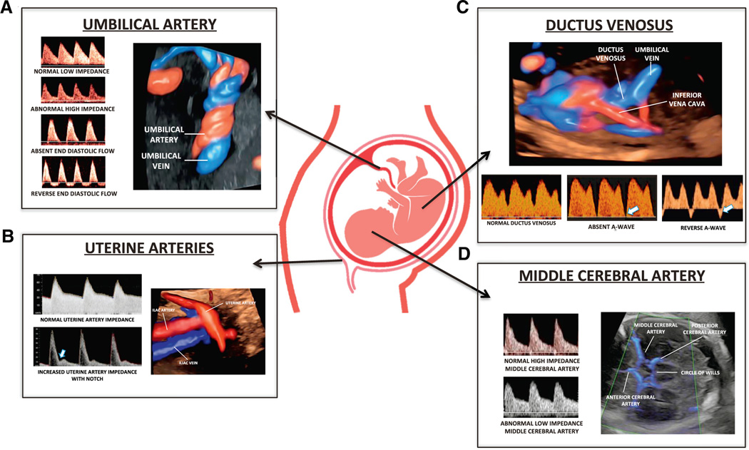
This study reviewed the literature about the diagnosis, antepartum surveillance, and time of delivery of fetuses suspected to be small for gestational age or growth restricted. Several guidelines have been issued by major professional organizations, including the International Society of Ultrasound in Obstetrics and Gynecology and the Society for Maternal-Fetal Medicine. The differences in recommendations, in particular about Doppler velocimetry of the ductus venosus and middle cerebral artery, have created confusion among clinicians, and this review has intended to clarify and highlight the available evidence that is pertinent to clinical management. A fetus who is small for gestational age is frequently defined as one with an estimated fetal weight of <10th percentile. This condition has been considered syndromic and has been frequently attributed to fetal growth restriction, a constitutionally small fetus, congenital infections, chromosomal abnormalities, or genetic conditions. Small for gestational age is not synonymous with fetal growth restriction, which is defined by deceleration of fetal growth determined by a change in fetal growth velocity. An abnormal umbilical artery Doppler pulsatility index reflects an increased impedance to flow in the umbilical circulation and is considered to be an indicator of placental disease. The combined finding of an estimated fetal weight of <10th percentile and abnormal umbilical artery Doppler velocimetry has been widely accepted as indicative of fetal growth restriction. Clinical studies have shown that the gestational age at diagnosis can be used to subclassify suspected fetal growth restriction into early and late, depending on whether the condition is diagnosed before or after 32 weeks of gestation. The early type is associated with umbilical artery Doppler abnormalities, whereas the late type is often associated with a low pulsatility index in the middle cerebral artery. A large randomized clinical trial indicated that in the context of early suspected fetal growth restriction, the combination of computerized cardiotocography and fetal ductus venosus Doppler improves outcomes, such that 95% of surviving infants have a normal neurodevelopmental outcome at 2 years of age. A low middle cerebral artery pulsatility index is associated with an adverse perinatal outcome in late fetal growth restriction; however, there is no evidence supporting its use to determine the time of delivery. Nonetheless, an abnormality in middle cerebral artery Doppler could be valuable to increase the surveillance of the fetus at risk. We propose that fetal size, growth rate, uteroplacental Doppler indices, cardiotocography, and maternal conditions (ie, hypertension) according to gestational age are important factors in optimizing the outcome of suspected fetal growth restriction.

Keywords: Disproportionate Intrauterine Growth Intervention Trial at Term; Doppler velocimetry; Prospective Observational Trial to Optimize Pediatric Health; Trial of Umbilical and Fetal Flow in Europe; abdominal circumference; cardiotocography; cesarean delivery; ductus venosus; fetal biometry; fetal death; fetal distress; fetal growth; longitudinal; middle cerebral artery; neurodevelopmental outcome; randomized controlled trial; short-term variation; small for gestational age; systematic review; umbilical artery Doppler; umbilical artery pH; uterine artery.


Conflict of interest statement

The other authors report no conflict of interest.

Figures

FIGURE 1
FIGURE 1. Perinatal mortality according to birthweight percentile
Adapted from Vasak et al.
FIGURE 2
FIGURE 2. Patterns of fetal growth in SGA and FGR
The figure illustrates 3 growth velocity pattern scenarios: (1) small for gestational age (blue circles), (2) suspected FGR with an EFW of <3rd percentile (red triangles), and (3) deceleration of growth velocity (orange diamonds). If clinical judgment is based only on time point evaluation of the fetal size, then the 2 last examinations (arrows: the blue circles and red triangles) would be both classified as FGR and the orange diamonds would be classified as appropriate for gestational age. However, the blue circles indicate a fetus with a maintained growth velocity, which indicates a constitutionally small fetus, whereas the orange diamonds indicate a fetus with a deceleration of growth velocity, although above the 10th percentile. The study by Sovio et al showed that in fetuses with a birthweight of <10th percentile (blue circles and red triangles), only fetuses with the AC growth velocity in lowest deciles (red triangles) have a significantly higher risk of adverse outcome than the fetuses in the control group; moreover, there was no difference between fetuses with a birthweight of <10th percentile and maintained growth velocity (blue circles) and fetuses in the control group in terms of adverse outcomes. To identify suspected FGR, Doppler evaluation of uteroplacental-fetal circulation is important (Figure 3). AC, abdominal circumference; EFW, estimated fetal weight; FGR, fetal growth restriction; GA, gestational age.
FIGURE 3
FIGURE 3. Uteroplacental-fetal vascular components evaluated with Doppler velocimetry
The figure represents the main uteroplacental-fetal vascular components that can be evaluated with Doppler velocimetry and that play a role in the diagnosis, surveillance, and/or time of delivery of fetuses with suspected FGR. A, The umbilical arteries reflect the impedance to blood flow on the fetal side of the placenta. With increasing GA, umbilical impedance becomes progressively reduced. With progressive damage of the chorionic villi, the umbilical artery impedance will increase until absent end-diastolic flow and reversed end-diastolic flow supervene. The umbilical artery PI does not correlate with fetal hypoxemia. B, The uterine artery blood flow reflects the resistance to the blood flow at the maternal side of the placenta. Similarly, to the umbilical artery uterine impedance decreases with GA in physiological states, whereas the resistance to the blood flow remains elevated in abnormal placental invasion and is predictive of FGR especially when associated with preeclampsia. C, The ductus venosus represents a fetal shunt that carries oxygenated blood at high velocity from the umbilical vein, through the foramen ovale into the left atrium and then left ventricle of the heart; the crista dividens separates this oxygenated blood from the deoxygenated blood of the inferior vena cava that passes through the right ventricle and systemic circulation. Physiologically, it appears tri-phasic with low impedance during the a-wave. An absent or reversed ductus venosus a-wave is an expression of an increased end-diastolic intracardiac pressure because of increased resistance in afterload and/or an expression of progressive dilatation of the ductus venosus to increase the delivery of the oxygenated blood directly to the myocardium and fetal brain. Changes in the ductus venosus waveform are associated with increased risk of perinatal mortality and morbidity. D, The middle cerebral artery normally shows high impedance to blood flow. Cerebral blood flow redistribution is a fetal adaptive response to hypoxemia and/or hypercapnia and can be identified as low impedance in the middle cerebral artery. For further discussion on Doppler velocimetry and indices, please refer to the recently published International Society of Ultrasound in Obstetrics and Gynecology guideline by Bhide et al. FGR, fetal growth restriction; GA, gestational age; PI, pulsatility index.
FIGURE 4
FIGURE 4. Different clinical and biophysical characteristics of early and late suspected FGR
The figure illustrates the different clinical and biophysical characteristics of early and late suspected FGR. Early suspected FGR (<32 weeks of gestation) is a rare condition and is characterized by placental insufficiency and reduced placental vascular perfusion on maternal (uterine arteries) and fetal interface (umbilical arteries). The fetus is usually very small, and profound Doppler changes might be present, with absent or reverse ductus venosus a-wave, which are associated with an increased risk of perinatal mortality and morbidity. The main challenge is the management because of prematurity and/or maternal hypertension (70% of cases). Late suspected FGR (>32 weeks of gestation) is a frequent condition caused by placental dysfunction. Vascular perfusion at the maternal and fetal interface is usually normal. The fetus is not necessarily very small, and signs of cerebral blood flow redistribution (low impedance in the middle cerebral artery and/or altered ratio to the umbilical artery) might be the only Doppler sign. The main challenge is the diagnosis. AEDF, absent end-diastolic flow; BPP, biophysical profile; cCTG, computerized cardiotocography; FGR, fetal growth restriction; REDF, reversed end-diastolic flow; STV, short-term variation; UA, umbilical artery.
FIGURE 5
FIGURE 5. Schematic representation of the TRUFFLE randomization and the “safety net”
The inclusion criteria were singleton fetus between 26 0/7 and 31 6/7 weeks of gestation with an estimated fetal weight >500 g, an abdominal circumference <10th percentile, and an umbilical artery >95th percentile, with a normal ductus venosus pulsatility index and short-term variation. Chromosomal and congenital anomalies constituted an exclusion criteria. TRUFFLE, Trial of Umbilical and Fetal Flow in Europe.
FIGURE 6
FIGURE 6. Main characteristics of computerized cardiotocography and STV
The figure summarizes the main characteristics of computerized cardiotocography and STV. An example in which STV is useful is represented. fHRV, fetal heart rate variability; STV, short-term variation.

References

    1. McCowan LM, Figueras F, Anderson NH. Evidence-based national guidelines for the management of suspected fetal growth restriction: comparison, consensus, and controversy. Am J Obstet Gynecol 2018;218:S855–68. - PubMed
    1. Chauhan SP, Gupta LM, Hendrix NW, Berghella V; American College of Obstetricians and Gynecologists. Intrauterine growth restriction: comparison of American College of Obstetricians and Gynecologists practice bulletin with other national guidelines. Am J Obstet Gynecol 2009;200:409.e1–6. - PubMed
    1. Society for Maternal-Fetal Medicine, Martins JG, Biggio JR, Abuhamad A. Society for Maternal-Fetal Medicine Consult Series #52: diagnosis and management of fetal growth restriction: (replaces clinical guideline number 3, April 2012). Am J Obstet Gynecol 2020;223: B2–17. - PubMed
    1. Lees CC, Stampalija T, Baschat A, et al. ISUOG Practice Guidelines: diagnosis and management of small-for-gestational-age fetus and fetal growth restriction. Ultrasound Obstet Gynecol 2020;56:298–312. - PubMed
    1. Gordijn SJ, Beune IM, Thilaganathan B, et al. Consensus definition of fetal growth restriction: a Delphi procedure. Ultrasound Obstet Gynecol 2016;48:333–9. - PubMed

Publication types