

Unveiling the potential of melt electrowriting in regenerative dental medicine
- PMID: 35026478
- PMCID: PMC11046422
- DOI: 10.1016/j.actbio.2022.01.010
Unveiling the potential of melt electrowriting in regenerative dental medicine
Abstract
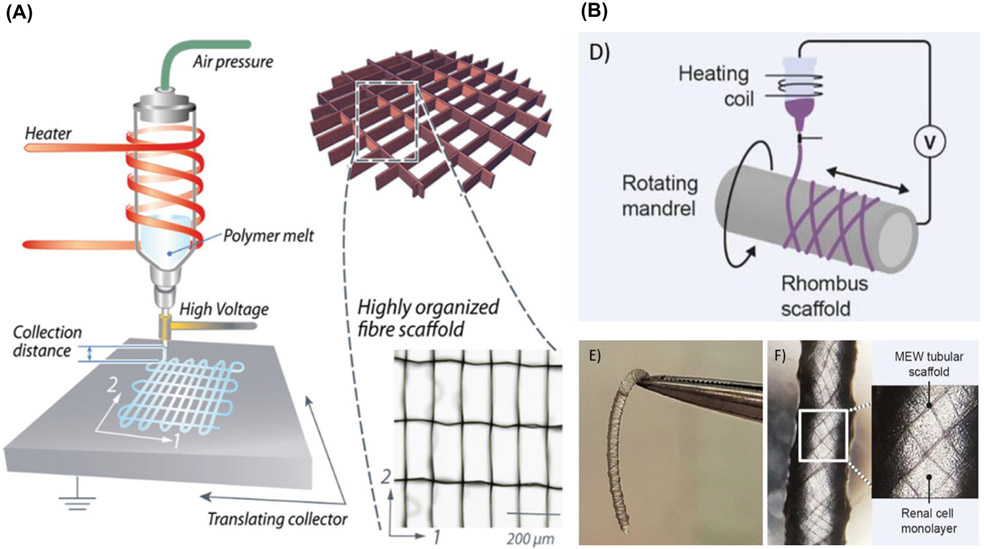
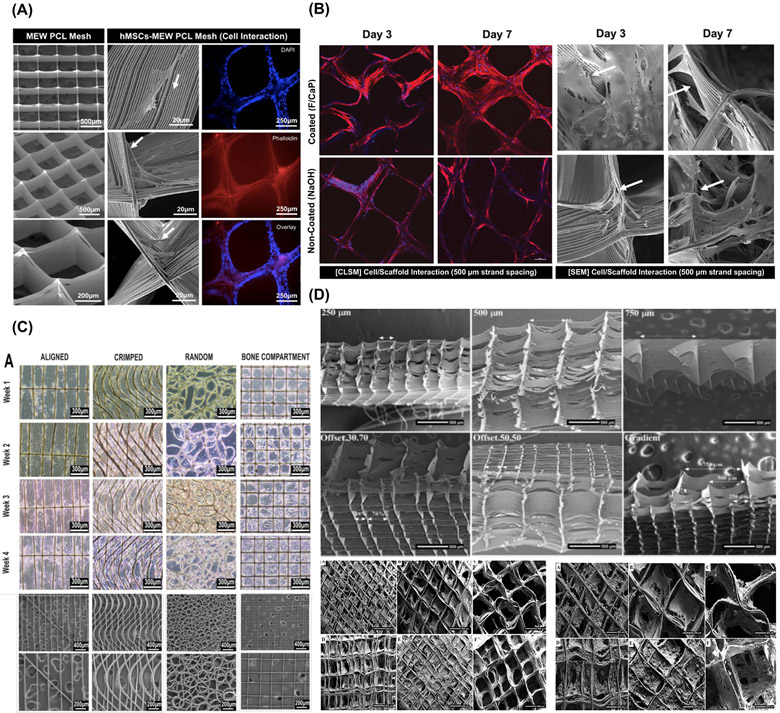
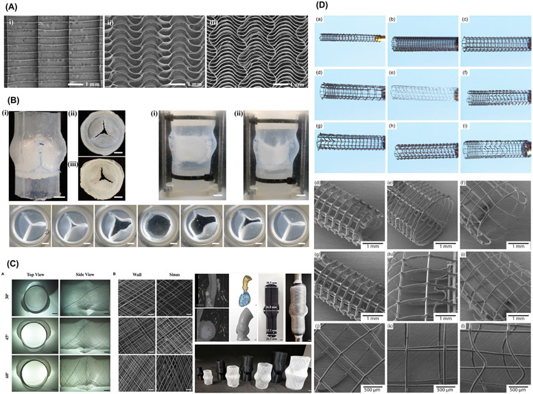
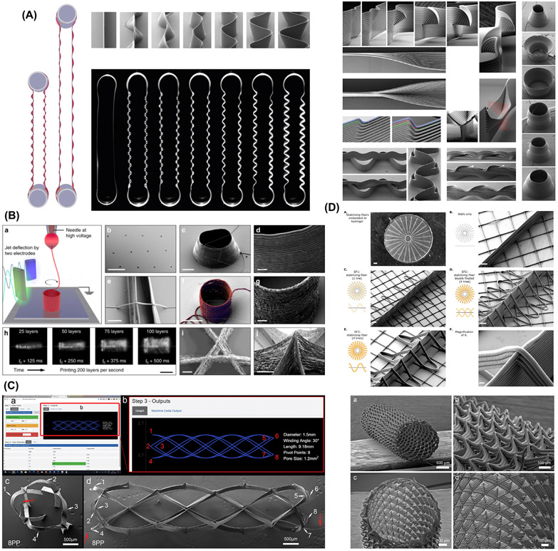
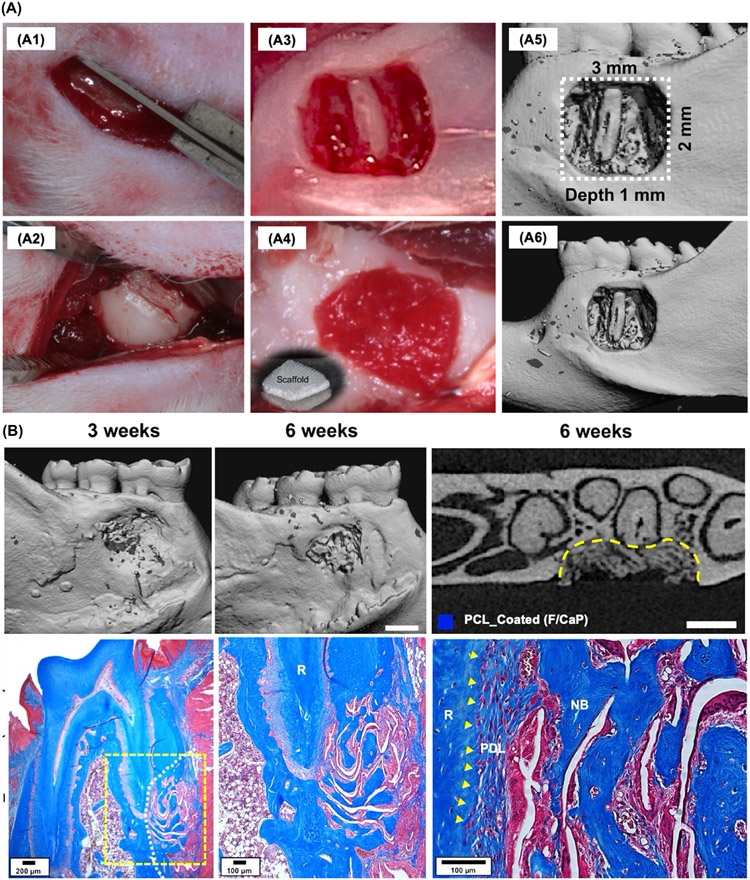
For nearly three decades, tissue engineering strategies have been leveraged to devise effective therapeutics for dental, oral, and craniofacial (DOC) regenerative medicine and treat permanent deformities caused by many debilitating health conditions. In this regard, additive manufacturing (AM) allows the fabrication of personalized scaffolds that have the potential to recapitulate native tissue morphology and biomechanics through the utilization of several 3D printing techniques. Among these, melt electrowriting (MEW) is a versatile direct electrowriting process that permits the development of well-organized fibrous constructs with fiber resolutions ranging from micron to nanoscale. Indeed, MEW offers great prospects for the fabrication of scaffolds mimicking tissue specificity, healthy and pathophysiological microenvironments, personalized multi-scale transitions, and functional interfaces for tissue regeneration in medicine and dentistry. Excitingly, recent work has demonstrated the potential of converging MEW with other AM technologies and/or cell-laden scaffold fabrication (bioprinting) as a favorable route to overcome some of the limitations of MEW for DOC tissue regeneration. In particular, such convergency fabrication strategy has opened great promise in terms of supporting multi-tissue compartmentalization and predetermined cell commitment. In this review, we offer a critical appraisal on the latest advances in MEW and its convergence with other biofabrication technologies for DOC tissue regeneration. We first present the engineering principles of MEW and the most relevant design aspects for transition from flat to more anatomically relevant 3D structures while printing highly-ordered constructs. Secondly, we provide a thorough assessment of contemporary achievements using MEW scaffolds to study and guide soft and hard tissue regeneration, and draw a parallel on how to extrapolate proven concepts for applications in DOC tissue regeneration. Finally, we offer a combined engineering/clinical perspective on the fabrication of hierarchically organized MEW scaffold architectures and the future translational potential of site-specific, single-step scaffold fabrication to address tissue and tissue interfaces in dental, oral, and craniofacial regenerative medicine. STATEMENT OF SIGNIFICANCE: Melt electrowriting (MEW) techniques can further replicate the complexity of native tissues and could be the foundation for novel personalized (defect-specific) and tissue-specific clinical approaches in regenerative dental medicine. This work presents a unique perspective on how MEW has been translated towards the application of highly-ordered personalized multi-scale and functional interfaces for tissue regeneration, targeting the transition from flat to anatomically-relevant three-dimensional structures. Furthermore, we address the value of convergence of biofabrication technologies to overcome the traditional manufacturing limitations provided by multi-tissue complexity. Taken together, this work offers abundant engineering and clinical perspectives on the fabrication of hierarchically MEW architectures aiming towards site-specific implants to address complex tissue damage in regenerative dental medicine.
Keywords: 3D printing; Biofabrication; Dentistry; Melt electrowriting; Regeneration; Scaffolds.
Copyright © 2022 Acta Materialia Inc. Published by Elsevier Ltd. All rights reserved.
Conflict of interest statement
Declaration of Competing Interest The authors declare no competing financial interest or with respect to the authorship and/or publication of this article.
Figures


Similar articles
-
Recent advances in non-planar collectors for melt electrowriting (MEW): creating physiologically relevant scaffold structures for tissue engineering.Prog Biomed Eng (Bristol). 2025 Aug 14;7(4). doi: 10.1088/2516-1091/adf78b. Prog Biomed Eng (Bristol). 2025. PMID: 40759155 Review.
-
Converging functionality: Strategies for 3D hybrid-construct biofabrication and the role of composite biomaterials for skeletal regeneration.Acta Biomater. 2021 Sep 15;132:188-216. doi: 10.1016/j.actbio.2021.03.008. Epub 2021 Mar 10. Acta Biomater. 2021. PMID: 33713862 Review.
-
Melt electrowriting of PLA, PCL, and composite PLA/PCL scaffolds for tissue engineering application.Sci Rep. 2022 Nov 19;12(1):19935. doi: 10.1038/s41598-022-24275-6. Sci Rep. 2022. PMID: 36402790 Free PMC article.
-
Simultaneous Micropatterning of Fibrous Meshes and Bioinks for the Fabrication of Living Tissue Constructs.Adv Healthc Mater. 2019 Apr;8(7):e1800418. doi: 10.1002/adhm.201800418. Epub 2018 Jun 17. Adv Healthc Mater. 2019. PMID: 29911317 Free PMC article.
-
Combining multi-scale 3D printing technologies to engineer reinforced hydrogel-ceramic interfaces.Biofabrication. 2020 Feb 19;12(2):025014. doi: 10.1088/1758-5090/ab69d9. Biofabrication. 2020. PMID: 31918421 Free PMC article.
Cited by
-
Recent Advances in Scaffolds for Guided Bone Regeneration.Biomimetics (Basel). 2024 Mar 1;9(3):153. doi: 10.3390/biomimetics9030153. Biomimetics (Basel). 2024. PMID: 38534838 Free PMC article. Review.
-
Influences of Process Parameters of Near-Field Direct-Writing Melt Electrospinning on Performances of Polycaprolactone/Nano-Hydroxyapatite Scaffolds.Polymers (Basel). 2022 Aug 19;14(16):3404. doi: 10.3390/polym14163404. Polymers (Basel). 2022. PMID: 36015663 Free PMC article.
-
A holistic model for melt electrowritten three-dimensional structured materials based on residual charge.Int J Bioprint. 2022 Dec 28;9(2):656. doi: 10.18063/ijb.v9i2.656. eCollection 2023. Int J Bioprint. 2022. PMID: 37065672 Free PMC article.
-
Melt electrowriting of bioglass-laden poly(ε-caprolactone) scaffolds for bone regeneration.J Mater Chem B. 2025 Mar 20;13(12):3864-3875. doi: 10.1039/d4tb02835j. J Mater Chem B. 2025. PMID: 39992649 Free PMC article.
-
Tissue-specific melt electrowritten polymeric scaffolds for coordinated regeneration of soft and hard periodontal tissues.Bioact Mater. 2022 Apr 22;19:268-281. doi: 10.1016/j.bioactmat.2022.04.013. eCollection 2023 Jan. Bioact Mater. 2022. PMID: 35574052 Free PMC article.
References
-
- Petrovic V, Zivkovic P, Petrovic D, Stefanovic V, Craniofacial bone tissue engineering, Oral Surg. Oral Med. Oral Pathol. Oral Radiol 114 (2012) e1–e9. - PubMed
-
- Bottino MC, Thomas V, Schmidt G, Vohra YK, Chu T-MG, Kowolik MJ, Janowski GM, Recent advances in the development of GTR/GBR membranes for periodontal regeneration–a materials perspective, Dent. Mater 28 (2012) 703–721. - PubMed
Publication types
MeSH terms
Grants and funding
LinkOut - more resources
Full Text Sources
Research Materials
