. 2022 Jan;10(1):e003461.
doi: 10.1136/jitc-2021-003461.

Large-scale manufacturing and characterization of CMV-CD19CAR T cells

Affiliations

Large-scale manufacturing and characterization of CMV-CD19CAR T cells

Xiuli Wang et al. J Immunother Cancer. 2022 Jan.

Abstract

Background: Adoptive transfer of CD19-specific chimeric antigen receptor (CD19CAR) T cells can induce dramatic disease regression in patients with B cell malignancies. CD19CAR T cell therapy may be limited by insufficient engraftment and persistence, resulting in tumor relapse. We previously demonstrated a proof of principle that cytomegalovirus (CMV)-specific T cells can be isolated and enriched prior to CD19CAR transduction to produce CMV-CD19CAR T cells, and that these CMV-CD19CAR T cells can be expanded in vivo through CMV vaccination, resulting in better tumor control in a murine model. Here we developed a clinical platform for generating CMV-CD19CAR T cells.

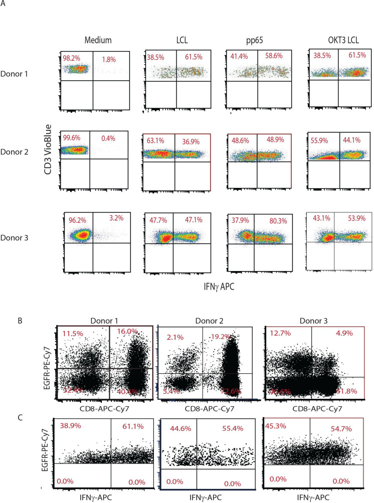
Methods: Peripheral blood mononuclear cells (PBMCs) collected from CMV-seropositive healthy donors were stimulated with a good manufacturing practices-grade PepTivator overlapping CMVpp65 peptide pool and enriched for CMV-responsive interferon γ (IFNγ)+T cells using IFNγ Catchmatrix, within the CliniMACS Prodigy Cytokine Capture System (Miltenyi Biotec). Resulting CMV-specific T cells were transduced with a lentiviral vector encoding a second generation CD19R:CD28:ζ/EGFRt CAR and expanded with interleukin 2 (IL-2) and IL-15 for 15 days before characterization.

Results: CMV-specific T cells were enriched from 0.8%±0.5 of input PBMC to 76.3%±11.6 in nine full-scale qualification runs (absolute yield of 4.2±3.3×10⁶ IFNγ+T cells from an input of 1×10⁹ PBMCs). Average CD19CAR transduction efficiency of CMV-specific T cells was 27.0%±14.2 in the final products, which underwent rapid expansion, resulting in a total cell dose of 6.2±0.9 × 10⁶ CD19CAR-tranduced T cells with CMV specificity (ie, functionally bispecific). CMV-CD19CAR T cells were polyclonal, expressed memory markers but had low expression of exhaustion markers, responded to both CD19 and CMVpp65 stimulation with rapid proliferation and exhibited antigen-specific effector functions against both CD19-expressing tumors and CMVpp65 antigen. The final products passed release criteria for clinical use.

Conclusions: We demonstrated the feasibility of our large-scale platform for generating CMV-CD19CAR T cells for clinical application. We plan to initiate a clinical trial at City of Hope using CMV-CD19CAR T cells for patients with intermediate/high-grade B cell non-Hodgkin's lymphoma immediately after autologous hematopoietic cell transplantation followed by vaccination with a novel CMV vaccine based on Modified Vaccinia Ankara (Triplex) 28 days and 56 days post-T cell infusion.

Keywords: adoptive; cell engineering; chimeric antigen; hematologic neoplasms; immunotherapy; receptors; vaccination.


Conflict of interest statement

Competing interests: None declared.

Figures

Figure 1
Figure 1
Clinical scale CliniMACS Prodigy immunomagnetic selection of cytomegalovirus (CMV)-specific T cells. (A) Development of clinically feasible platform for derivation of CMV-CD19CAR T cells. (B) Leukapheresis products were obtained from CMV-immune healthy donors and peripheral blood mononuclear cells (PBMCs) were isolated and purified by density gradient centrifugation over Ficoll-Paque. PBMCs (1×10⁹) were loaded on the closed and automated CliniMACS Prodigy cytokine capture system (CCS) system (Miltenyi Biotec) and CMV-specific T cells were isolated. The percentage of live, purified interferon γ (IFNγ) positive cells (black) over pre-enrichment (red) from three representative donors are depicted. (C) Phenotypic characterization of IFNγ-positive gated cells are presented. Data from three representative donors are presented. APC, allophycocyanin; FITC, fluorescein; PE, phycoerythrin.
Figure 2
Figure 2
Generation of cytomegalovirus (CMV)-specific CD19 chimeric antigen receptor (CAR) T cells. (A) One day after isolation, CMV-specific T cells were transduced with a second-generation CD19CAR/CD28/EGFRt lentiviral vector with IgG4 Fc hinge region mutations (L235E; N297Q) at MOI 3 in the presence of 50 U/mL IL-2, 1 ng/mL IL-15, and 100 µg/mL protamine sulfate. The transduced CMV-CD19CAR T cells formed clusters as seen under light microscopy in 10 × magnification. (B) Total viable cell count was performed every other day for 14 days to assess in vitro expansion. (C) The proportion of CAR +cells in the products after 14 days of in vitro expansion was analyzed with flow cytometry. EGFR +cells after subtraction of isotype control (red) are presented. Representative data from multiple donors are presented.
Figure 3
Figure 3
Phenotypic characterization of cytomegalovirus (CMV)-specific CD19CAR T cells. (A) The final products were stained with antibodies against indicated immune receptors. Per cent positive cells (means±SD) under chimeric antigen receptor (CAR) +population from three different T cell products are presented. (B) CMV-CD19CAR T cells were labeled with CellTrace Violet dye (CTV) and co-cultured with 8000-cGy-irradiated lymphoblastoid cell line (LCL) cells and 3700 cGy-irradiated, autologous CMVpp65-pepmix-pulsed peripheral blood mononuclear cells (PBMCs) at 1:1 ratio for 7 days. Dilution of CTV was analyzed by flow cytometry. Per cent positive cells (means±SD) under CAR +population from three different donor-derived T cell products are depicted. Replicates were conducted for each product. (C) To simultaneously quantify specific mRNA levels and both cell surface and intracellular proteins, CMV-CD19CAR T cells were incubated overnight with either pp65pepmix-loaded autologous PBMC to stimulate the CMV-specific T cell receptor (TCR) (pp65 stim) or LCL cells to stimulate the CD19CAR (CAR stim). Cells were subsequently stained with Erbitux and anti-CD3 antibodies to detect CAR +and CD3+cells. After fixation, cells were permeabilized with PrimeFlow RNA permeabilization buffer in the presence of RNAase inhibitors followed by intracellular interferon γ (IFNγ) staining. Next day, the cells were labeled with RNA probes including genes that are related T cell persistence (telomerase Tert, IL-2), memory (KLF2, TCF7, Lef1), homing (CCR7, ITAG5, ITAG3), and activation/exhaustion (PDCD1). Gene expression on gated EGFR (CAR+) and intracellular IFNγ double positive cells were analyzed with flow cytometry (10,000 cells were acquired for analysis).
Figure 4
Figure 4
Dual functionality of cytomegalovirus (CMV)--specific CD19CAR T cells against CD19 and CMV pp65 antigens. (A) Propagated CMV-CD19CAR T cells were stimulated overnight with CD19 +tumor (lymphoblastoid cell line (LCL)) and CMVpp65pepmix-pulsed PBMC. OKT3 LCL and medium were used as positive and negative controls, respectively. The frequency of intracellular interferon γ (IFNγ) in CD3 +T cells from three representative CMV-CD19CAR T cell products are depicted. (B) The composition of T cell subsets in the final CMV-CD19CAR T cell products within the CD3 +population and (C) the relative proportion of IFNγ+cells within the chimeric antigen receptor (CAR) + populations from three different donors are presented.
Figure 5
Figure 5
Cytolytic potency and effector cytokine secretion by cytomegalovirus (CMV)-specific CD19CAR T cells. (A) CMV-CD19CAR T cells were co-cultured with GFP+ target cells (lymphoblastoid cell line (LCL)) at different effector:target ratios for 3 days and remaining eGFP+ tumor cells were analyzed by flow cytometry. KG1a (CD19-) and OKT3 LCL were used as negative and positive controls, respectively. Cytotoxicity was normalized to the tumor only cultures. Data from three different donor-derived CMV-CD19CAR T cells are presented. (B) To test the proliferative capacity against CD19+ (LCL) and CMVpp65 targets, CMV-CD19CAR T cells were labeled with CellTrace Violet dye (CTV) and co-cultured with 8000-cGy-irradiated LCL cells and 3700 cGy-irradiated autologous CMVpp65-pepmix pulsed peripheral blood mononuclear cells (PBMCs) at 1:1 ratio for 7 days. Dilution of CTV was analyzed by flow cytometry. Data from two representative CMV-CD19CAR T cell products are depicted. (C) CMV-CD19CAR T cells (1×10⁵) were cocultured overnight in 96-well tissue culture plates with 1×10⁵ LCL, OKT3 LCL, KG1a, or autologous CMVpp65-pepmix pulsed PBMCs. Supernatants were collected and analyzed with the Luminex IS100 bead array technology for Human Cytokine 30-Plex according to the manufacturer’s instructions. Mean±SD from three different donors are presented. #: out of range above. ***p<0.001. **p<0.01, *p<0.05.
Figure 6
Figure 6
Cytomegalovirus (CMV)-specific CD19CAR T cells possess stronger effector function against CD19 +tumor compared with monospecific CD19CAR T cells. (A) CMV-CD19CAR T cells were also generated from the same donor and were expanded for 14 days. Both CD19CAR T cells and CMV-CD19CAR T cells were then stimulated with CD19+target cells (lymphoblastoid cell line (LCL)) overnight and intracellular interferon γ (IFNγ) was analyzed by flow cytometry. Data from three different donor-derived CD19CAR and CMV-CD19CAR T cells are presented. CMV-CD19 chimeric antigen receptor (CAR) T cells without stimulation were used as controls. (B) To generate conventional monospecific CD19CAR T cells, peripheral blood mononuclear cells (PBMCs) were stimulated with anti-CD3/CD28 and transduced with the same CD19CAR lentivirus and expanded for 14 days. CD19CAR T cells were then stimulated with CD19+target cells (LCL) overnight and intracellular IFNγ was analyzed by flow cytometry. Data from three different donor-derived CD19CAR T cells are presented. CD19CAR T cells without stimulation were used as controls.

References

    1. Walter EA, Greenberg PD, Gilbert MJ, et al. . Reconstitution of cellular immunity against cytomegalovirus in recipients of allogeneic bone marrow by transfer of T-cell clones from the donor. N Engl J Med 1995;333:1038–44. 10.1056/NEJM199510193331603 - DOI - PubMed
    1. Leen AM, Christin A, Myers GD, et al. . Cytotoxic T lymphocyte therapy with donor T cells prevents and treats adenovirus and Epstein-Barr virus infections after haploidentical and matched unrelated stem cell transplantation. Blood 2009;114:4283–92. 10.1182/blood-2009-07-232454 - DOI - PMC - PubMed
    1. Bollard CM, Kuehnle I, Leen A, et al. . Adoptive immunotherapy for posttransplantation viral infections. Biol Blood Marrow Transplant 2004;10:143–55. 10.1016/j.bbmt.2003.09.017 - DOI - PubMed
    1. Feuchtinger T, Opherk K, Bethge WA, et al. . Adoptive transfer of pp65-specific T cells for the treatment of chemorefractory cytomegalovirus disease or reactivation after haploidentical and matched unrelated stem cell transplantation. Blood 2010;116:4360–7. 10.1182/blood-2010-01-262089 - DOI - PubMed
    1. Dudley ME, Rosenberg SA. Adoptive-cell-transfer therapy for the treatment of patients with cancer. Nat Rev Cancer 2003;3:666–75. 10.1038/nrc1167 - DOI - PMC - PubMed

Publication types

Substances

LinkOut - more resources