. 2022 Jun;77(6):616-620.
doi: 10.1136/thoraxjnl-2020-216807. Epub 2022 Jan 13.

Inhaled corticosteroids reduce senescence in endothelial progenitor cells from patients with COPD

Affiliations

Inhaled corticosteroids reduce senescence in endothelial progenitor cells from patients with COPD

Koralia Paschalaki et al. Thorax. 2022 Jun.

Abstract

Cellular senescence contributes to the pathophysiology of chronic obstructive pulmonary disease (COPD) and cardiovascular disease. Using endothelial colony-forming-cells (ECFC), we have demonstrated accelerated senescence in smokers and patients with COPD compared with non-smokers. Subgroup analysis suggests that ECFC from patients with COPD on inhaled corticosteroids (ICS) (n=14; eight on ICS) exhibited significantly reduced senescence (Senescence-associated-beta galactosidase activity, p21CIP1), markers of DNA damage response (DDR) and IFN-γ-inducible-protein-10 compared with patients with COPD not on ICS. In vitro studies using human-umbilical-vein-endothelial-cells showed a protective effect of ICS on the DDR, senescence and apoptosis caused by oxidative stress, suggesting a protective molecular mechanism of action of corticosteroids on endothelium.

Keywords: COPD pharmacology.


Conflict of interest statement

Competing interests: Part of this work was funded by an academic AstraZeneca AB Project Grant.

Figures
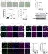
Figure 1
Figure 1
Reduced senescence and DNA damage response in patients with COPD on ICS). (A) Senescence-associated-β-galactosidase activity was assessed as a marker of cellular senescence in ECFC samples from healthy non-smokers (n=11), healthy smokers (n=6) and patients with COPD (n=14, 8 on ICS). ECFC from smokers and patients with COPD exhibited increased senescence compared with non-smokers; Kruskal-Wallis test followed by Dunn’s multiple comparison test (graph A - left panel). ECFC from patients with COPD on ICS exhibited reduced senescence compared with ECFC from patients with COPD not receiving ICS; Mann-Whitney U test (graph A - right panel); (scale bars 100 µm). (B) mRNA levels for p21 were measured by real-time PCR in ECFC from patients with COPD (n=11, 5 on ICS). Ribosomal protein L13a was used for normalisation. (C) p21 protein levels were quantified by Western blotting. α-tubulin was measured for normalisation (n=3 in each group). (D) Representative images of immunofluorescence staining of ECFC from COPD-ICS versus COPD-no ICS patients for p21 (cyan, left panel) and p16 (cyan, right panels). DAPI (blue) was used as nuclear staining and VE-cadherin (magenta) as an endothelial marker. (E) DNA damage was assessed by immunofluorescence staining for 53BP1 (green) and γ-H2AX (red) (n=3 in each group). DAPI (blue) was used as a nuclear marker and VE-cadherin (magenta) as an endothelial marker. The number of distinct nuclear immunofluorescent foci (see arrows) per nucleus was counted in at least 5 z-stack images and 20 cells, using a 63 × objective lens (scale bars = 20 µm). Mann-Whitney U test; ****P<0.0001; COPD, chronic obstructive pulmonary disease; ECFC, endothelial colony-forming cells; ICS, inhaled corticosteroids.
Figure 2
Figure 2
Corticosteroids may exert a protective effect against premature endothelial senescence caused by oxidative stress—reduced senescence associated secretory phenotype involving Interferon-gamma (IFN-γ) inducible-protein-10 (IP-10) in patients with chronic obstructive pulmonary disease (COPD) on inhaled corticosteroids. (A) Human-umbilical-vein-endothelial cells (HUVECs) were cultured in the presence or absence of increasing doses of budesonide (10−10−10−6mol/L) or control vehicle (DMSO). Following 1-hour pretreatment, HUVECs were exposed to 50 µM of H2O2 for 1.5 hours to induce stress-induced premature senescence. SA-β-gal activity was measured after 72 hours from H2O2 treatment; n=3 (scale bars 100 µm). (B) Apoptosis was quantified by measuring caspase-3/7 Glo activity after 24 hours from H2O2 treatment as described in (A); n=3 (samples in triplicate). (C) γ-H2AX protein after 24 hours from H2O2 treatment as described in (A); n=3. (A–C) Friedman test followed by Dunn’s multiple comparison test. (D) HUVECs were stained for 53BP1 and for DRAQ5 (nuclear marker) at 60 min, 240 min, 24 hours or 48 hours after exposure to H2O2 treatment. The number of 53BP1 positive cells and the number of foci per cell were quantified (scale bars 20 µm). (E) IL-8 and IFN-γ-inducible protein 10 (IP-10 or CXCL10) were measured in supernatant samples from ECFC cultures under baseline conditions from non-smokers (n=5) and patients with COPD (n=8; n=5 COPD-ICS) by a Luminex assay. Pearson correlation coefficient of IL-8 and IP-10 with SA-b-gal activity. Reduced expression of IP-10 was observed in samples from patients with COPD on ICS compared with patients with COPD not receiving ICS; Kruskal-Wallis test followed by Dunn’s multiple comparison test. (F) Immunofluorescence staining for IP-10 (green) and p21 (cyan). DAPI (blue) was used as a nuclear marker and VE-cadherin (magenta) as an endothelial marker. At least 5 z-stack images and 20 cells per ECFC sample were analysed for IP-10 and p21 using a 63 × objective lens in ECFC from COPD-ICS (n=4) and COPD-no ICS (n=3) (scale bars = 20 µm); Mann-Whitney U test; ****p<0.0001. DMSO, Dimethyl sulfoxide; ECFC, endothelial colony forming cells; ICS, inhaled corticosteroids; IL-8, interleukin-8.

References

    1. Barnes PJ, Baker J, Donnelly LE. Cellular senescence as a mechanism and target in chronic lung diseases. Am J Respir Crit Care Med 2019;200:556–64. 10.1164/rccm.201810-1975TR - DOI - PubMed
    1. Paschalaki KE, Randi AM. Recent advances in endothelial colony forming cells toward their use in clinical translation. Front Med 2018;5:295. 10.3389/fmed.2018.00295 - DOI - PMC - PubMed
    1. Paschalaki KE, Starke RD, Hu Y, et al. . Dysfunction of endothelial progenitor cells from smokers and chronic obstructive pulmonary disease patients due to increased DNA damage and senescence. Stem Cells 2013;31:2813–26. 10.1002/stem.1488 - DOI - PMC - PubMed
    1. Paschalaki KE, Zampetaki A, Baker JR, et al. . Downregulation of microRNA-126 augments DNA damage response in cigarette smokers and patients with chronic obstructive pulmonary disease. Am J Respir Crit Care Med 2018;197:665–8. 10.1164/rccm.201706-1304LE - DOI - PMC - PubMed
    1. Loke YK, Kwok CS, Singh S. Risk of myocardial infarction and cardiovascular death associated with inhaled corticosteroids in COPD. Eur Respir J 2010;35:1003–21. 10.1183/09031936.00095909 - DOI - PubMed

Publication types

MeSH terms

Substances