

Extension of human GCSF serum half-life by the fusion of albumin binding domain
- PMID: 35027593
- PMCID: PMC8758692
- DOI: 10.1038/s41598-021-04560-6
Extension of human GCSF serum half-life by the fusion of albumin binding domain
Abstract
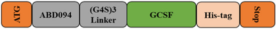
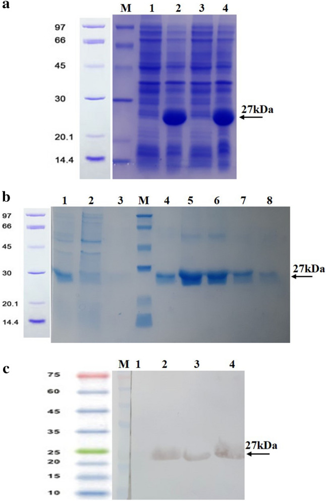
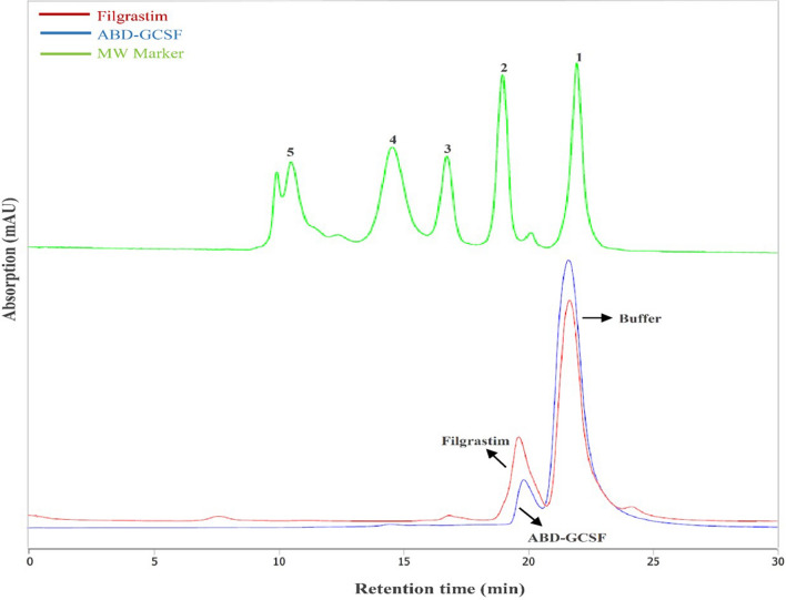
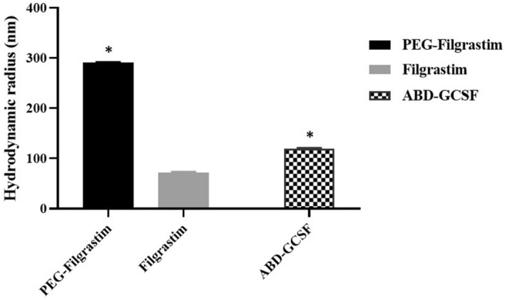
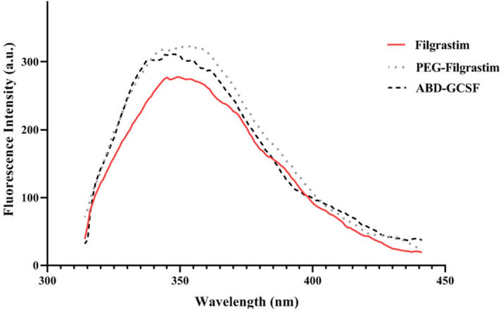
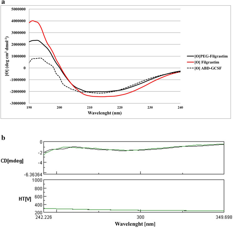
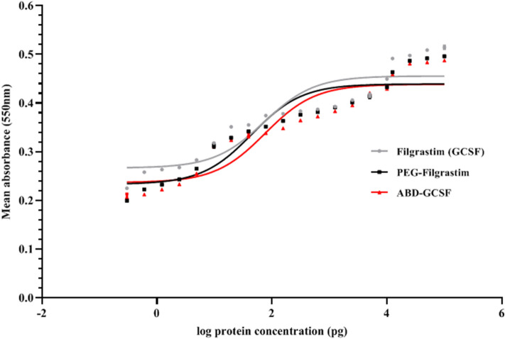
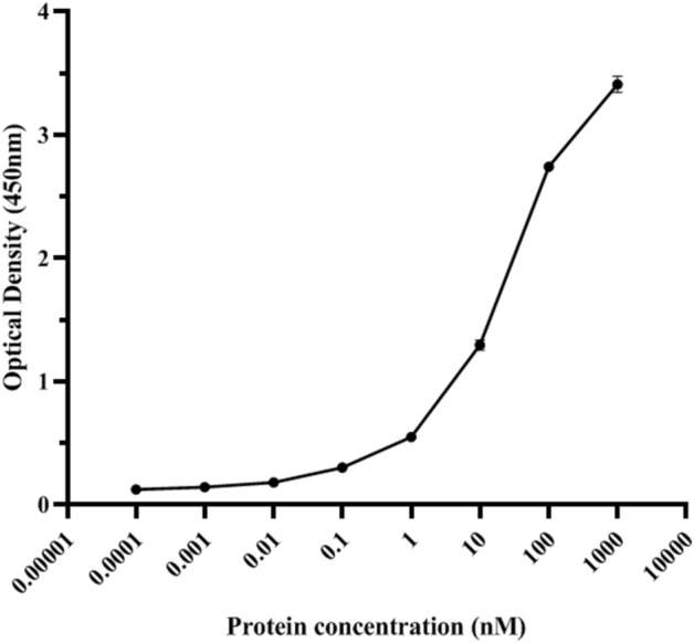
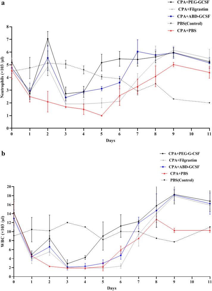
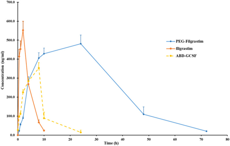
Granulocyte colony stimulating factor (GCSF) can decrease mortality of patients undergo chemotherapy through increasing neutrophil counts. Many strategies have been developed to improve its blood circulating time. Albumin binding domain (ABD) was genetically fused to N-terminal end of GCSF encoding sequence and expressed as cytoplasmic inclusion bodies within Escherichia coli. Biological activity of ABD-GCSF protein was assessed by proliferation assay on NFS-60 cells. Physicochemical properties were analyzed through size exclusion chromatography, circular dichroism, intrinsic fluorescence spectroscopy and dynamic light scattering. Pharmacodynamics and pharmacokinetic properties were also investigated in a neutropenic rat model. CD and IFS spectra revealed that ABD fusion to GCSF did not significantly affect the secondary and tertiary structures of the molecule. DLS and SEC results indicated the absence of aggregation formation. EC50 value of the ABD-GCSF in proliferation of NFS-60 cells was 75.76 pg/ml after 72 h in comparison with control GCSF molecules (Filgrastim: 73.1 pg/ml and PEG-Filgrastim: 44.6 pg/ml). Animal studies of ABD-GCSF represented improved serum half-life (9.3 ± 0.7 h) and consequently reduced renal clearance (16.1 ± 1.4 ml/h.kg) in comparison with Filgrastim (1.7 ± 0.1 h). Enhanced neutrophils count following administration of ABD-GCSF was comparable with Filgrastim and weaker than PEG-Filgrastim treated rats. In vitro and in vivo results suggested the ABD fusion as a potential approach for improving GCSF properties.
© 2022. The Author(s).
Conflict of interest statement
The authors declare no competing interests.
Figures
References
-
- Schneider A, Kuhn HG, Schäbitz WR. A role for G-CSF (Granulocyte-colony stimulating factor) in the central nervous system. Cell Cycle. 2005;4:1753–1757. - PubMed
-
- Tiwari K, et al. Refolding of recombinant human granulocyte colony stimulating factor: Effect of cysteine/cystine redox system. Indian. J. Biochem. Biophys. 2012;49:285–288. - PubMed
-
- Solaroglu I, Cahill J, Jadhav V, Zhang JH. A novel neuroprotectant granulocyte-colony stimulating factor. Stroke. 2006;37:1123–1128. - PubMed
Publication types
MeSH terms
Substances
LinkOut - more resources
Full Text Sources
