. 2022 Jan 13;12(1):688.
doi: 10.1038/s41598-021-03837-0.

Neuropathogenicity of non-viable Borrelia burgdorferi ex vivo

Affiliations

Neuropathogenicity of non-viable Borrelia burgdorferi ex vivo

Geetha Parthasarathy et al. Sci Rep. .

Abstract

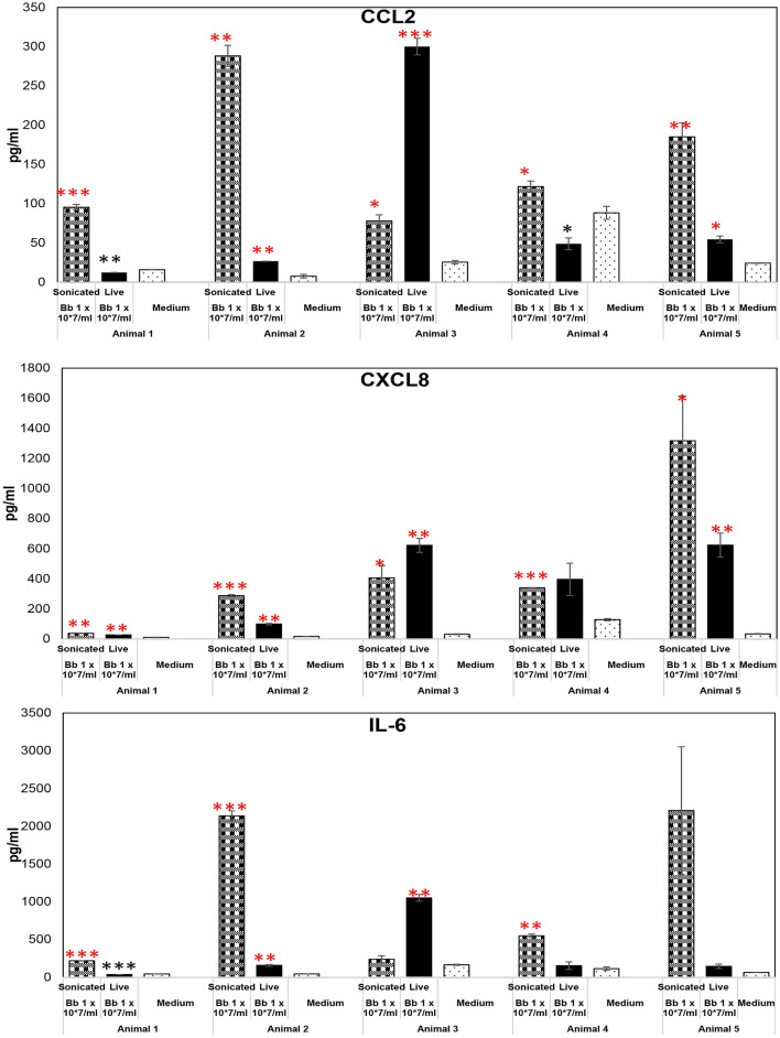
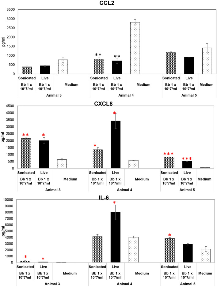
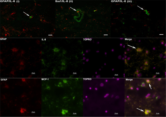
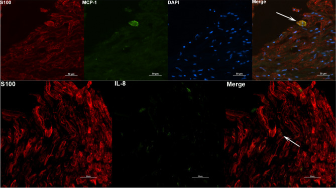
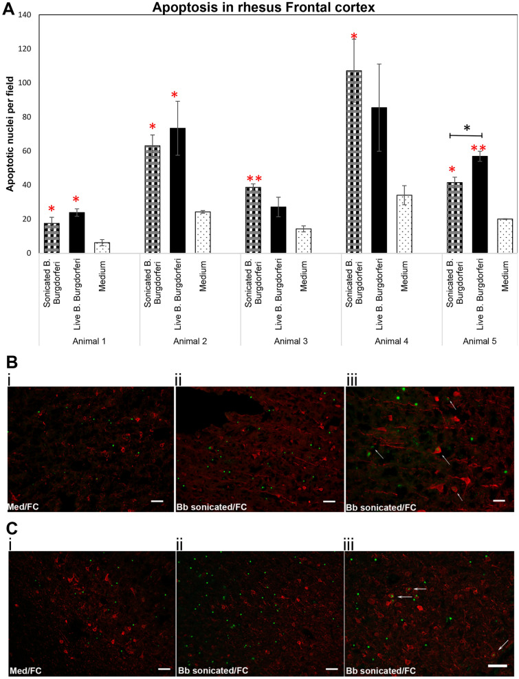
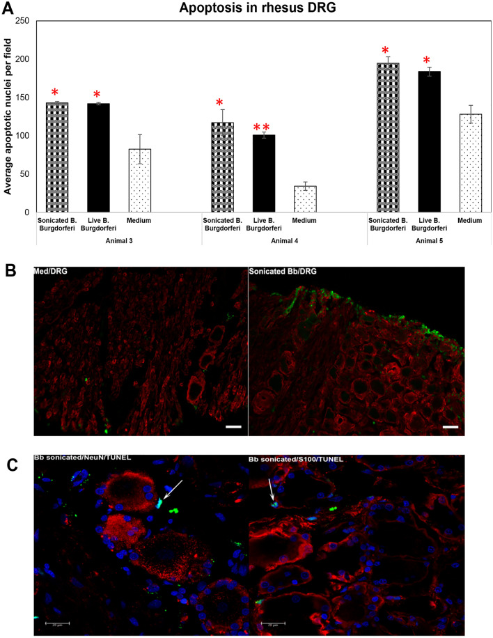
Even after appropriate treatment, a proportion of Lyme disease patients suffer from a constellation of symptoms, collectively called Post-Treatment Lyme Disease Syndrome (PTLDS). Brain PET scan of patients with PTLDS have demonstrated likely glial activation indicating persistent neuroinflammatory processes. It is possible that unresolved bacterial remnants can continue to cause neuroinflammation. In previous studies, we have shown that non-viable Borrelia burgdorferi can induce neuroinflammation and apoptosis in an oligodendrocyte cell line. In this follow-up study, we analyze the effect of sonicated remnants of B. burgdorferi on primary rhesus frontal cortex (FC) and dorsal root ganglion (DRG) explants. Five FC and three DRG tissue fragments from rhesus macaques were exposed to sonicated B. burgdorferi and analyzed for 26 inflammatory mediators. Live bacteria and medium alone served as positive and negative control, respectively. Tissues were also analyzed for cell types mediating inflammation and overall apoptotic changes. Non-viable B. burgdorferi induced significant levels of several inflammatory mediators in both FC and DRG, similar to live bacteria. However, the levels induced by non-viable B. burgdorferi was often (several fold) higher than those induced by live ones, especially for IL-6, CXCL8 and CCL2. This effect was also more profound in the FC than in the DRG. Although the levels often differed, both live and dead fragments induced the same mediators, with significant overlap between FC and DRG. In the FC, immunohistochemical staining for several inflammatory mediators showed the presence of multiple mediators in astrocytes, followed by microglia and oligodendrocytes, in response to bacterial remnants. Staining was also seen in endothelial cells. In the DRG, chemokine/cytokine staining was predominantly seen in S100 positive (glial) cells. B. burgdorferi remnants also induced significant levels of apoptosis in both the FC and DRG. Apoptosis was confined to S100 + cells in the DRG while distinct neuronal apoptosis was also detected in most FC tissues in response to sonicated bacteria. Non-viable B. burgdorferi can continue to be neuropathogenic to both CNS and PNS tissues with effects likely more profound in the former. Persistence of remnant-induced neuroinflammatory processes can lead to long term health consequences.


Conflict of interest statement

The authors declare no competing interests.

Figures

Figure. 1
Figure. 1
Non-viable B. burgdorferi mediates inflammatory mediator release from rhesus FC. The effect of live or sonicated B. burgdorferi on inflammatory mediator release from rhesus frontal cortex tissues is shown in this figure. Ex vivo cultured tissues were exposed to indicated treatments for 4 h. Supernatants were analyzed for CCL2, CXCL8 and IL-6. Results from tissues obtained from 5 different animals is shown. ***p < 0.001; **p < 0.01; *p < 0.05. Significantly increased levels over medium control is denoted by the red asterisks, while black asterisks indicate they were below medium levels. Bars represent standard deviation. Live bacteria were used as a positive control, while medium exposed tissues were used as a negative control throughout the study.
Figure 2
Figure 2
Effect of non-viable B. burgdorferi on inflammatory mediator production from the DRG. Rhesus DRG tissues were exposed to live or sonicated B. burgdorferi for 4 h. Supernatants were analyzed for CCL2, CXCL-8 and IL-6. Results from tissues obtained from 3 animals is shown. ***p < 0.001; **p < 0.01; *p < 0.05. Significantly increased levels over medium control is denoted by the red asterisks. Black asterisks represent significantly lower levels compared to medium control. Bars represent standard deviation.
Figure 3
Figure 3
Immunohistochemical staining for inflammatory mediators and brain cells. Tissue sections from Frontal cortex were stained for IL-6, IL-8 and MCP-1, along with cell type specific markers. Top panel (iiii) shows IL-6/ IL-8 (green) staining likely within endothelial cells. GFAP or Iba1 are stained in red. Bar represents 25 µm. IL-6 staining in GFAP + astrocytes (i), IL-8 staining in Iba1 + microglia (ii) are also seen, as yellow colocalization (arrows). In top panel (iii) the arrow points to IL-8 staining (green) in likely endothelial cells. Middle Panel shows IL-8 (green) stain in GFAP positive (red) cells co-localizing as yellow. The bottom panel similarly shows MCP-1 staining (green) in astrocytes stained with GFAP (red), resulting in yellow colocalization. Top panel is from Animal 1, middle panel from Animal 2 and bottom panel from Animal 3.
Figure 4
Figure 4
Identity of DRG cells responding to non-viable B. burgdorferi. DRG tissues were stained for immune mediators (green) and S100 (red). Positive staining for MCP-1 and IL-8 was seen in S100 positive cells, shown as yellow colocalizations (White arrows). Top panel is from Animal 3 tissue while the bottom panel is from Animal 4 DRG.
Figure 5
Figure 5
Non-viable B. burgdorferi mediates cell death in the rhesus FC. Rhesus frontal cortex tissues were exposed to live or sonicated B. burgdorferi for 4 h. TUNEL assays were carried out on cryosections as described in Materials and methods. Results from 5 different animal tissues is shown in (A). Bars represent standard deviation. Red asterisks indicate significantly higher levels of apoptosis compared to tissue sections exposed to medium alone. Black asterisk shows significantly higher level of apoptosis in tissues exposed to live B. burgdorferi compared to sonicated bacteria in that animal tissue. ***p < 0.001; **p < 0.01; *p < 0.05. Immunofluorescence photographs of TUNEL assays from Animal 1 (B) and Animal 2 (C). i and ii show the overall difference in apoptotic nuclei (green) between tissue sections exposed to Medium (i) and sonicated bacteria (ii). (iii) shows apoptotic nuclei within MAP2 stained (red) neurons indicated by white arrows. Bar represents 50 µm except for B iii (25 µm).
Figure 6
Figure 6
Apoptosis in the rhesus DRG in response to non-viable B. burgdorferi. Apoptosis data from three different rhesus DRG tissues in response to live or sonicated B. burgdorferi is shown (A). Red asterisks indicate statistically significant increases over medium controls. Bar represents standard deviation. (B) Immunofluorescence pictures (Animal 4) showing overall increase in apoptosis (green) in tissues exposed to sonicated B. burgdorferi in comparison to the tissue sections that received medium only. Bar represents 50 µm. (C) confocal images (Animal 5) showing colocalization (cyan, arrows) of TUNEL positive nuclei (green) with DAPI (blue), seen in tissue sections also stained for neurons (NeuN, red) or glial cells (S100, red). Positive apoptotic nuclei are seen only in S100 positive or non-neuronal areas.

References

    1. Kugeler KJ, Schwartz AM, Delorey MJ, Mead PS, Hinckley AF. Estimating the frequency of lyme disease diagnoses, United States, 2010–2018. Emerg. Infect. Dis. 2021;27:616–619. - PMC - PubMed
    1. Wormser GP, Dattwyler RJ, Shapiro ED, Halperin JJ, Steere AC, Klempner MS, Krause PJ, Bakken JS, Strle F, Stanek G, et al. The clinical assessment, treatment, and prevention of lyme disease, human granulocytic anaplasmosis, and babesiosis: Clinical practice guidelines by the Infectious Diseases Society of America. Clin. Infect. Dis. 2006;43:1089–1134. - PubMed
    1. Rebman AW, Bechtold KT, Yang T, Mihm EA, Soloski MJ, Novak CB, Aucott JN. The clinical, symptom, and quality-of-life characterization of a well-defined group of patients with posttreatment lyme disease syndrome. Front. Med. 2017;4:224. - PMC - PubMed
    1. Rebman AW, Aucott JN. Post-treatment lyme disease as a model for persistent symptoms in lyme disease. Front. Med. 2020;7:57. - PMC - PubMed
    1. Aucott JN, Rebman AW, Crowder LA, Kortte KB. Post-treatment Lyme disease syndrome symptomatology and the impact on life functioning: Is there something here? Qual. Life Res. 2013;22:75–84. - PMC - PubMed

Publication types

MeSH terms