Review
. 2021 Jun 30;52(2):e4064808.
doi: 10.25100/cm.v52i2.4808. eCollection 2021 Apr-Jun.

Damage control in abdominal vascular trauma

Affiliations
Review

Damage control in abdominal vascular trauma

Alberto García et al. Colomb Med (Cali). .

Abstract

In patients with abdominal trauma who require laparotomy, up to a quarter or a third will have a vascular injury. The venous structures mainly injured are the vena cava (29%) and the iliac veins (20%), and arterial vessels are the iliac arteries (16%) and the aorta (14%). The initial approach is performed following the ATLS principles. This manuscript aims to present the surgical approach to abdominal vascular trauma following damage control principles. The priority in a trauma laparotomy is bleeding control. Hemorrhages of intraperitoneal origin are controlled by applying pressure, clamping, packing, and retroperitoneal with selective pressure. After the temporary bleeding control is achieved, the compromised vascular structure must be identified, according to the location of the hematomas. The management of all lesions should be oriented towards the expeditious conclusion of the laparotomy, focusing efforts on the bleeding control and contamination, with a postponement of the definitive management. Their management of vascular injuries includes ligation, transient bypass, and packing of selected low-pressure vessels and bleeding surfaces. Subsequently, the unconventional closure of the abdominal cavity should be performed, preferably with negative pressure systems, to reoperate once the hemodynamic alterations and coagulopathy have been corrected to carry out the definitive management.

En pacientes con trauma de abdomen que requieren laparotomía, hasta una cuarta o tercera parte, habrán sufrido una lesión vascular. Las estructuras venosas principalmente lesionadas son la vena cava y las iliacas, y de vasos arteriales, son las iliacas y la aorta. El abordaje de este tipo de heridas vasculares se puede ser difícil en el contexto de un paciente hemodinámicamente inestable ya que requiera medidas rápidas que permita controlar la exanguinación del paciente. El objetivo de este manuscrito es presentar el abordaje del trauma vascular abdominal de acuerdo con la filosofía de cirugía de control de daños. La primera prioridad en una laparotomía por trauma es el control de la hemorragia. Las hemorragias de origen intraperitoneal se controlan con compresión, pinzamiento o empaquetamiento, y las retroperitoneales con compresión selectiva. Posterior al control transitorio de la hemorragia, se debe identificar la estructura vascular comprometida, de acuerdo con la localización de los hematomas. El manejo de las lesiones debe orientarse a la finalización expedita de la laparotomía, enfocado en el control de la hemorragia y contaminación, con aplazamiento del manejo definitivo. Lo pertinente al tratamiento de las lesiones vasculares incluyen la ligadura, derivación transitoria y el empaquetamiento de vasos seleccionados de baja presión y de superficies sangrantes. Posteriormente se debe realizar el cierre no convencional de la cavidad abdominal, preferiblemente con sistemas de presión negativa, para consecutivamente reoperar una vez corregidas las alteraciones hemodinámicas y la coagulopatía para realizar el manejo definitivo.

Keywords: Vascular system injuries; amputation; anastomosis, surgical; aorta, abdominal; compartment syndromes; dissection; femoral vein; hematoma; hemodynamics; ischemia; laparotomy; mesenteric artery, superior; mesenteric veins; peritoneal cavity; portal vein; renal veins; reperfusion; vena cava, inferior; viscera.


Conflict of interest statement

Conflict of Interest: The authors declare that they have no conflict of interest.

Figures

Figure 1
Figure 1. Surgical management of abdominal vascular trauma. 1. During the preoperative period the occlusion can be done using a Resuscitative Endovascular Balloon Occlusion of the Aorta (REBOA). 2. Elements for prevention and management of hypothermia, rapid infusion, blood recovery, and autotransfusion, vascular access, procedures in different cavities and monitoring are included. 3. Consider autotransfusion. 4. Due to persistent hypotension or the need for proximal vascular control in arterial lesions. 5. According to the location of the hematoma, the site of bleeding, the trajectory of the wound and other injured organs. 6. List of vessels that can be ligated are in the Table 5. 7. Closure of the fascia or skin increases the risk of abdominal hypertension. 8. Non-emergent reoperation should be done as soon as hypothermia, coagulopathy, and perfusion are adjusted in patients with transient shunts or vascular ligation.
Figure 2
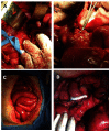
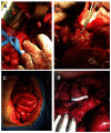
Figure 2. Penetrating trauma to the right iliac artery. A) Transient bleeding control with local pressure. B) Definitive vascular control with atraumatic clamps
Figure 3
Figure 3. Clamping of the supraceliac aorta. A) Entering the lesser sac. The left hepatic lobe has been pushed back, the gastrohepatic ligament (Ghl) has been divided with a blunt maneuver with the index finger. The body of the pancreas (Pa) and the fibers of the diaphragmatic hiatus (DC) are observed. Li Liver. St stomach B) Guided by the pulsations of the aorta, the surgeon longitudinally dissects the fibers of the diaphragm at the hiatus (D). He surrounds the vessel on each side and clamps it between his index and middle fingers. Occasionally, some diaphragm fibers must be separated with an electrosurgical device. It is not recommended to completely surround the aorta during this maneuver. Ao Supraceliac Aorta. C) Aortic clamp placement. The surgeon keeps his fingers embracing the aorta. He advances the aortic clamp until he feels the resistance of the prevertebral fascia. D) Without removing the fingers, the clamp is closed, and the complete occlusion of the vessel is verified.
Figure 4
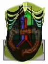
Figure 4. Location of retroperitoneal hematomas. 1. Central Medial 1A. Central supra-mesocolic.1 B. Central infra-mesocolic. 2. Flank. 2A. Peri-renal flank. 2B. Flank, pericolic. 3. Pelvic. 4. Portal. 5. Retro-hepatic
Figure 5
Figure 5. Mattox maneuver. A) Identification of the peritoneal reflection line of the descending colon, where the avascular plane that allows access to the retroperitoneum will be dissected. B) Identification of the peritoneal reflection line of the splenic flexure of the colon, spleen and stomach, to perform the medial rotation of these organs. C) By sharp and blunt dissection, the descending colon, the splenic flexure of the colon (Co), the spleen, and the stomach are mobilized medially. As it is advanced posteriorly in the retroperitoneum (R), the pancreas and kidney are mobilized. D) The lesion was located between the celiac trunk (CT) and the superior mesenteric artery. In addition to medial rotation of the viscera, the exposure required sectioning the fibers of the diaphragm and completely dissecting the firm plexus surrounding the aorta and the origin of its branches. Ao Aorta, St stomach, Spl spleen. E) Lesion located in the aorta (Ao) above the left renal artery (Re). SM superior mesenteric artery, K Kidney, Ur ureter. F) Exposure of the supraceliac and visceral aorta (Ao). A lesion of the celiac trunk (CT) has been managed with ligation. The remnants of the plexus surrounding the aorta can be seen. MS superior mesenteric artery, Re left renal artery.
Figure 6
Figure 6. Injury to the suprarenal aorta and its branches. A) Injury between the inferior mesenteric artery and the renal artery. Note the medial rotation of the viscera on the left side. The injured segment has been replaced by a dacron graft. B) Celiac trunk injury. Note the medial rotation of the viscera on the left side. The injured vessel was handled with ligature (yellow arrow).
Figure 7
Figure 7. Exposure and control of an infrarenal aortic injury with medial mobilization of the left colon.
Figure 8
Figure 8. Catell Braasch maneuver. A) Hematoma in zones 2 and 3 on the right side. The arrows indicate the peritoneal reflection, where the incision would be made to access the retroperitoneum. The possibility of surrounding the cecum and directing the incision at the base of the mesentery is indicated, allowing the elevation of the right colon and the small intestine, to expose in addition to the vena cava, the right iliac vessels and the infra-renal aorta. B) Identification of the peritoneal reflection line of the ascending colon. C) The surgeon has mobilized the hepatic flexure of the colon (Co) and is entering the retroperitoneum posterior to the duodenum (Du). St Stomach D) Mobilization of the hepatic flexure of the colon (Co) and exposure of the first three duodenal portions (Du) and the head of the pancreas (Pa). Li liver, Gb gallbladder, St stomach. E) Mobilization of the duodenum (Du) and the head of the pancreas (Pa), to expose the suprarenal vein cava (Ca). Li liver, Gb gallbladder, St stomach. F) Mobilization of the right colon (Co) and duodenum (Du), to expose the entire vena cava (Ca). Li liver, Gb gallbladder, Ap caecal appendix.
Figure 9
Figure 9. Trauma of the infrarenal vena cava. A) Exposure of the vein with medial mobilization of the right colon. B) Infrarenal vena cava ligation.
Figure 10
Figure 10. Patient with exsanguinating suprahepatic hemorrhage and injury to the right lower lobe. Wound in the suprahepatic area (segment IV) with hemorrhage that was not controlled with clamping of the hepatic hilum. Due to the need to resolve the intrathoracic bleeding, the surgeon packed the liver injury achieving control of the hemorrhage. A) Chest X-ray, Day 1. Laparotomy pads are observed on the chest and abdomen. B) Chest X-ray, Day 3, After chest unpacking and closure and partial abdominal unpacking. C) Chest X-ray, Day 10, after completing abdominal unpacking and closing the fascia.
Figure 11
Figure 11. Wound on the medial area of the retrohepatic cava. Hepatic exclusion was performed by intravascular occlusion of the aorta in zone 1, of the vena cava in its retrohepatic portion, and clamping of the hepatic hilum. The left liver lobe has been mobilized to the right and the surgeon's finger controls the bleeding, in preparation for its suture (arrow)
Figure 12
Figure 12. Placement of a temporary shunt in a lesion of the right iliac artery. A). Proximally and distally repaired artery. B) The graft (LeVin 16 Fr probe segment) has been installed sideways. C) The graft has been completely installed. Notice the firm ties that fix it. Courtesy Dr. Diego Rivera.
Figure 13
Figure 13. Blunt trauma with multiple intestinal and mesentery injuries. Severe instability requiring aortic clamping. A). Ligated distal end of the superior mesenteric vein. B) Proximal stump ligated. C) Day 5. Six reoperations were require due to progressive necrosis of the small intestine. Massively swollen and congestive intestine. D) A mesenteric-caval bypass was performed. After this the intestinal congestion resolved and the intestine could be anastomosed in subsequent procedures.
Figura 1
Figura 1. Manejo quirúrgico del trauma vascular abdominal. 1. Durante el preoperatorio y la oclusión puede hacerse mediante un catéter de oclusión aortica endovascular (REBOA). 2. Se incluyen los elementos para prevención y manejo de la hipotermia, infusión rápida, recuperación de sangre y autotransfusión, accesos vasculares, procedimientos en diferentes cavidades y monitoria. 3. Considere autotransfusión. 4. Por hipotensión persistente o necesidad de control vascular proximal en lesiones arteriales. 5. De acuerdo con la localización del hematoma, el sitio de la hemorragia, el recorrido de la herida y otros órganos lesionados. 6. Listado de vasos que se pueden ligar en la Tabla 5. 7. El cierre de la fascia o la piel aumentan el riesgo de hipertensión abdominal. 8. La reintervención no urgente debe hacerse tan pronto la hipotermia, la coagulopatía y la alteración de la perfusión estén corregidos en pacientes con derivación transitorias o ligadura vascular.
Figura 2
Figura 2. Trauma penetrante de arteria iliaca derecha. A) Control transitorio de la hemorragia con compresión local. B) Control vascular definitivo con pinzas atraumáticas
Figura 3
Figura 3. Pinzamiento de la aorta supraceliaca. A) Ingreso a la transcavidad de los epiplones. El lóbulo hepático izquierdo ha sido rechazado hacia arriba, el ligamento gastrohepático (LGH) ha sido seccionado con una maniobra roma con el dedo índice. Se observan el cuerpo del páncreas (Pa) y las fibras del hiato diafragmático (HD). Hi Hígado. Es Estómago. B) Orientado por la pulsación aórtica el cirujano diseca longitudinalmente las fibras del diafragma en el hiato (D). Rodea el vaso en cada uno de sus lados y lo aprisiona entre sus dedos índice y medio. Ocasionalmente deben seleccionarse algunas fibras del diafragma con electro bisturí. No se recomienda rodear completamente la aorta durante esta maniobra. Ao Aorta supraceliaca. C. Colocación de la pinza aórtica. El cirujano mantiene sus dedos abrazando la aorta. Avanza las ramas de la pinza aórtica hasta sentir la resistencia de la fascia prevertebral. D) Sin retirar los dedos se cierra la pinza y se verifica la oclusión completa del vaso.
Figura 4
Figura 4. Localización de los hematomas retroperitoneales. 1. Centro Medial 1A. Central supramesocólico. 1B. Central inframesocólico 2. Flanco 2A. Flanco peri-renal. 2B. Flanco pericólico. 3. Pélvico. 4. Portal. 5. Retro hepático
Figura 5
Figura 5. Maniobra de Mattox. A) Identificación de la línea de reflexión peritoneal del colon descendente, donde se ingresará al plano avascular que permite acceder al retroperitoneo. B) Identificación de la línea reflexión peritoneal del ángulo esplénico del colon, el bazo y el estómago, para realizar la rotación medial de estos órganos. C) Mediante disección aguda y roma se movilizan medialmente el colon descendente, el ángulo esplénico del colon (Co), el bazo y el estómago. En la medida en que se avanza posteriormente en el retroperitoneo (R) se movilizan el páncreas y el riñón. D) La lesión se ubicaba entre el tronco celíaco (Tc) y la arteria mesentérica superior. Además de la rotación medial de las vísceras, la exposición exigió seccionar las fibras del diafragma y disecar completamente el plexo firme que rodea la aorta y el origen de sus ramas. Ao Aorta, Es estómago, Ba bazo. E) Lesión localizada en la aorta (Ao) arriba de la arteria renal izquierda (Re). MS arteria mesentérica superior, Ri riñón, Ur uréter. F) Exposición de la aorta supracelíaca y visceral (Ao). Una lesión del tronco celíaco (TC)ha sido controlada manejada con ligadura. Pueden observarse los remanentes del plexo que rodea la aorta. MS arteria mesentérica superior, Re Arteria renal izquierda.
Figura 6
Figura 6. Lesión de la arteria aorta suprarrenal y sus ramas. A) Lesión entre la arteria mesentérica inferior y la renal. Se observa la rotación medial de las vísceras del lado izquierdo. El segmento lesionado ha sido reemplazado por un injerto de dacrón. B) Lesión del tronco celiaco. Se observa la rotación medial de las vísceras del lado izquierdo. El vaso lesionado fue manejado con ligadura (flecha amarilla)
Figura 7
Figura 7. Exposición y control de una herida de la aorta infrarrenal con movilización medial del colon izquierdo
Figura 8
Figura 8. Maniobra de Catell Braash. A) Hematoma en zonas 2 y 3 en lateral derecho. Las flechas señalan la reflexión peritoneal, donde se practicaría la incisión para acceder al retroperitoneo. Se indica la posibilidad de rodear el ciego y dirigir la incisión en la base del mesenterio, permitiendo la elevación del colon derecho y el intestino delgado, para exponer además de la vena Cava, los vasos ilíacos derechos y la aorta infra renal. B) Identificación de la línea de reflexión peritoneal del colon ascendente. C) El cirujano ha movilizado el ángulo hepático del colon (Co) y está ingresando al retroperitoneo posterior al duodeno (Du). D) Movilización del ángulo hepático del colon (Co) y exposición de las tres primeras porciones duodenales (Du) y la cabeza del páncreas (Pa). Hi hígado, Vb vesícula biliar, Es estómago. E) Movilización del duodeno (Du) y la cabeza del páncreas (Pa), para exponer la vena Cava suprarrenal (Ca). Hi hígado, Vb vesícula biliar, Es estómago. F) Movilización de el colon derecho (Co) y el duodeno (Du), para exponer a la cava en su totalidad (Ca). Hi hígado, Vb vesícula biliar, Ap apéndice cecal.
Figura 9
Figura 9. Trauma de la vena cava infrarrenal. A) Exposición de la vena con movilización medial del colon derecho. B) Ligadura de la vena cava infrarrenal
Figura 10
Figura 10. Paciente con hemorragia suprahepática exanguinante y herida del lóbulo pulmonar inferior derecho. Herida en área suprahepática (segmento IV) con hemorragia que no controló con pinzamiento del hilio hepático. Debido a la necesidad de resolver el sangrado intratorácico el cirujano empaquetó la lesión hepática con control de la hemorragia. A) Radiografía de tórax, día 1. Se observan las compresas en el tórax y el abdomen. B) Radiografía de tórax, día 3, después de desempaquetamiento y cierre del tórax y desempaquetamiento parcial del abdomen. C) Radiografía de tórax, día 10, después de completar el desempaquetamiento abdominal y cerrar la fascia
Figura 11
Figura 11. Herida de la cara medial de la cava retrohepática. Se practicó la exclusión hepática mediante la oclusión intravascular de la Aorta en la zona 1, de la vena Cava en su porción retrohepática y el pinzamiento del hilio hepático. El lóbulo hepático izquierdo ha sido movilizado hacia la derecha y el dedo del cirujano controla la herida, en preparación para su sutura (flecha).
Figura 12
Figura 12. Colocación de una derivación transitoria en una lesión de la arteria iliaca derecha. A) Arteria reparada proximal y distalmente. B) El puente (segmento de sonda LeVin 16 Fr) ha sido instalado completamente. Observe las ligaduras firmes que lo fijan. Cortesía Dr. Diego Rivera
Figura 13
Figura 13. Trauma cerrado con múltiples lesiones intestinales y en el mesenterio. Inestabilidad grave con requerimiento de pinzamiento aórtico. A) Cabo distal de la vena mesentérica superior ligada. B) Cabo proximal ligado. C) Día 5. Ha requerido 6 reintervenciones por necrosis progresiva del intestino delgado. Intestino masivamente edematizado y congestivo. D) Se practicó una derivación mesentérico-cava. Después de esto la congestión intestinal se resolvió y el intestino pudo ser anastomosado en procedimientos posteriores

References

    1. 1. Branco BC, Musonza T, Long MA, Chung J, Todd SR, Wall MJ, et al. Survival trends after inferior vena cava and aortic injuries in the United States. J Vasc Surg. 2018; 68(6):1880-1888. doi:10.1016/j.jvs.2018.04.033. - PubMed
    2. Branco BC, Musonza T, Long MA, Chung J, Todd SR, Wall MJ. Survival trends after inferior vena cava and aortic injuries in the United States. J Vasc Surg. 2018;68(6):1880–1888. doi: 10.1016/j.jvs.2018.04.033. - DOI - PubMed
    1. 2. De Bakey ME, Simeone FA. Battle Injuries of the Arteries in World War II : An Analysis of 2,471 Cases. Ann Surg. 1946; 123(4): 534-579 Doi: 10.1097/00000658-194604000-00005. - PMC - PubMed
    2. De Bakey ME, Simeone FA. Battle Injuries of the Arteries in World War II An Analysis of 2,471 Cases. Ann Surg. 1946;123(4):534–579. doi: 10.1097/00000658-194604000-00005. - DOI - PMC - PubMed
    1. 3. Rich NM, Baugh JH, Hughes CW. Acute arterial injuries in Vietnam: 1,000 cases. J Trauma. 1970; 10(5):359-369. Doi: 10.1097/00005373-197005000-00001. - PubMed
    2. Rich NM, Baugh JH, Hughes CW. Acute arterial injuries in Vietnam 1,000 cases. J Trauma. 1970;10(5):359–369. doi: 10.1097/00005373-197005000-00001. - DOI - PubMed
    1. 4. Patel JA, White JM, White PW, Rich NM, Rasmussen TE. A contemporary, 7-year analysis of vascular injury from the war in Afghanistan. J Vasc Surg. 2018; 68(6):1872-1879. Doi: 10.1016/j.jvs.2018.04.038. - PubMed
    2. Patel JA, White JM, White PW, Rich NM, Rasmussen TE. A contemporary, 7-year analysis of vascular injury from the war in Afghanistan. J Vasc Surg. 2018;68(6):1872–1879. doi: 10.1016/j.jvs.2018.04.038. - DOI - PubMed
    1. 5. Mattox KL, Feliciano DV, Burch J, Beall AC, Jr Jordan GL, Jr De Bakey ME. Five thousand seven hundred sixty cardiovascular injuries in 4459 patients. Epidemiologic evolution 1958 to 1987. Ann Surg. 1989; 209(6): 698-705. Doi: 10.1097/00000658-198906000-00007. - PMC - PubMed
    2. Mattox KL, Feliciano DV, Burch J, Beall AC, Jr Jordan GL. Jr De Bakey ME Five thousand seven hundred sixty cardiovascular injuries in 4459 patients Epidemiologic evolution 1958 to 1987. Ann Surg. 1989;209(6):698–705. doi: 10.1097/00000658-198906000-00007. - DOI - PMC - PubMed