. 2021 Sep;32(3):121-133.
doi: 10.7171/jbt.21-3203-007.

Direct detection of SARS-CoV-2 RNA using high-contrast pH-sensitive dyes

Affiliations

Direct detection of SARS-CoV-2 RNA using high-contrast pH-sensitive dyes

Timothy A Brown et al. J Biomol Tech. 2021 Sep.

Abstract

The worldwide coronavirus disease 2019 pandemic has had devastating effects on health, healthcare infrastructure, social structure, and economics. One of the limiting factors in containing the spread of this virus has been the lack of widespread availability of fast, inexpensive, and reliable methods for testing of individuals. Frequent screening for infected and often asymptomatic people is a cornerstone of pandemic management plans. Here, we introduce 2 pH-sensitive "LAMPshade" dyes as novel readouts in an isothermal Reverse Transcriptase Loop-mediated isothermal AMPlification amplification assay for severe acute respiratory syndrome coronavirus 2 RNA. The resulting JaneliaLAMP assay is robust, simple, inexpensive, and has low technical requirements, and we describe its use and performance in direct testing of contrived and clinical samples without RNA extraction.

Keywords: LAMPshade; RT-LAMP; Si-fluorescein; carbofluorescein.


Figures

FIGURE 1
FIGURE 1
LAMP and LAMPshade dyes. A) Schematic of LAMP assays for detection of SARS-CoV-2. Bst 2.0 is a strand-displacing DNA polymerase (NEB) (B) pH absorbance/fluorescence response curves. C) Dye photo properties including maximum excitation and emission wavelengths, extinction coefficient, quantum yield, pKa, and Hill coefficient. LSM; LAMPshade Magenta, LSV; LAMPshade Violet
FIGURE 2
FIGURE 2
Colorimetric dye pH response and LAMP readouts with various pH indicators. A) Images of solutions of LAMPshade Magenta, LAMPshade Violet, and Phenol Red at varying pH values ranging from pH 9.0 to 6.25 in 0.25-U increments, left to right. B) Images of negative (−) and positive (+) jLAMP reactions under normal light. C) Images under handheld UV (365 nm) light using LAMPshade Magenta and Violet.
FIGURE 3
FIGURE 3
jLAMP assay optimization. A) Workflow of jLAMP assay. B) Comparison of HCoV-229E viral RNA detected after RNA purification or directly from samples in either VTM or H2O. Data represent averages of 4 replicates showing standard deviation bars. C) Effects of heat and freeze-thaw treatments on RNA recovery in direct qRT-PCR sample assays. Heat-inactivated (HI) samples were heated to 65°C for 30 min. Data represent averages of triplicate samples with standard deviation bars.
FIGURE 4
FIGURE 4
Sensitivity and specificity of jLAMP. A) Limits of detection of jLAMP in various VTM spiked with HI-SARS-CoV-2. Data were compiled from multiple independent experiments. B) jLAMP sensitivity in UTM and VTM at low viral copy numbers. C) False-positive (no viral RNA) frequency in different swab sample transport media: VTM, UTM, or saline. Positive controls (1) contained HI-SARS-CoV-2 at ∼260 copies. The single positive in saline is marked with an asterisk. One sample outof 60 yields a 1.66% false-positive percentage. Positive reactions were determined by lack of color detection and comparison to controls by visual inspection against a white background.
FIGURE 5
FIGURE 5
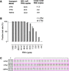
jLAMP assay optimization. A) jLAMP sensitivity in COVID-19–negative NP swab samples. In total, 13 COVID-19 negative samples were spiked with serially diluted heat-inactivated SARS-CoV-2. Percent positive samples are plotted against the corresponding RNA copy values; LAMP reactions were performed with LAMPshade Magenta. Each bar represents the percent positivity rate for 13 samples in duplicate. B) Saliva sample testing for LAMP performance. In total, 8 normal saliva samples were spiked with 23 copies of heat-inactivated SARS-CoV-2 (n = 10). Percent positive for each saliva sample is displayed relative to a water control.
FIGURE 6
FIGURE 6
COVID-19 patient nasal swab quantification and jLAMP test results. SARS-CoV-2–positive NP swabs in UTM were LAMP tested (n = 30). Each sample was assayed in triplicate, and all data points are shown in the plot. Each data point is assigned a sample number (x axis) ranked by Ct value. qRT-PCR Ct values obtained from the CDC N gene primer set are reported based on assays after heat inactivation. LAMP positive samples are represented by clear circles, and negative samples are displayed as red.
FIGURE 7
FIGURE 7
LAMP master mix storage stability and integrity controls. A) Tube labels with 2-fold serial dilutions of SARS-CoV-2 from 92.8 to 2.9 copies: top, unfrozen control; middle, −20°C storage for 2 wk; bottom, −20°C storage for 4 wk. B) LAMP assays for RPP30 and 18S rRNA as sample integrity controls. RPP30 (RNaseP subunit) and 18S rRNA LAMP primers were tested on HEK293 mammalian cells at 20, 10, and 2 cells per tube in duplicate.

References

    1. Notomi T, Okayama H, Masubuchi H, Yonekawa T, Watanabe K, Amino N et al. Loop-mediated isothermal amplification of DNA. Nucleic Acids Res . 2000;28:E63. doi: 10.1093/nar/28.12.e63. - DOI - PMC - PubMed
    1. Wong Y-P, Othman S, Lau Y-L, Radu S, Chee H-Y. Loop-mediated isothermal amplification (LAMP): a versatile technique for detection of micro-organisms. J Appl Microbiol . 2018;124:626–643. doi: 10.1111/jam.13647. - DOI - PMC - PubMed
    1. Parida M, Posadas G, Inoue S, Hasebe F, Morita K. Real-time reverse transcription loop-mediated isothermal amplification for rapid detection of West Nile virus. J Clin Microbiol . 2004;42:257–263. doi: 10.1128/JCM.42.1.257-263.2004. - DOI - PMC - PubMed
    1. Mori Y, Nagamine K, Tomita N, Notomi T. Detection of loop-mediated isothermal amplification reaction by turbidity derived from magnesium pyrophosphate formation. Biochem Biophys Res Commun . 2001;289:150–154. doi: 10.1006/bbrc.2001.5921. - DOI - PubMed
    1. Tanner NA, Zhang Y, Evans TC., Jr Visual detection of isothermal nucleic acid amplification using pH-sensitive dyes. Biotechniques . 2015;58:59–68. doi: 10.2144/000114253. - DOI - PubMed

Publication types