. 2021 Jun 15:3:133-145.
doi: 10.1016/j.crstbi.2021.05.003. eCollection 2021.

Rewards of divergence in sequences, 3-D structures and dynamics of yeast and human spliceosome SF3b complexes

Affiliations

Rewards of divergence in sequences, 3-D structures and dynamics of yeast and human spliceosome SF3b complexes

Arangasamy Yazhini et al. Curr Res Struct Biol. .

Abstract

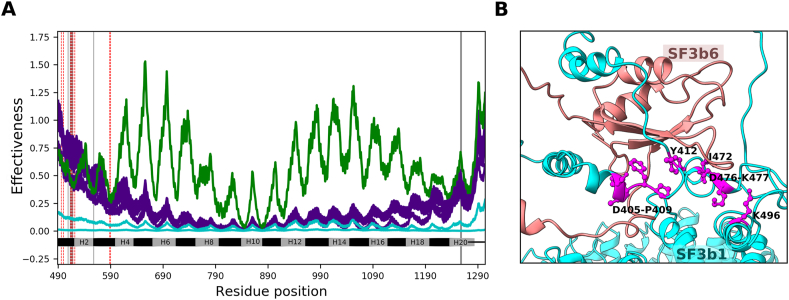
The evolution of homologous and functionally equivalent multiprotein assemblies is intriguing considering sequence divergence of constituent proteins. Here, we studied the implications of protein sequence divergence on the structure, dynamics and function of homologous yeast and human SF3b spliceosomal subcomplexes. Human and yeast SF3b comprise of 7 and 6 proteins respectively, with all yeast proteins homologous to their human counterparts at moderate sequence identity. SF3b6, an additional component in the human SF3b, interacts with the N-terminal extension of SF3b1 while the yeast homologue Hsh155 lacks the equivalent region. Through detailed homology studies, we show that SF3b6 is absent not only in yeast but in multiple lineages of eukaryotes implying that it is critical in specific organisms. We probed for the potential role of SF3b6 in the spliceosome assembled form through structural and flexibility analyses. By analysing normal modes derived from anisotropic network models of SF3b1, we demonstrate that when SF3b1 is bound to SF3b6, similarities in the magnitude of residue motions (0.86) and inter-residue correlated motions (0.94) with Hsh155 are significantly higher than when SF3b1 is considered in isolation (0.21 and 0.89 respectively). We observed that SF3b6 promotes functionally relevant 'open-to-close' transition in SF3b1 by enhancing concerted residue motions. Such motions are found to occur in the Hsh155 without SF3b6. The presence of SF3b6 influences motions of 16 residues that interact with U2 snRNA/branchpoint duplex and supports the participation of its interface residues in long-range communication in the SF3b1. These results advocate that SF3b6 potentially acts as an allosteric regulator of SF3b1 for BPS selection and might play a role in alternative splicing. Furthermore, we observe variability in the relative orientation of SF3b4 and in the local structure of three β-propeller domains of SF3b3 with reference to their yeast counterparts. Such differences influence the inter-protein interactions of SF3b between these two organisms. Together, our findings highlight features of SF3b evolution and suggests that the human SF3b may have evolved sophisticated mechanisms to fine tune its molecular function.

Keywords: Allostery; BPS, branch-point sequence; Bact, activated B spliceosome assembly; Cryo-EM structure; Cryo-EM, cryo-electron microscopy; DOPE, discrete optimized protein energy; NMA, normal mode analysis; PDB, protein data bank; Protein dynamics; RMSD, root mean square deviation; RRM, RNA recognition motif; SF3b complex; SF3b1; SF3b1SF3b6−bound, SF3b1 bound to SF3b6; SF3b1iso, SF3b1 in isolation; SIP, square inner product; Spliceosome.


Conflict of interest statement

The authors declare that they have no known competing financial interests or personal relationships that could have appeared to influence the work reported in this paper.

Figures

Image 1
Graphical abstract
Fig. 1
Fig. 1
SF3b complex in yeast and human. A) Shown is the surface representation of yeast (left) and human (right) SF3b complexes. Each protein component is colored differently, with labels pointing to their location in both complexes. A cross symbol indicates the absence of SF3b6. B) Cartoons depicting Pfam domains present in the yeast (top) and human (bottom) SF3b proteins. The length scale of the cartoon reflects protein sequence length. Domain names and boundaries are marked for SF3b proteins that are labelled as per human nomenclature. Pfam domains referred as SF3b1 (Pfam id: PF08920) and SAP (Pfam id: PF02037) are assigned uniquely to human SF3b1 and SF3b2 proteins, respectively.
Fig. 2
Fig. 2
Comparison of residue fluctuation profiles among Hsh155 (grey), SF3b1 in isolation (SF3b1iso, maroon) and SF3b1 bound to SF3b6 (SF3b1SF3b6-bound, green). Results are shown only for the common region that includes 20 HEAT repeats and C-terminus. A) Residue mean square fluctuations are normalized by the maximum residue fluctuation observed in the protein anisotropic elastic network model and residues corresponding to sequence insertions/deletions are excluded from the comparison. B) Collectivity of top 20 normal modes (left panel) and vector representation (right panel) of the magnitude and direction of residue motions of SF3b1iso (maroon) and SF3b1SF3b6−bound (green) in the first (left) and second (right) global motions. In the collectivity profile, ‘open-to-close’ and ‘twist’ movements are labelled.
Fig. 3
Fig. 3
SF3b binding modulates inter-residue correlated motions in the SF3b1. A) Heatmaps show difference in cross-correlation matrices between SF3b1iso and SF3b1SF3b6−bound that represents the strength of correlated motions of Cα atoms for all residue pairs. Left panel shows difference in the cross-correlations matrices of ‘open-to-close’ and ‘twist’ movements (upper triangle) as well as difference in the cross-correlation matrices of 20 modes between SF3b1iso and SF3b1SF3b6−bound (lower triangle). B) and C) show difference in the cross-correlation values of SF3b6 binding site with pre-mRNA binding sites and U2 snRNA binding sites between SF3b1iso and SF3b1SF3b6−bound. Positive value (>0) indicates stronger correlation for a given residue pair in the SF3b1SF3b6−bound compared to SF3b1iso while negative value (<0) indicates stronger correlation in the SF3b1iso compared to SF3b1SF3b6−bound.
Fig. 4
Fig. 4
Comparison of inter-residue correlation matrices between Hsh155 and SF3b1iso/SF3b1SF3b6-bound/SF3b1 (without N-terminal). A) Lower triangle indicates difference in the cross-correlation matrices between Hsh155 and SF3b1iso. Upper triangle indicates difference in the cross-correlation matrices between Hsh155 and SF3b1SF3b6−bound. Black boxes highlight notable differences in the strength of residue correlations. B) Shown in the upper triangle is difference in the cross-correlation matrices between Hsh155 and SF3b1 (without N-terminal). In both figures, positive value indicates residue correlation is stronger in SF3b1iso/SF3b1SF3b6−bound/SF3b1 without N-terminal, whereas negative value indicates the residue correlation is stronger in Hsh155. Regions corresponding to 20 HEAT repeats are labelled by numerical numbers.
Fig. 5
Fig. 5
Effect of perturbation of SF3b6 binding site on SF3b1. A) shown are effectiveness profiles of SF3b6 binding residues (purple lines), effectiveness profiles of residues (Arg390 and Ser400) adjacent to SF3b6 binding sites, considered as background signal (cyan lines) and effectiveness profile of Lys700 (green line). Regions of 20 heat repeats are labelled. B) Spatial position of selected SF3b6 binding residues (shown as ball and stick representation in magenta) in the SF3b1 structure whose perturbations exhibit higher effect on the SF3b1 than the background signal as shown in the left panel (A).
Fig. 6
Fig. 6
Structure comparison of yeast and human SF3b complexes. A) Superposition of yeast and human SF3b complexes. Color represents different constituent proteins of SF3b and homologous proteins are shown in gradients of the same color. Shown within the box is the interaction of SF3b14b/Rds3 with pre-mRNA that succeeds U2 snRNA/BPS duplex. Cys61 that coordinated with the Zn atom is colored blue (referred yeast structure, PDB code: 5GM6). B) Shown are structural differences between yeast and human SF3b proteins. Regions with Cα distance above 2.0 ​Å between equivalent residues are highlighted by thickness as well as color of cartoon representation. Thickened region indicates regions with structural differences when superposed in isolation. Color red indicates structural differences observed when superposed along with the whole SF3b complex. Given below the cartoon representations are Cα RMSD values of homologous protein structures when superposed in isolation (left) and with the entire complex (right) separated by ‘,’.
Fig. 7
Fig. 7
Local structural variability in SF3b3/Rse1. A) Centroids corresponding to BPA, BPB and BPC domains of human SF3b3 (maroon)/yeast Rse1 (grey) are shown with details on centroid distance between respective domains of two homologues and inter-domain angles among three domains. Centroids were defined from multiple structure superposition. For structure superposition, 9 available structures were used for SF3b3 and 5 available structures were used for Rse1. B) Regions with difference in the conformation of secondary structures between SF3b3 (red) and Rse1 (blue) are highlighted and symbol ‘∗’ indicates 15aa long helix insertion in Rse1. C) Shown are interactions between C-terminal tail of SF3b1 (navy) and SF3b3 (orange, top panel). Sequence insertion is highlighted in red and interacting residues are shown in ball and stick representation. In bottom panel, the topologically equivalent regions in yeast are highlighted (magenta). D) Cartoon representation of SF3b3 (orange) associated with SF3b5 (pink) and Ysf3 (blue). In the right panel, bottom figure shows the superposed structure of Ysf3 on to SF3b5 and residues that have short contacts with SF3b3 are highlighted in ball and stick representation. Top figure depicts a short contact observed between Cα atoms of Ala201 in SF3b3 and Gly76 in Ysf3.

References

    1. Alsafadi S., Houy A., Battistella A., Popova T., Wassef M., Henry E., Tirode F., Constantinou A., Piperno-Neumann S., Roman-Roman S., et al. Cancer-associated SF3B1 mutations affect alternative splicing by promoting alternative branchpoint usage. Nat. Commun. 2016;7:1–12. - PMC - PubMed
    1. Atilgan C., Atilgan A.R. Perturbation-response scanning reveals ligand entry-exit mechanisms of ferric binding protein. PLoS Comput. Biol. 2009;5 - PMC - PubMed
    1. Atilgan A.R., Durell S.R., Jernigan R.L., Demirel M.C., Keskin O., Bahar I. Anisotropy of fluctuation dynamics of proteins with an elastic network model. Biophys. J. 2001;80:505–515. - PMC - PubMed
    1. Bai R., Wan R., Yan C., Lei J., Shi Y. Structures of the fully assembled Saccharomyces cerevisiae spliceosome before activation. Science. 2018;360:1423–1429. - PubMed
    1. Berman H.M., Battistuz T., Bhat T.N., Bluhm W.F., Bourne P.E., Burkhardt K., Feng Z., Gilliland G.L., Iype L., Jain S., et al. The protein data bank. Acta Crystallogr. Sect. D Biol. Crystallogr. 2002;58:899–907. - PubMed

LinkOut - more resources