Review
. 2022 Feb;23(2):202-217.
doi: 10.3348/kjr.2021.0417. Epub 2022 Jan 4.

Pulmonary Arteriovenous Malformation and Its Vascular Mimickers

Affiliations
Review

Pulmonary Arteriovenous Malformation and Its Vascular Mimickers

Hyoung Nam Lee et al. Korean J Radiol. 2022 Feb.

Abstract

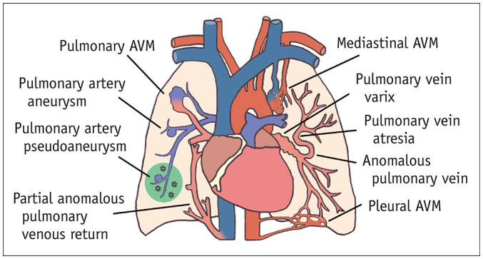
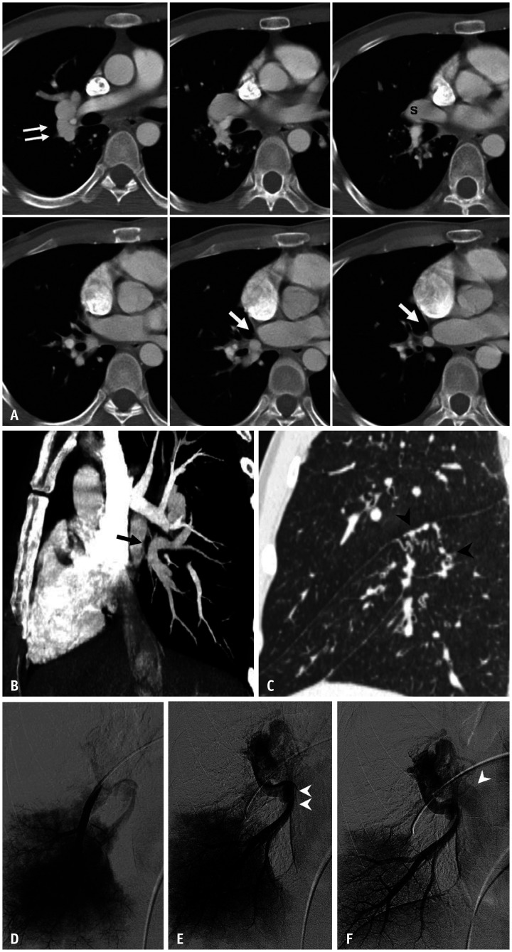
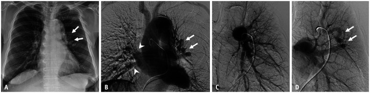
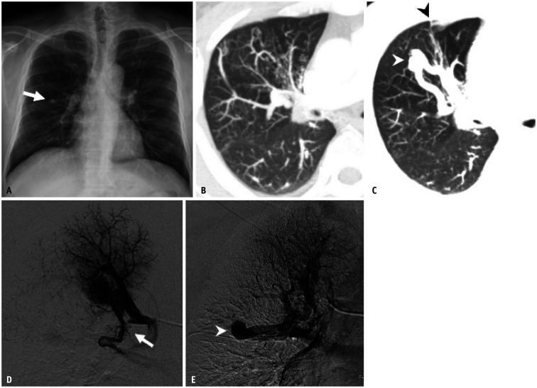
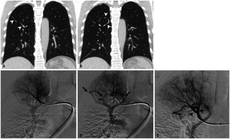
Pulmonary arteriovenous malformation (AVM) is a congenital vascular disease in which interventional radiologists can play both diagnostic and therapeutic roles in patient management. The diagnosis of pulmonary AVM is simple and can usually be made based on CT images. Endovascular treatment, that is, selective embolization of the pulmonary artery feeding the nidus of the pulmonary AVM, and/or selectively either the nidus or draining vein, has become a first-line treatment with advances in interventional devices. However, some vascular diseases can simulate pulmonary AVMs on CT and pulmonary angiography. This subset can confuse interventional radiologists and referring physicians. Vascular mimickers of pulmonary AVM have not been widely known and described in detail in the literature, although some of these require surgical correction, while others require regular follow-up. This article reviews the clinical and radiologic features of pulmonary AVMs and their mimickers.

Keywords: Aneurysm; Arteriovenous malformations; Diagnosis, differential; Lung; Pleura; Scimitar syndrome; Stenosis, pulmonary vein; Thoracic wall.


Conflict of interest statement

The authors have no potential conflicts of interest to disclose.

Figures

Fig. 1
Fig. 1. Illustration of the vascular mimickers of pulmonary AVM.
AVM = arteriovenous malformation
Fig. 2
Fig. 2. A 19-year-old male patient with hereditary hemorrhagic telangiectasia.
A. Chest radiograph shows a well-circumscribed round opacity (arrow) in the lower third of the left hemithorax. B-D. Axial enhanced chest CT images demonstrate large arteriovenous malformation (arrow) with a feeding artery in the left lower lobe. Numerous, small arteriovenous malformations (arrowheads) are also identified in both lung fields. E, F. Arterial-phase images from the pulmonary angiogram reveal the feeding artery (arrow) and rapid opacification of the draining vein (arrowheads). G. Delayed venous-phase image shows opacification of the normal pulmonary veins. H, I. Contrast-enhanced brain CT and contrast-enhanced T1-weighted MR image demonstrate two small enhancing lesions (arrows) located at the left lateral ventricle temporal horn and midbrain, which suggest brain arteriovenous malformation. J, K. Left cerebral and vertebral angiograms confirm two small arteriovenous malformations (arrows).
Fig. 3
Fig. 3. A 49-year-old female patient with hereditary hemorrhagic telangiectasia who had a mediastinal arteriovenous malformation.
A. Chest radiograph shows no definite abnormality. B, C. Axial contrast-enhanced CT images with lung and mediastinal window settings demonstrate prominent draining veins (double arrows) at the periphery of the right middle lobe and along the mediastinal pleura (arrows). D, E. Selective angiograms of the truncus anterior (white arrow) and right interlobar pulmonary artery (black arrow) show no discernible vascular abnormalities. F, G. Feeding arteries originate from the hypertrophied right internal mammary and inferior phrenic arteries. Note also the large draining vein (arrowheads), which communicates with the right pulmonary artery (arrow). A nidus is seen at the beginning of the draining veins.
Fig. 4
Fig. 4. A 19-year-old female patient with pleural arteriovenous malformation who underwent pulmonary artery embolization six years earlier.
A. Chest radiograph shows markedly enlarged vascular markings at the right lower lung zone. Multiple plugs from prior embolization procedures were also noted. B-G. Axial and coronal contrast-enhanced CT images with a mediastinal window setting demonstrate engorged vascular structures along the diaphragmatic pleural surface (arrowheads) and extremely dilated right inferior pulmonary veins. The feeding arteries originated from the hypertrophied right intercostal (black arrow) and inferior phrenic arteries (white arrow). H-K. Thoracic aortogram and selective angiograms of the right intercostal and inferior phrenic arteries reveal pleural arteriovenous malformation and early drainage through dilated right inferior pulmonary veins (arrowheads).
Fig. 5
Fig. 5. An anomalous pulmonary vein caused by lobar pulmonary vein atresia in a 32-year-old male patient.
Adapted from Lee et al. Korean J Radiol 2011;12:395-399 [30]. A. Serial axial CT images show a low attenuation linear structure (arrows) between the right inferior pulmonary vein and left atrium, as well as a prominent right superior pulmonary vein(s). An abnormally enlarged vascular structure (double arrow) near the right pulmonary artery, suggesting the presence of collateral drainage of the interrupted inferior pulmonary vein, was also noted. B. The reformatted oblique sagittal CT image shows the atretic right inferior pulmonary vein (arrow). C. The reformatted coronal image with the lung setting reveals multiple tortuous and dot-like collaterals (arrowheads) in the superior segment of the right lower lobe crossing the major fissure. D-F. Venous-phase images of the right interlobar selective pulmonary angiogram show no connection between the left atrium and the right inferior pulmonary vein (double arrowhead), which is draining into the superior pulmonary vein (arrowhead) through the collaterals.
Fig. 6
Fig. 6. An anomalous pulmonary vein in a 75-year-old female patient who presented with persistent blood-tinged sputum.
Adapted from Hyun et al. Cardiovasc Intervent Radiol 2014;37:835-838, with permission of the Springer Nature [4]. A. Chest radiographs show a tortuous abnormal vascular structure (arrows) at the left parahilar area. B. Venous-phase image in the main pulmonary angiogram shows a tortuous and dilated abnormal vessel in the left parahilar area, which drains into the left atrium through an anomalous pulmonary vein (arrows). The right superior and inferior pulmonary veins drain normally into the left atrium (arrowheads). C. Early arterial-phase image of the left upper lobar selective pulmonary angiogram shows no early draining vein. D. An anomalous venous structure (arrows) is seen in the late-venous phase.
Fig. 7
Fig. 7. An anomalous pulmonary vein and segmental pulmonary vein atresia in a 43-year-old male patient who presented with persistent cough and sputum production.
Adapted from Lee et al. Korean J Radiol 2011;12:395-399 [30]. A. Chest radiograph shows a single nodular opacity (arrow) in the right mid-lung zone. B, C. Slab axial images with the lung window setting show tortuous and dot-like collaterals and an aneurysmal dilatation of vascular structure (white arrowhead) crossing the minor fissure (black arrowhead) in the anterior segment of the right upper lobe. D, E. Venous-phase images in the right apical segmental selective pulmonary angiogram show that the apical segmental pulmonary vein is interrupted (arrow). Venous drainage of the affected segment detours via pulmonary vein varix associated with anomalous pulmonary vein (arrowhead).
Fig. 8
Fig. 8. A 61-year-old female with an anomalous pulmonary vein at the segmental level.
A, B. Coronal CT images demonstrate several tortuous vascular structures (arrowheads) in the right upper lobe. C, D. Selective angiograms at the truncus anterior show normal pulmonary arteries, but abnormal drainage of the right upper lobe apical segment (arrows) through anomalous pulmonary veins. E. In the angiogram with a catheter wedged into the segmental pulmonary artery, the anomalous pulmonary vein mimics a nidus and the early draining vein (arrowheads) of arteriovenous malformation.
Fig. 9
Fig. 9. Partial anomalous pulmonary venous drainage involving the left superior pulmonary vein in a 38-year-old female.
A. Chest radiograph shows no definite abnormality. B, C. Coronal CT image and volume-rendered image demonstrate that a vertical anomalous left superior pulmonary vein (arrows) courses lateral to the aortic arch prior to draining into the left brachiocephalic vein. D. Serial axial CT images show that the anomalous left superior pulmonary vein (arrows) drains into the left brachiocephalic vein.
Fig. 10
Fig. 10. Pulmonary artery aneurysm in a 68-year-old male.
A. Chest radiograph shows a rounded shadow (arrowheads) superimposed onto the enlarged cardiac silhouette. B-E. Enhanced CT images in the lung and mediastinal window settings reveal an aneurysm (arrows) arising from the left lower lobe segmental pulmonary artery (arrowhead). F. Digital subtraction angiography image with the catheter tip in the aneurysmal sac (arrow) shows a segmental branch of the left lower lobe pulmonary artery (arrowhead), but no draining veins. G. Post-embolization angiogram depicts complete packing of the segmental pulmonary artery proximal and distal to the aneurysm with coils.
Fig. 11
Fig. 11. Pulmonary artery pseudoaneurysm in a 65-year-old male with lung cancer undergoing chemotherapy.
A. Chest radiograph shows a thin, radiolucent crescent-shaped structure (arrowheads) superimposed onto the enlarged cardiac silhouette. B, C. Axial and coronal CT images reveal an air crescent adjacent to a pseudoaneurysm (arrowheads) with surrounding consolidation. D, E. Selective angiogram of the left lower lobe pulmonary artery demonstrates the pseudoaneurysm (arrows) arising from the segmental branch. F. Completion angiogram after coil embolization shows the absence of pseudoaneurysm filling.
Fig. 12
Fig. 12. Pulmonary artery ectasia in a 56-year-old female patient with hemoptysis.
A. Chest radiographs show mild cardiomegaly and prominent bilateral pulmonary vascular markings. B, C. Axial images in the lung window setting show dilatation of the pulmonary trunk to a maximum diameter of 36 mm with dilated and beaded peripheral pulmonary arteries (arrows). A ventricular septal defect is also noted (arrowhead). D-F. Arterial-phase images of the selective bilateral pulmonary angiogram reveal diffuse dilatation of the pulmonary trunk and branches.

References

    1. Hanson JM, Wood AM, Seymour R, Petheram IS. Anomalous unilateral single pulmonary vein: two cases mimicking arteriovenous malformations and a review of the literature. Australas Radiol. 2005;49:246–251. - PubMed
    1. Tokunaga K, Kubo T, Yamaoka T, Isoda H, Togashi K. Can the “pine-needle sign” on computed tomography be used to differentiate pulmonary arteriovenous malformation from its mimics? Analysis based on dynamic contrast-enhanced chest computed tomography in adults. Eur J Radiol. 2017;95:314–318. - PubMed
    1. Gill SS, Roddie ME, Shovlin CL, Jackson JE. Pulmonary arteriovenous malformations and their mimics. Clin Radiol. 2015;70:96–110. - PubMed
    1. Hyun D, Do YS, Lim SJ, Park HS, Park KB. Anomalous unilateral single pulmonary vein mimicking pulmonary arteriovenous malformation. Cardiovasc Intervent Radiol. 2014;37:835–838. - PubMed
    1. Lin CT, Zimmerman SL, Mitchell SE, Fishman EK. Pulmonary venous anomalies causing misdiagnosis of pulmonary arteriovenous malformations. Clin Imaging. 2018;47:96–100. - PubMed

Publication types

MeSH terms

Supplementary concepts