Review
. 2022 Mar;34(11):e2106456.
doi: 10.1002/adma.202106456. Epub 2022 Feb 3.

Understanding Nanomaterial-Liver Interactions to Facilitate the Development of Safer Nanoapplications

Affiliations
Review

Understanding Nanomaterial-Liver Interactions to Facilitate the Development of Safer Nanoapplications

Jiulong Li et al. Adv Mater. 2022 Mar.

Abstract

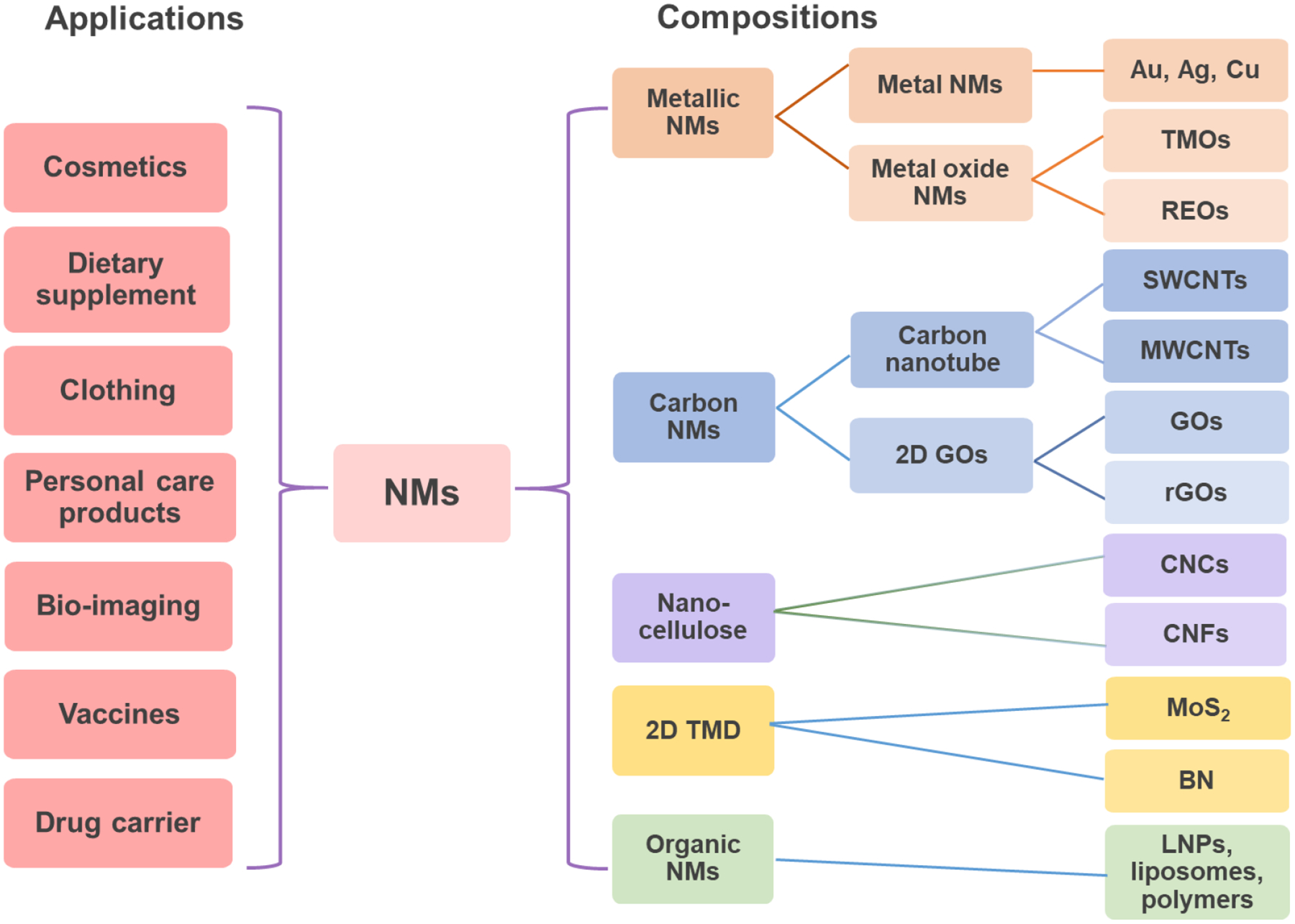
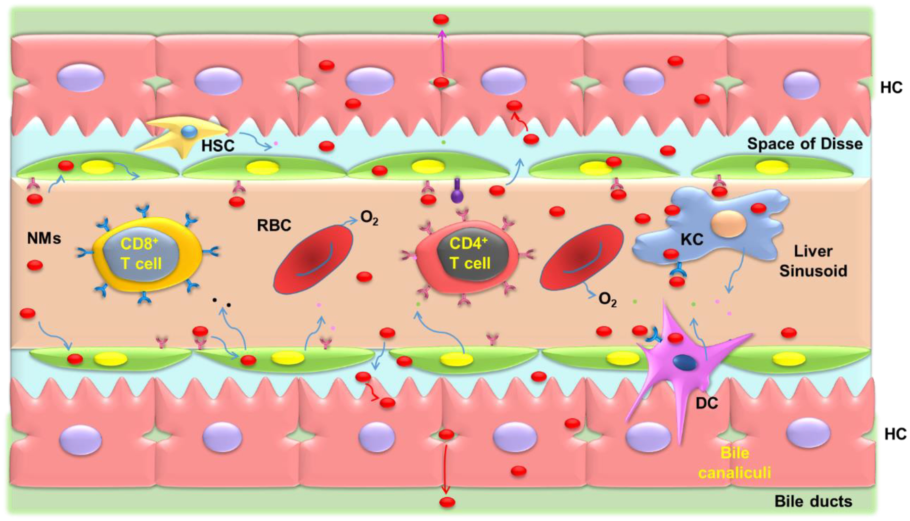
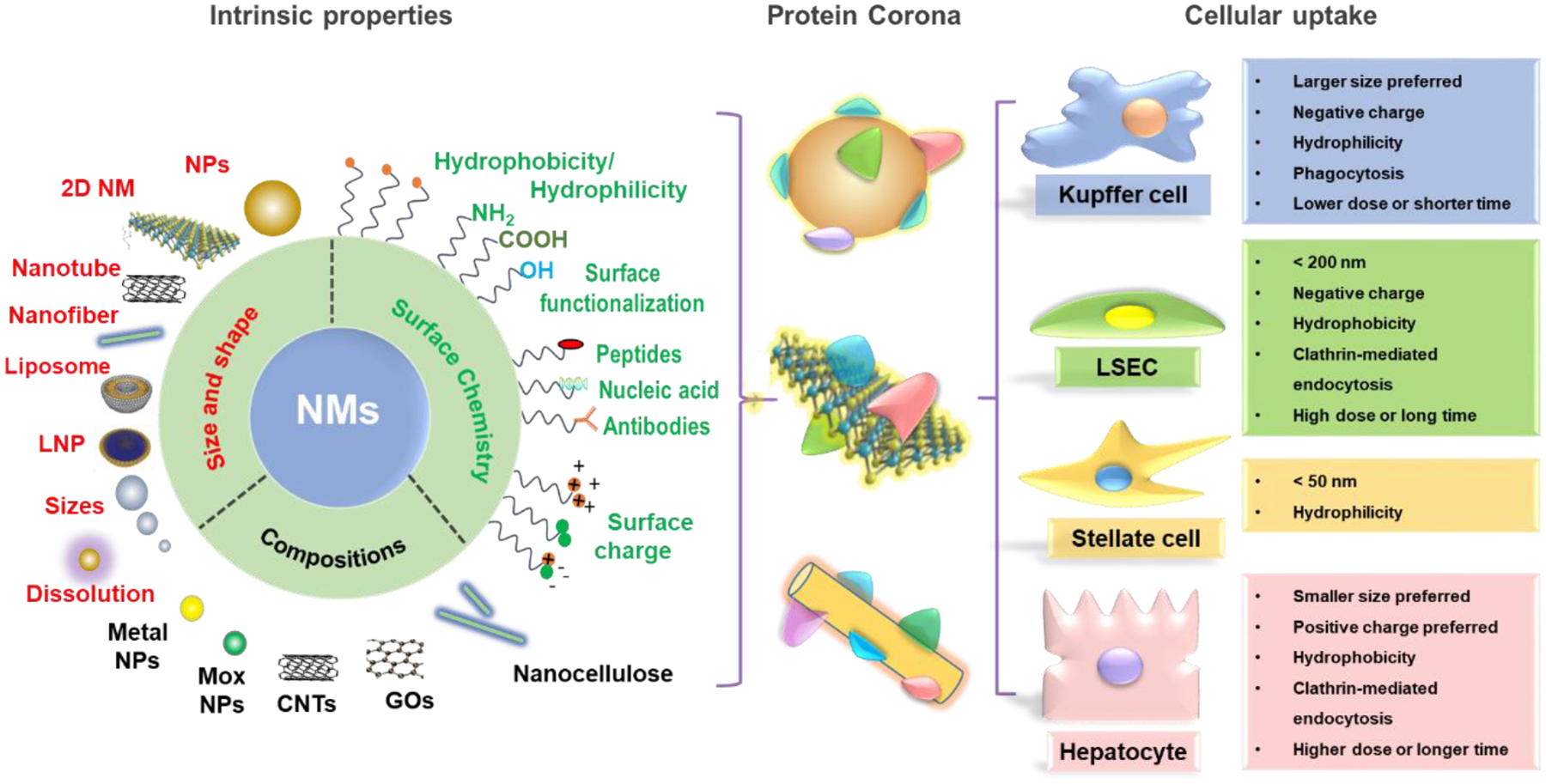
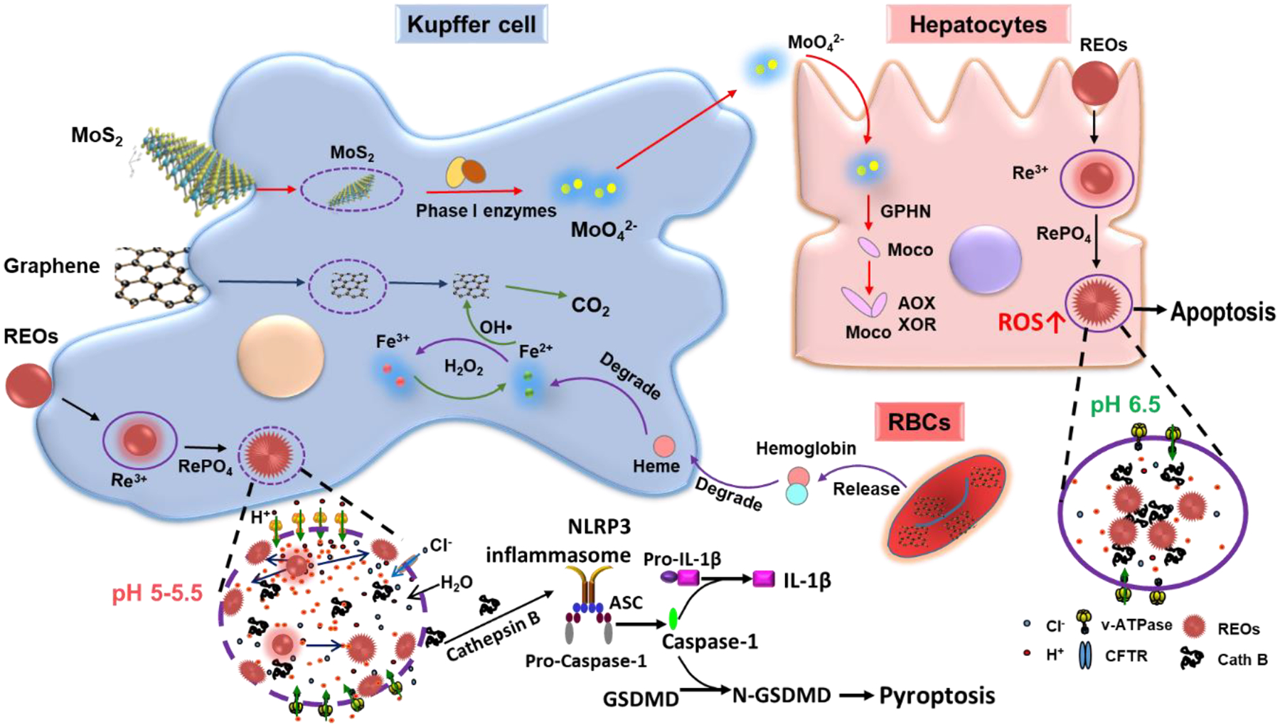
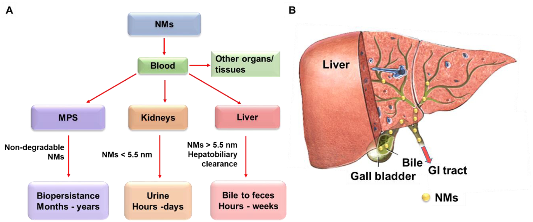
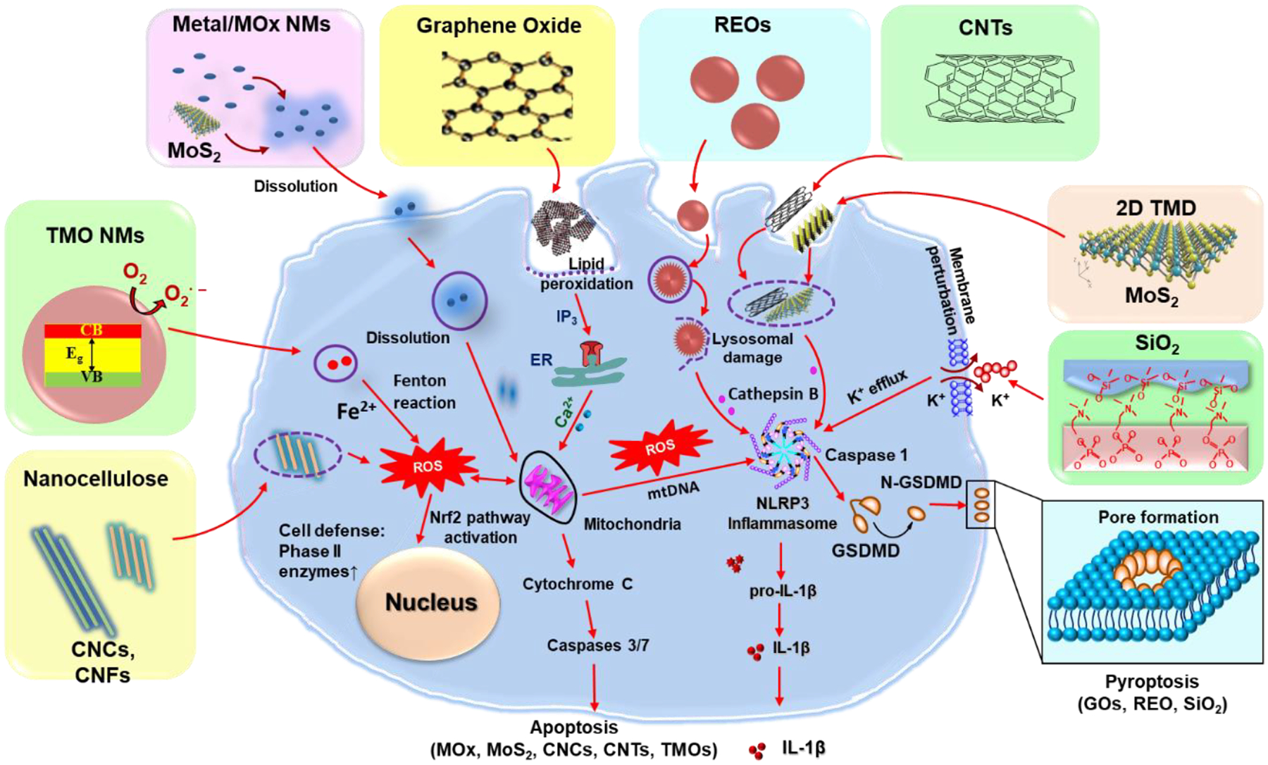
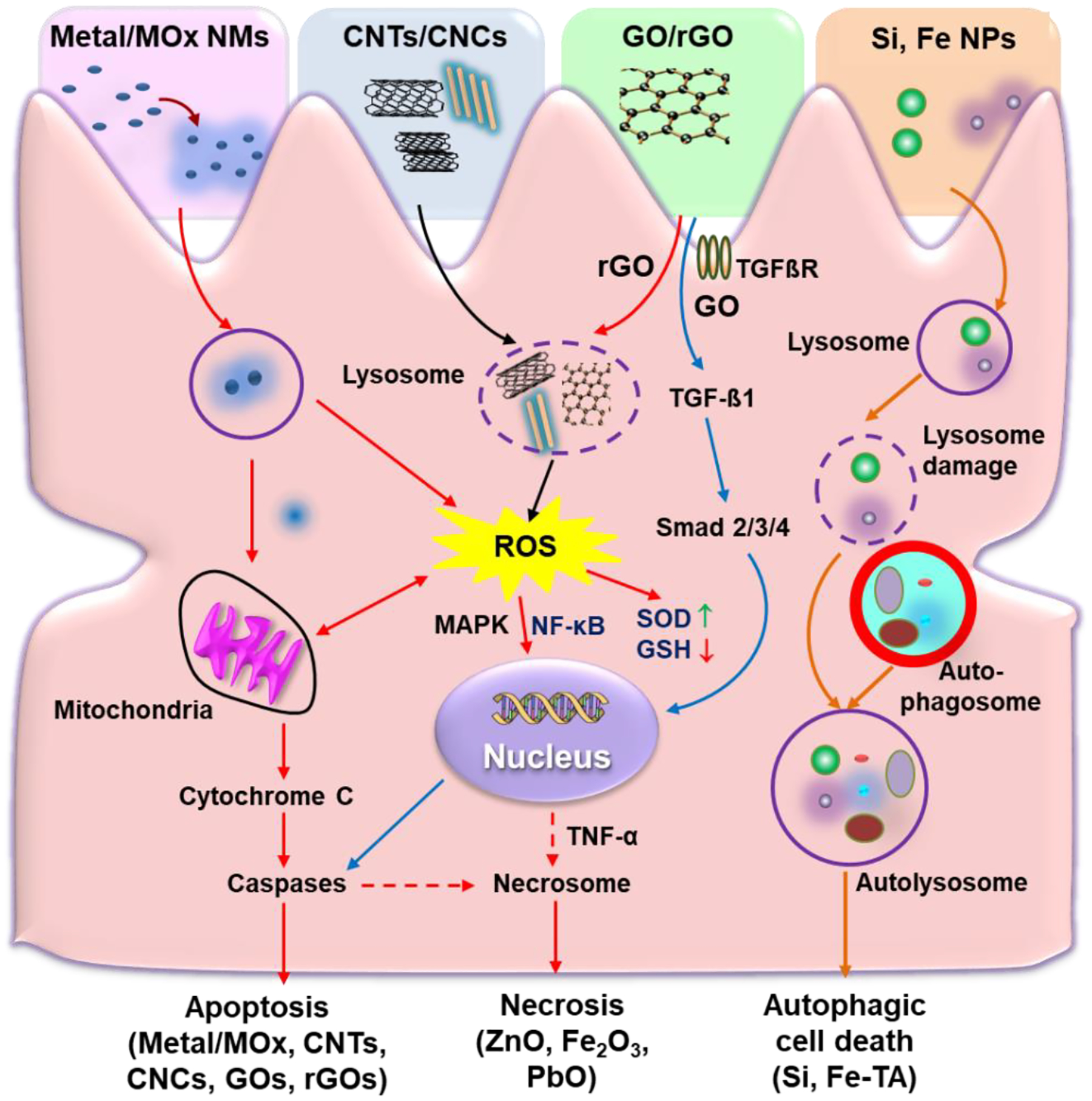
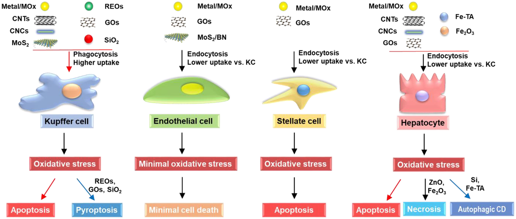
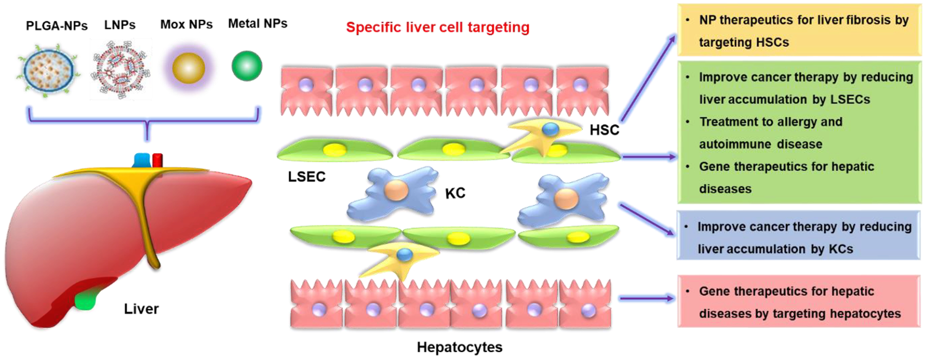
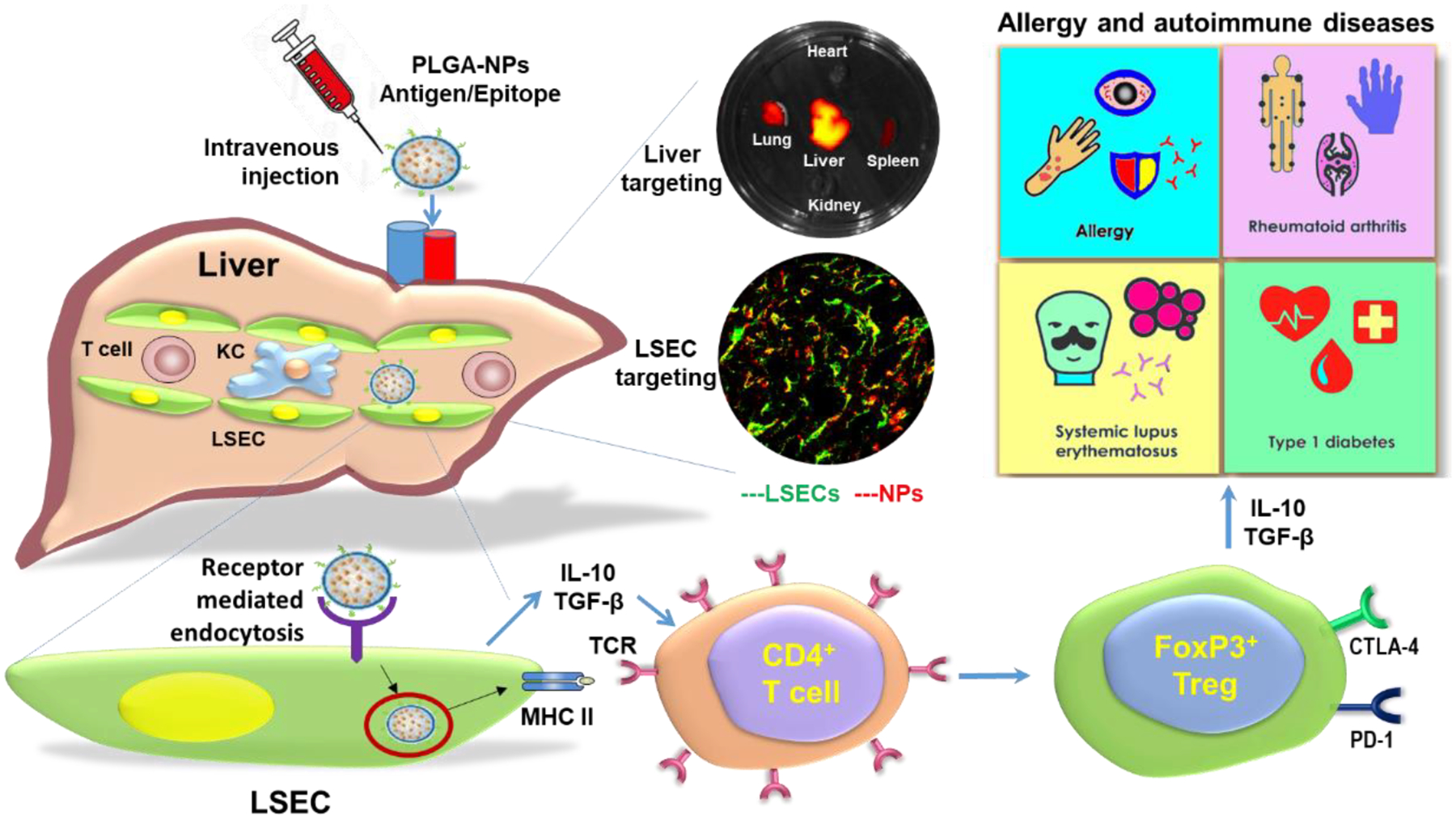
Nanomaterials (NMs) are widely used in commercial and medical products, such as cosmetics, vaccines, and drug carriers. Exposure to NMs via various routes such as dermal, inhalation, and ingestion has been shown to gain access to the systemic circulation, resulting in the accumulation of NMs in the liver. The unique organ structures and blood flow features facilitate the liver sequestration of NMs, which may cause adverse effects in the liver. Currently, most in vivo studies are focused on NMs accumulation at the organ level and evaluation of the gross changes in liver structure and functions, however, cell-type-specific uptake and responses, as well as the molecular mechanisms at cellular levels leading to effects at organ levels are lagging. Herein, the authors systematically review diverse interactions of NMs with the liver, specifically on major liver cell types including Kupffer cells (KCs), liver sinusoidal endothelial cells (LSECs), hepatic stellate cells (HSCs), and hepatocytes as well as the detailed molecular mechanisms involved. In addition, the knowledge gained on nano-liver interactions that can facilitate the development of safer nanoproducts and nanomedicine is also reviewed.

Keywords: cell-type-specific uptake; mode of action; nano-liver interaction; nanomedicine; nanosafety.


Figures

Figure 1.
Figure 1.
Major applications and compositions of nanomaterials (NMs) interacted with the liver. NMs have been widely applied in industrial, commercial, and medical fields, which include bio-imaging, vaccines, and drug carriers. NMs have diverse compositions, which include metallic NMs including metal (Ag and Au nanoparticles) and metal oxide (MOx) NMs including transition-metal oxides (TMOs, e.g., SiO2, Co3O4, and Mn2O3) and rare-earth oxides (REOs, e.g., Gd2O3, La2O3, and Y2O3) NPs, carbon NMs including one-dimensional carbon nanotubes (CNTs) and two-dimensional (2D) graphene-based NPs (graphene oxide, GO, and reduced graphene oxide, rGO), cellulose nanocrystal (CNC) and cellulose nanofiber (CNF), 2D transition metal dichalcogenide (TMD) including MoS2 and BN, and organic NMs including lipid NPs, liposomes, and polymer NPs.
Figure 2.
Figure 2.
Structural and functional organization of the liver. A) Anatomy of the liver and its blood supply. The vessel (red) represents the hepatic artery that delivers oxygenated blood from the general circulation. The vessel (blue) represents the hepatic portal vein that delivers deoxygenated blood from the small intestine containing nutrients. The vessel (green) represents the bile duct that carries bile from the liver and gallbladder to the duodenum. B) Schematic of a liver lobule in a hexagonal shape with rows of hepatocytes radiating out from the central vein towards the portal triad. C) Schematic demonstrates the blood flow of the liver via the portal vein and hepatic artery through the sinusoids to the central vein. A lobule could be divided into three zones, zone 1 (periportal), zone 2 (transition zone), and zone 3 (pericentral) based on oxygen gradient from high to low. D) Schematic sinusoids that receive blood from terminal branches of the hepatic artery and portal vein at the periphery of lobules and drain into central veins (red arrow), and the bile ducts that carry bile from the liver and gallbladder to the duodenum (green arrow). Sinusoids are lined with endothelial cells and flanked by plates of hepatocytes. E) Schematic shows the cross-section of a liver lobule and the flow direction of blood and bile. F) Spatial map to demonstrate flow velocities within the virtual sinusoid network. The red and yellow colors indicate a greater flow velocity while the blue color represents a lower flow velocity. Color bar units indicate μm/s. Figures 2A–B are reproduced under terms of the CC-BY license.[35] Copyright 2018, Øie et al., published by De Gruyter; Figure 2C is reproduced with permission.[44] Copyright 2010, Nature Publishing Group; Figure 2D is reproduced under terms of the CC-BY license.[39] Copyright 2005, Frevert et al., published by PLOS; Figures 2E is courtesy of Bio Ninja (https://ib.bioninja.com.au/options/option-d-human-physiology/d3-functions-of-the-liver/liver-structure.html) and used with permission; Figure 2F is reproduced under terms of the CC-BY license.[40] Copyright 2018, Fu et al., published by PLOS.
Figure 3.
Figure 3.
The major cell types in the liver. It includes the parenchymal hepatocytes, which occupy about 60–80% of the total number of liver cells, and the non-parenchymal cells occupying approximately 20–40% of the total number. In the non-parenchymal cell, it consists of the liver sinusoidal endothelial cells (approximately 50% of the total number of non-parenchymal cells), phagocytic Kupffer cells (approximately 20%), lymphocytes (approximately 25%), biliary cells (approximately 5%), and hepatic stellate cells (approximately 1–8%). In the lymphocytes, it includes the T lymphocytes (approximately 63%), natural killer (NK) cells (approximately 31%), B lymphocytes (approximately 6%), and less than 1% of dendritic cells (DCs). In the T lymphocytes, it contains the conventional T cells, including CD4+ T cells and CD8+ T cells, and the unconventional T cells, including natural killer T (NKT) cells, TCRγδ T cells, and others.
Figure 4.
Figure 4.
NMs able to induce or exacerbate liver disorders. For example, the silver, gold, and silicon NPs exacerbated the liver disorders including liver fibrosis and steatosis; Fe3O4, ZnO, and CuO NPs induced liver damage by triggering higher levels of liver enzyme release; heavy metal NMs including Cd, Ag, and ZnO aggravated hepatic chemical injury induced by environmental toxins; CNTs induced hepatic steatosis and liver injury, and graphene-based NMs induced hepatic dysfunction or liver functional zonation changes.
Figure 5.
Figure 5.
Interactions of NM uptake and elimination in the liver during systematic circulation after NMs exposure. As NMs move along the sinusoid, they will come into contact with T cells, Kupffer cells, sinusoidal endothelial cells, and DC cells. Depending on their physicochemical properties, NMs have better access through fenestrae to enter the space of Disse and contact with and hepatocytes. The smaller NMs may transcytose through the hepatocytes and enter the bile duct through bile canaliculi.
Figure 6.
Figure 6.
NMs intrinsic properties affect corona composition and cellular uptake in the liver. The major intrinsic properties of NMs include size, shape, surface chemistry, and composition, which will determine the corona composition and cellular uptake by the major liver cells. Larger size, negatively charged or hydrophilic NMs are preferentially swallowed by KCs via phagocytosis; NMs less than 200 nm or with negative surface charge or hydrophobicity tend to be taken up by endothelial cells through clathrin-mediated endocytosis with a high exposure dose or long time. NMs less than 50 nm or hydrophilic NMs could be captured by stellate cells. Smaller NMs with positive surface charge or hydrophobic NMs are preferentially taken up by hepatocytes through clathrin-mediated endocytosis.
Figure 7.
Figure 7.
Schematic to demonstrate the transformation and metabolic processes of NMs in the liver. For example, MoS2 degrade into MoO42− by phase I enzymes in KCs, which can be used for biosynthesis of molybdenum cofactors (Moco) in hepatocytes.[93] Few layer graphene is degraded into CO2 by the OH• generated through the Fenton reaction in KCs, which originated from the degradation of released hemoglobin from the damaged RBCs by graphene into hemes, and the differential transformation of REOs in KCs and hepatocytes due to different levels of acidification in the phagolysosomes of macrophages (pH 5–5.5) vs hepatocytes (pH 6.5). The intense lysosomal acidification in KCs is driven by v-ATPase on lysosomal membranes, creating a high concentration of protons near the lysosomal membrane, driving the transformation of REOs and the formation of sea urchin structures on the lysosomal membrane by stripping phosphate groups from the phospholipids, leading to lysosomal membrane damage, NLRP3 inflammasome activation and pyroptosis in KCs. The same transformation also happens in hepatocytes, however, only in the interior of lysosomes, which will not lead to lysosomal damage. The Figure is reproduced with permission.[99] Copyright 2021, American Chemical Society.
Figure 8.
Figure 8.
Main pathways for NMs clearance. A) NMs are cleared through hepatobiliary, renal, and mononuclear phagocyte systems. NMs circulate in the blood to reach organs or tissues, including MPS, liver, and kidneys. The non-degradable NMs are more likely to be taken up and retained by the MPS for months to years. The NMs less than 5.5 nm are cleared from the kidneys by renal clearance and eliminated in feces within hours to days. The NMs larger than 5.5 nm can be cleared from the liver by hepatobiliary clearance within hours to weeks. B) The hepatobiliary clearance is performed through interactions among the hepatic ducts, bile, gallbladder, common bile duct, duodenum, gastrointestinal tract, and feces. Figure 8 is reproduced with permission.[49] Copyright 2016, Elsevier B.V.
Figure 9.
Figure 9.
NMs-induced various cellular responses in KCs. This includes the metal or metal oxide (MOx) or transition-metal oxide (TMO) NMs, e.g., Ag, CuO, Co3O4, or Mn2O3, induce apoptosis due to their dissolution and shedding of toxic ions, bandgap energy, and oxidative stress; REOs (e.g., Gd2O3, La2O3, and Y2O3) and GOs induced pyroptosis in KCs. For REOs, the transformation from sphere to sea urchin-shaped and the formation of rare-earth phosphate (REPO4) structures on the lysosomal membrane, where RE(III) ions strip phosphate from the phospholipids on a lysosomal membrane and induce lysosomal damage, cathepsin B release, leading to NLRP3 inflammasome activation and GSDMD-mediated pyroptosis; the phagocytized GOs-induced NADPH oxidase activation and lipid peroxidation, triggering PLC activation that leads to calcium flux, mitochondrial ROS generation, and NLRP3 inflammasome activation, resulting in IL-1β production as well as subsequent pyroptosis; for fumed SiO2, the activation of NLRP3 inflammasome is involved in the pathway premised on K+ efflux resulting from the plasma membrane perturbation after SiO2 binding. Moreover, 2D TMD, CNCs, and CNTs induce ROS-mediated apoptosis and inflammatory responses in KCs after their internalization.
Figure 10.
Figure 10.
NMs-induced cellular responses in hepatocytes. Metallic NMs (e.g., Au, Ag, and ZnO NPs) could induce oxidative stress-mediated inflammatory responses, apoptosis, or necrosis; CNTs or CNCs could induce oxidative stress-mediated inflammatory responses and apoptotic cell death; rGOs could induce apoptosis via the nuclear factor kappa B (NF-κB) and oxidative stress pathways, while GOs induce apoptosis via the TGFβ1-mediated signaling pathway; Si and Fe-TA NPs could induce autophagic cell death.
Figure 11.
Figure 11.
Cell type-specific responses induced by various NMs in the liver cells. High-level uptake of metal, MOx, CNTs, CNCs, MoS2, etc., and REOs, GOs, SiO2, etc., through phagocytosis, could induce apoptosis or pyroptosis in KCs, respectively. Low-level uptake of metal, MOx, GOs, MoS2, BN, etc., through endocytosis, did not induce significant cell death in LSECs. However, the lower uptake of metal, MOx, GOs, etc., through endocytosis, has been shown to induce apoptosis in stellate cells. Similarly, hepatocytes have lower uptake of metal, MOx, CNTs, CNCs, GOs, Fe-TA, etc. through endocytosis, which could induce cell death via apoptosis, necrosis, or autophagic cell death.
Figure 12.
Figure 12.
Schematic to demonstrate the utilization of nano-liver interactions to treat diseases based on cell-type-specific uptake or cellular response behaviors in the liver. This includes utilizing LNPs to treat liver fibrosis by targeting HSCs, utilizing metal NPs to improve cancer therapeutic efficacy by reducing LSEC or KC uptake, utilizing PLGA-NPs to treat allergy and autoimmune diseases by targeting LSEC, as well as using LNPs to improve gene therapy efficacy for hepatic diseases by targeting LSECs or hepatocytes. The Figure is reproduced with permission.[220] Copyright 2016, Elsevier B.V.
Figure 13.
Figure 13.
Schematic to demonstrate the treatment of allergy and autoimmune diseases by targeting natural tolerogenic LSEC. The intravenous injected PLGA-NPs attached ApoBP ligand delivering the antigens are epitopes to LSECs in the liver through endocytic uptake. Antigen processing and presentation to naive CD4+ T-cells are capable to generate Foxp3+ Tregs that are recruited to the site of pathology, where they exert immunosuppressive effects on allergy and autoimmune diseases, including allergy, rheumatoid arthritis, systemic lupus erythematosus, and type 1 diabetes. The middle panel shows the liver targeting and LSEC targeting determined by the representative ex vivo IVIS images and confocal images, respectively. The Figure is reproduced with permission.[206] Copyright 2020, American Chemical Society.

References

    1. Baig N, Kammakakam I, Falath W, Materials Advances 2021, 2, 1821.
    1. Smith BR, Gambhir SS, Chem. Rev 2017, 117, 901. - PubMed
    1. Valsami-Jones E, Lynch I, Science 2015, 350, 388. - PubMed
    1. Hochella M, Mogk DW, Ranville J, Allen IC, Luther GW, Marr LC, Mcgrail BP, Murayama M, Qafoku NP, Rosso KM, Science 2019, 363, 1414. - PubMed
    1. Khin MM, Nair AS, Babu VJ, Murugan R, Ramakrishna S, Energ. Environ. Sci 2012, 5, 8075.