. 2022 Feb 1;163(2):e368-e381.
doi: 10.1097/j.pain.0000000000002344.

Conditional knockout of CRMP2 in neurons, but not astrocytes, disrupts spinal nociceptive neurotransmission to control the initiation and maintenance of chronic neuropathic pain

Affiliations

Conditional knockout of CRMP2 in neurons, but not astrocytes, disrupts spinal nociceptive neurotransmission to control the initiation and maintenance of chronic neuropathic pain

Lisa Boinon et al. Pain. .

Abstract

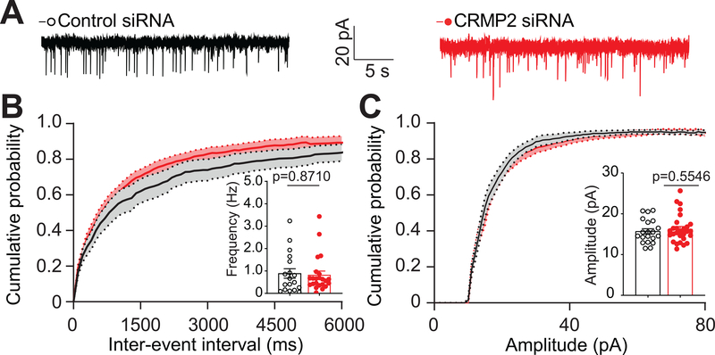
Mechanistic studies principally focusing on primary afferent nociceptive neurons uncovered the upregulation of collapsin response mediator protein 2 (CRMP2)-a dual trafficking regulator of N-type voltage-gated calcium (Cav2.2) as well as Nav1.7 voltage-gated sodium channels-as a potential determinant of neuropathic pain. Whether CRMP2 contributes to aberrant excitatory synaptic transmission underlying neuropathic pain processing after peripheral nerve injury is unknown. Here, we interrogated CRMP2's role in synaptic transmission and in the initiation or maintenance of chronic pain. In rats, short-interfering RNA-mediated knockdown of CRMP2 in the spinal cord reduced the frequency and amplitude of spontaneous excitatory postsynaptic currents, but not spontaneous inhibitory postsynaptic currents, recorded from superficial dorsal horn neurons in acute spinal cord slices. No effect was observed on miniature excitatory postsynaptic currents and inhibitory postsynaptic currents. In a complementary targeted approach, conditional knockout of CRMP2 from mouse neurons using a calcium/calmodulin-dependent protein kinase II alpha promoter to drive Cre recombinase expression reduced the frequency and amplitude of spontaneous excitatory postsynaptic currents, but not miniature excitatory SCss. Conditional knockout of CRMP2 from mouse astrocytes using a glial fibrillary acidic protein promoter had no effect on synaptic transmission. Conditional knockout of CRMP2 in neurons reversed established mechanical allodynia induced by a spared nerve injury in both male and female mice. In addition, the development of spared nerve injury-induced allodynia was also prevented in these mice. Our data strongly suggest that CRMP2 is a key regulator of glutamatergic neurotransmission driving pain signaling and that it contributes to the transition of physiological pain into pathological pain.


Figures

Fig 1.
Fig 1.. siRNA-mediated knockdown of CRMP2 decreases the frequency and amplitude of spontaneous excitatory postsynaptic currents (sEPSCs) in the Substantia gelatinosa region of the lumbar dorsal horn.
A. Schematic representation of the method used for slice electrophysiology and the analysis of CRMP2 expression in the DRGs and SCs of rat pups. Two days following intrathecal injections, DRGs and SCs of a first cohort of each treatment group were removed for western blot analyses, while spinal cords of a second cohort of each treatment group were dissected for slice recordings. B. Representative immunoblots showing the expression of CRMP2 in the DRG and the SDH of rats from each treatment group. Samples were harvested 2 days following injections. βIII-Tubulin is used as a loading control. C. Bar graph with scatter plot showing decreased CRMP2 expression in DRG and SDH of rats injected with the siRNA targeting CRMP2 (CRMP2 siRNA) compared to control (n=3 per condition). D. Representative traces of sEPSC recordings from substantia gelatinosa (SG) neurons transfected with control siRNA or siRNA targeting CRMP2 (CRMP2 siRNA). E. Cumulative distribution of sEPSC inter-event intervals recorded from SG neurons transfected with CRMP2 siRNA revealed a rightward shift towards longer inter-event intervals compared to control. Inset: Bar graph with scatter plot showing decreased sEPSC frequency compared to control. F. A cumulative distribution of sEPSC amplitudes revealed a rightward shift toward smaller amplitudes for neurons transfected with CRMP2 siRNA. Inset: Bar graph with scatter plot showing decreased sEPSC amplitude compared to control. Data are expressed as means ± SEM. Unpaired t-test with Welch’s. For full statistical analyses, see Table 1. sEPSC: spontaneous Excitatory Postsynaptic Currents, SG: Substantia Gelatinosa, DRG: Dorsal Root Ganglion, SC: Spinal Cord, SDH: Spinal Dorsal Horn.
Fig 2.
Fig 2.. siRNA-mediated knockdown of CRMP2 does not affect the frequency or amplitude of spontaneous inhibitory postsynaptic currents (sIPSCs) in the Substantia gelatinosa region of the lumbar dorsal horn.
A. Representative traces of sIPSC recordings from SG neurons from the indicated groups. B. Cumulative distribution of sIPSC inter-event intervals and bar graph with scatter plot summary of sIPSC frequencies. No significant change was observed. C. Cumulative distribution and bar graph with scatter plot summary of sIPSC amplitudes. No significant change was observed. Data are expressed as means ± SEM. Non-parametric Mann-Whitney test. For full statistical analyses, see Table 1. sIPSC: spontaneous Inhibitory Postsynaptic Currents, SG: Substantia Gelatinosa.
Fig 3.
Fig 3.. siRNA-mediated knockdown of CRMP2 does not affect miniature excitatory or inhibitory postsynaptic currents in the lumbar dorsal horn.
A. Representative traces of mEPSC recordings from SG neurons transfected either with a control siRNA or an siRNA targeting CRMP2 (CRMP2 siRNA). Cumulative distribution of mEPSC inter-event intervals B, and amplitudes C. Insets are bar graph with scatter plot summaries of mEPSC frequencies and amplitudes as indicated. No significant change was observed. D. Representative traces of mIPSC recordings from SG neurons from the indicated groups. Cumulative distribution of mIPSC inter-event intervals E and amplitudes F. Insets are bar graph with scatter plot summaries of mIPSC frequencies and amplitudes. No significant change was observed between the conditions. Data are expressed as ± SEM. Unpaired t-test with Welch’s correction. See Table 1. for full statistical analyses. mEPSC: miniature Excitatory Postsynaptic Currents, mIPSC: miniature Inhibitory Postsynaptic Currents, SG: Substantia Gelatinosa.
Fig 4.
Fig 4.. Validation of conditional knockout of CRMP2 in mouse dorsal root ganglia (DRG) and spinal dorsal horn (SDH).
A. Representative immunoblots showing the expression of CRMP2 in the DRG and SDH of mice with either a control AAV (CaMKIIα-GFP) or an AAV deleting CRMP2 in CaMKIIα+ neurons (CaMKIIα-CRE). Samples were harvested 7 days following injections. βIII-Tubulin is used as a loading control. B. Bar graph with scatter plot showing decreased CRMP2 expression in DRG and SDH of mice injected with the AAV allowing for expression of the recombinase Cre under the control of the CaMKII promoter (CaMKII-CRE) compared to control. Data are expressed as mean ± SEM. Unpaired t-test with Welch’s correction. For full statistical analyses, see Table 1. DRG: Dorsal Root Ganglion, SDH: Spinal Dorsal Horn.
Fig 5.
Fig 5.. Conditional knockout of CRMP2 in mouse neurons reduces the frequency and amplitude of spontaneous excitatory postsynaptic currents (sEPSCs) but does not affect miniature excitatory postsynaptic currents (mEPSCs) in the lumbar dorsal horn.
A. Representative traces of sEPSC recordings from SG neurons transduced with a control AAV (CaMKIIα-GFP) or an AAV allowing for expression of the recombinase Cre under the control of the CaMKII promoter (CaMKIIα-CRE). B. Cumulative distribution of sEPSC inter-event intervals, and summary of sEPSC frequencies recorded from SG neurons from mice injected with CaMKIIα-CRE AAV revealed a right shift towards longer inter-event intervals compared to control, as well as a significant decrease in the frequency (inset) compared to control. C. Cumulative distribution and summary of sEPSC amplitudes from mice transfected with CaMKII-CRE revealed a left shift toward smaller amplitudes compared to control, as well as a significant decrease in amplitude (inset) compared to control. D. Representative traces of mEPSC recordings from SG neurons transduced with the indicated AAV. Cumulative distributions of mEPSC inter-event intervals E, and amplitudes F., and bar graph with scatter plot summaries of mEPSC frequencies and amplitudes. No significant change was observed. Data are expressed as ± SEM. Unpaired t-test with Welch’s correction. For full statistical analyses, see Table 1. sEPSC: spontaneous Excitatory Postsynaptic Currents, mEPSC: miniature Excitatory Postsynaptic Currents, SG: Substantia Gelatinosa, CaMKIIα: Calcium/Calmodulin-dependent protein Kinase IIα.
Fig 6.
Fig 6.. CRMP2 expressed in astrocytes does not play a role in spinal transmission.
A. Representative traces of sEPSC recordings from SG neurons from either control (GFAP-GFP) or CRMP2 deleted (GFAP-CRE) mice. B. Cumulative distribution of sEPSC inter-event intervals and bar graph with scatter plot summary of sEPSC frequencies (inset). Deleting CRMP2 in spinal astrocytes did not affect these parameters. C. Cumulative distribution and bar graph with scatter plot summary of sEPSC amplitudes (inset). Deletion of CRMP2 in spinal astrocytes had no effect. Data are expressed as means ± SEM. Nonparametric Mann-Whitney test. For full statistical analyses, see Table 1. sEPSC: spontaneous Excitatory Postsynaptic Currents, SG; Substantia Gelatinosa, GFAP: Glial Fibrillary Acidic Protein.
Fig 7.
Fig 7.. Conditional knockout of neuronal CRMP2 in mice reverses mechanical allodynia in the spared nerve injury (SNI) model of chronic neuropathic pain without affecting physiological nociception.
A. Paw withdrawal and B. Tail-flick latency at 52°C in naïve male and female mice injected with either CaMKII-GFP (Control) or CaMKII-CRE (CRMP2 knockout in CaMKII+ neurons). No significant change was observed in the response latency between the indicated groups for both male and female mice. Nonparametric Mann-Whitney test. CRMP2f/f mice with a spared nerve injury (SNI) were injected with either CaMKII-GFP or CaMKII-CRE AAV over 35 days. Timeline (C), time course (D – males; F – females) and area under the curve (E – males; G – females) are shown. SNI elicited mechanical allodynia 15 days after surgery. Conditional knockout of CRMP2 in CaMKII+ neurons reversed mechanical allodynia compared to control. Data are shown as mean ± SEM and were analyzed by nonparametric two-way analysis of variance where time was the within-subject factor and treatment was the between-subject factor (post hoc: Sidak) (D, F) and by nonparametric Mann-Whitney test (E, G). The experiments were analyzed by an investigator blinded to the treatment. P values of comparisons between treatments are as indicated; for full statistical analyses, see Table 1. Blue arrows: AAV injection, SNI: Spared Nerve Injury.
Fig 8.
Fig 8.. Conditional knockout of CRMP2 in CaMKIIα+ neurons prevents mechanical allodynia in mice with a spared nerve injury (SNI).
CRMP2f/f mice were injected with either CaMKIIα-eGFP or CaMKIIα-GFP-Cre AAV before spared nerve injury (SNI). Timeline (A), time course (B – males; D females) and area under the curve (C – males; E – females) are shown. SNI elicited mechanical allodynia 7 days after surgery in control mice. Conditional knockout of CRMP2 in CaMKII+ neurons prevented mechanical allodynia in male mice (p=0.00264), and reversed mechanical allodynia compared to control in both male and female CRMP2f/f mice. Data are shown as mean ± SEM and were analyzed by non-parametric two-way analysis of variance where time was the within-subject factor and treatment was the between-subject factor (post hoc: Sidak) (B, D) and by non-parametric Mann-Whitney test (C, E). The experiments were analyzed by an investigator blinded to the treatment. P values of comparison between treatments are as indicated, for full statistical analyses see Table 1. Blue arrows: AAV injection, SNI: Spared Nerve Injury

References

    1. Arimura N, Kimura T, Nakamuta S, Taya S, Funahashi Y, Hattori A, Shimada A, Menager C, Kawabata S, Fujii K, Iwamatsu A, Segal RA, Fukuda M, Kaibuchi K. Anterograde transport of TrkB in axons is mediated by direct interaction with Slp1 and Rab27. Dev Cell 2009;16(5):675–686. - PubMed
    1. Brittain JM, Chen L, Wilson SM, Brustovetsky T, Gao X, Ashpole NM, Molosh AI, You H, Hudmon A, Shekhar A, White FA, Zamponi GW, Brustovetsky N, Chen J, Khanna R. Neuroprotection against traumatic brain injury by a peptide derived from the collapsin response mediator protein 2 (CRMP2). The Journal of biological chemistry 2011;286(43):37778–37792. - PMC - PubMed
    1. Brittain JM, Piekarz AD, Wang Y, Kondo T, Cummins TR, Khanna R. An atypical role for collapsin response mediator protein 2 (CRMP-2) in neurotransmitter release via interaction with presynaptic voltage-gated calcium channels. The Journal of biological chemistry 2009;284(45):31375–31390. - PMC - PubMed
    1. Castillo C, Martinez JC, Longart M, Garcia L, Hernandez M, Carballo J, Rojas H, Matteo L, Casique L, Escalona JL, Rodriguez Y, Rodriguez J, Hernandez D, Balbi D, Villegas R. Extracellular Application of CRMP2 Increases Cytoplasmic Calcium through NMDA Receptors. Neuroscience 2018;376:204–223. - PubMed
    1. Chan KY, Jang MJ, Yoo BB, Greenbaum A, Ravi N, Wu WL, Sanchez-Guardado L, Lois C, Mazmanian SK, Deverman BE, Gradinaru V. Engineered AAVs for efficient noninvasive gene delivery to the central and peripheral nervous systems. Nat Neurosci 2017;20(8):1172–1179. - PMC - PubMed

Publication types