Medial patellofemoral ligament reconstruction: A review
- PMID: 35029909
- PMCID: PMC8735765
- DOI: 10.1097/MD.0000000000028511
Medial patellofemoral ligament reconstruction: A review
Abstract
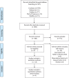
Introduction: Reconstruction of the medial patellofemoral ligament (MPFL) is an effective surgical method for the treatment of lateral patellar instability. At present, there is not much controversies regarding the femoral attachment, however, the controversies regarding patellar attachment versus attachment, number of graft strands, tension, isometry and so on. The following electronic databases will be searched: PubMed, the Cochrane Library, Embase, Web of Science, Medline. We will consider articles published between database initiation and March 2021. MPFL in the subject heading will be included in the study. Language is limited to English. Research selection, data extraction, and research quality assessment were independently completed by 2 researchers.
Conclusions: MPFL reconstruction is a reliable technique for the treatment of patellofemoral instability. The Schöttle point is still the mainstream method for locating the femoral attachment, the patellar attachment for single-bundle is located at the junction of the proximal one third and the distal two third of the longitudinal axis of the patella. For double-bundles, one is located in the proximal one third of the medial patellar edge and another is in the center of the patellar edge. Meanwhile, the adjustment of graft tension during operation is very important.
Copyright © 2022 the Author(s). Published by Wolters Kluwer Health, Inc.
Conflict of interest statement
The authors have no conflicts of interest to disclose.
Figures


References
-
- Fithian DC, Paxton EW, Stone ML, et al. . Epidemiology and natural history of acute patellar dislocation. Am J Sports Med 2004;32:1114–21. - PubMed
-
- Desio SM, Burks RT, Bachus KN. Soft tissue restraints to lateral patellar translation in the human knee. Am J Sports Med 1998;26:59–65. - PubMed
-
- Testa EA, Camathias C, Amsler F, et al. . Surgical treatment of patellofemoral instability using trochleoplasty or MPFL reconstruction: a systematic review. Knee Surg Sports Traumatol Arthrosc 2017;25:2309–20. - PubMed
-
- Warren LF, Marshall JL. The supporting structures and layers on the medial side of the knee: an anatomical analysis. J Bone Joint Surg 1979;61:56–62. - PubMed
-
- Reider B, Marshall JL, Koslin B, et al. . The anterior aspect of the knee joint. J Bone Joint Surg 1981;63:351–6. - PubMed
Publication types
MeSH terms
LinkOut - more resources
Full Text Sources

