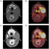
PET metabolic tumor volume as a new prognostic factor in childhood rhabdomyosarcoma
- PMID: 35030176
- PMCID: PMC8759649
- DOI: 10.1371/journal.pone.0261565
PET metabolic tumor volume as a new prognostic factor in childhood rhabdomyosarcoma
Abstract
Purpose: Childhood RMS is a rare malignant disease in which evaluation of tumour spread at diagnosis is essential for therapeutic management. F-18 FDG-PET imaging is currently used for initial RMS disease staging.
Materials and methods: This multicentre retrospective study in six French university hospitals was designed to analyse the prognostic accuracy of MTV at diagnosis for patients with RMS between 1 January 2007 and 31 October 2017, for overall (OS) and progression-free survival (PFS). MTV was defined as the sum of the primitive tumour and the largest metastasis, where relevant, with a 40% threshold of the primary tumour SUVmax. Additional aims were to define the prognostic value of SUVmax, SUVpeak, and bone lysis at diagnosis.
Results: Participants were 101 patients with a median age of 7.4 years (IQR [4.0-12.5], 62 boys), with localized disease (35 cases), regional nodal spread (43 cases), or distant metastases (23). 44 patients had alveolar subtypes. In a univariate analysis, a MTV greater than 200 cm3 was associated with OS (HR = 3.47 [1.79;6.74], p<0.001) and PFS (HR = 3.03 [1.51;6.07], p = 0.002). SUVmax, SUVpeak, and bone lysis also influenced OS (respectively p = 0.005, p = 0.004 and p = 0.007) and PFS (p = 0.029, p = 0.019 and p = 0.015). In a multivariate analysis, a MTV greater than 200 cm3 was associated with OS (HR = 2.642 [1.272;5.486], p = 0.009) and PFS (HR = 2.707 [1.322;5.547], p = 0.006) after adjustment for confounding factors, including SUVmax, SUVpeak, and bone lysis.
Conclusion: A metabolic tumor volume greater than 200 cm3, SUVmax, SUVpeak, and bone lysis in the pre-treatment assessment were unfavourable for outcome.
Conflict of interest statement
The authors have declared that no competing interests exist.
Figures


References
-
- Howlader N, Noone AM, Krapcho M, Miller D, Brest A, Yu M, et al.. (eds). SEER Cancer Statistics Review, 1975–2017, National Cancer Institute. Bethesda, MD, https://seer.cancer.gov/csr/1975_2017/, based on November 2019 SEER data submission, posted to the SEER web site, April 2020.
Publication types
MeSH terms
Substances
LinkOut - more resources
Full Text Sources

