. 2022 Apr 1:249:118865.
doi: 10.1016/j.neuroimage.2021.118865. Epub 2022 Jan 12.

Functional connectome of arousal and motor brainstem nuclei in living humans by 7 Tesla resting-state fMRI

Affiliations

Functional connectome of arousal and motor brainstem nuclei in living humans by 7 Tesla resting-state fMRI

Kavita Singh et al. Neuroimage. .

Abstract

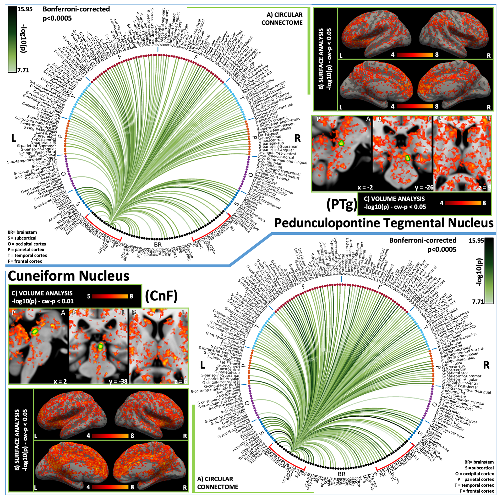
Brainstem nuclei play a pivotal role in many functions, such as arousal and motor control. Nevertheless, the connectivity of arousal and motor brainstem nuclei is understudied in living humans due to the limited sensitivity and spatial resolution of conventional imaging, and to the lack of atlases of these deep tiny regions of the brain. For a holistic comprehension of sleep, arousal and associated motor processes, we investigated in 20 healthy subjects the resting-state functional connectivity of 18 arousal and motor brainstem nuclei in living humans. To do so, we used high spatial-resolution 7 Tesla resting-state fMRI, as well as a recently developed in-vivo probabilistic atlas of these nuclei in stereotactic space. Further, we verified the translatability of our brainstem connectome approach to conventional (e.g. 3 Tesla) fMRI. Arousal brainstem nuclei displayed high interconnectivity, as well as connectivity to the thalamus, hypothalamus, basal forebrain and frontal cortex, in line with animal studies and as expected for arousal regions. Motor brainstem nuclei showed expected connectivity to the cerebellum, basal ganglia and motor cortex, as well as high interconnectivity. Comparison of 3 Tesla to 7 Tesla connectivity results indicated good translatability of our brainstem connectome approach to conventional fMRI, especially for cortical and subcortical (non-brainstem) targets and to a lesser extent for brainstem targets. The functional connectome of 18 arousal and motor brainstem nuclei with the rest of the brain might provide a better understanding of arousal, sleep and accompanying motor functions in living humans in health and disease.

Keywords: 7Tesla; Arousal network; Brainstem; Human functional connectome; Motor network.


Conflict of interest statement

Declaration of Competing Interest The authors declare no conflicts of interest.

Figures

Figure 1.
Figure 1.
(A). Connectivity matrix, of all arousal and motor nuclei. We show the 2D connectivity matrix of the arousal-motor brainstem nuclei at p < 0.0005 Bonferrroni corrected threshold. (B). 2D circular connectome of all arousal and motor nuclei. We show the region-based 2D-functional connectome at the group level of the arousal-motor brainstem nuclei at p < 0.0005 Bonferrroni corrected threshold. List of abbreviations: (brainstem nuclei used as seeds are marked with red brackets: Median Raphe nucleus (MnR), Periaqueductal Gray (PAG), Substantia Nigra-subregion1 (SN1), Substantia Nigra-subregion2 (SN2), Red nucleus-subregion1 (RN1), Red Nucleus-subregion2 (RN2), Mesencephalic Reticular formation (mRt), Cuneiform (CnF), Pedunculotegmental nuclei (PTg), Isthmic Reticular formation (isRt), Laterodorsal Tegmental Nucleus- Central Gray of the rhombencephalon (LDTg-CGPn), Pontine Reticular Nucleus, Oral Part- Pontine Reticular Nucleus Caudal Part (PnO-PnC), Locus Coeruleus (LC), Subcoeruleus nucleus (SubC), Inferior Olivary Nucleus (ION), Caudal-Rostral Linear Raphe (CLi-RLi), Dorsal raphe (DR), and Paramedian Raphe nucleus (PMnR). Additional brainstem nuclei used as targets: Superior Colliculus (SC), Inferior Colliculus (IC), Ventral Tegmental Area-Parabrachial Pigmented Nucleus (VTA-PBP), Microcellular Tegmental Nucleus-Parabigeminal nucleus (MiTg-PBG), Lateral Parabrachial Nucleus (LPB), Medial Parabrachial Nucleus (MPB), Vestibular nuclei complex (Ve), Parvicellular reticular nucleus alpha-part (PCRtA), Superior Olivary Complex (SOC), Superior Medullary Reticular formation (sMRt), Viscero-Sensory Motor nuclei complex (VSM), Inferior Medullary Reticular formation (iMRt), Raphe Magnus (RMg), Raphe Obscurus (ROb) and Raphe Pallidus (RPa). Abbreviations used: ASS:Assortativity, RC 21, RC 108:, SYN:Synchornization, HIER: Hiererichy, CLUST: clustering cofficient, cPL: Characteristic Path length, gEFF:global efficiency, lEFF: local efficiency.
Figure 2.
Figure 2.. Global graph measures and laterality index of all arousal and motor nuclei.
We display the global graph measures separately for (i) brainstem to brainstem connections, and for (ii) cortical to cortical connections; in (iii) we display the brainstem nuclei laterality index. List of brainstem nuclei abbreviations: Median Raphe nucleus (MnR), Periaqueductal Gray (PAG), Substantia Nigra-subregion1 (SN1), Substantia Nigra-subregion2 (SN2), Red nucleus-subregion1 (RN1), Red Nucleus-subregion2 (RN2), Mesencephalic Reticular formation (mRt), Cuneiform (CnF), Pedunculotegmental nuclei (PTg), Isthmic Reticular formation (isRt), Laterodorsal Tegmental Nucleus- Central Gray of the rhombencephalon (LDTg-CGPn), Pontine Reticular Nucleus, Oral Part- Pontine Reticular Nucleus Caudal Part (PnO-PnC), Locus Coeruleus (LC), Subcoeruleus nucleus (SubC), Inferior Olivary Nucleus (ION), Caudal-Rostral Linear Raphe (CLi-RLi), Dorsal raphe (DR), and Paramedian Raphe nucleus (PMnR). Abbreviations used: ASS:Assortativity, RC 21, RC 108:, SYN:Synchornization, HIER: Hiererichy, CLUST: clustering cofficient, cPL: Characteristic Path length, gEFF:global efficiency, lEFF: local efficiency.
Figure 3.
Figure 3.. 2D circular connectome (A), voxel-based connectivity maps in the cortex (B) and subcortex (C) of (top) MnR and (bottom) PMnR nucleus.
MnR showed significant connections with arousal network regions such as hypothalamus, thalamus, along with arousal brainstem nuclei (PMnR, CnF and PnO-PnC). Similarly, PMnR showed significant connectivity with several arousal regions including the cortex, forebrain, hypothalamus and thalamus, along with all major brainstem arousal nuclei (MnR, PAG, SN1, DR, CLi-RLi, mRt, CnF, isRt, VTA, LPB-MPB, LDTG-CGPn, PnO-PnC), except for LC, SN2, PTg. List of abbreviations: (brainstem nuclei used as seeds are marked with red brackets: Median Raphe nucleus (MnR), Periaqueductal Gray (PAG), Substantia Nigra-subregion1 (SN1), Substantia Nigra-subregion2 (SN2), Red nucleus-subregion1 (RN1), Red Nucleus-subregion2 (RN2), Mesencephalic Reticular formation (mRt), Cuneiform (CnF), Pedunculotegmental nuclei (PTg), Isthmic Reticular formation (isRt), Laterodorsal Tegmental Nucleus- Central Gray of the rhombencephalon (LDTg-CGPn), Pontine Reticular Nucleus, Oral Part- Pontine Reticular Nucleus Caudal Part (PnO-PnC), Locus Coeruleus (LC), Subcoeruleus nucleus (SubC), Inferior Olivary Nucleus (ION), Caudal-Rostral Linear Raphe (CLi-RLi), Dorsal raphe (DR), and Paramedian Raphe nucleus (PMnR). Additional brainstem nuclei used as targets: Superior Colliculus (SC), Inferior Colliculus (IC), Ventral Tegmental Area-Parabrachial Pigmented Nucleus (VTA-PBP), Microcellular Tegmental Nucleus-Parabigeminal nucleus (MiTg-PBG), Lateral Parabrachial Nucleus (LPB), Medial Parabrachial Nucleus (MPB), Vestibular nuclei complex (Ve), Parvicellular reticular nucleus alpha-part (PCRtA), Superior Olivary Complex (SOC), Superior Medullary Reticular formation (sMRt), Viscero-Sensory Motor nuclei complex (VSM), Inferior Medullary Reticular formation (iMRt), Raphe Magnus (RMg), Raphe Obscurus (ROb) and Raphe Pallidus (RPa).
Figure 4.
Figure 4.. 2D circular connectome (A), voxel based connectivity maps in the cortex (B) and subcortex (C) of (top) PAG and (bottom) DR.
PAG showed strong connectivity with arousal regions (basal forebrain, hypothalamus, thalamus, VTA-PBP, LC, CnF, LDTg-CGPn, and mRt), REM-sleep brainstem nuclei (SubC, sMRt, iMRt), autonomic and limbic regions (hypothalamus, prefrontal cortex, insular cortex, cingulate cortex, LPB, MPB, VSM, and RPa), yet it was not connected with the amygdala. DR showed high connectivity with all cortical regions except occipital regions. It showed expected connectivity with other arousal network nuclei except MnR although it showed connectivity with PMnR. List of abbreviations: (brainstem nuclei used as seeds are marked with red brackets: Median Raphe nucleus (MnR), Periaqueductal Gray (PAG), Substantia Nigra-subregion1 (SN1), Substantia Nigra-subregion2 (SN2), Red nucleus-subregion1 (RN1), Red Nucleus-subregion2 (RN2), Mesencephalic Reticular formation (mRt), Cuneiform (CnF), Pedunculotegmental nuclei (PTg), Isthmic Reticular formation (isRt), Laterodorsal Tegmental Nucleus- Central Gray of the rhombencephalon (LDTg-CGPn), Pontine Reticular Nucleus, Oral Part- Pontine Reticular Nucleus Caudal Part (PnO-PnC), Locus Coeruleus (LC), Subcoeruleus nucleus (SubC), Inferior Olivary Nucleus (ION), Caudal-Rostral Linear Raphe (CLi-RLi), Dorsal raphe (DR), and Paramedian Raphe nucleus (PMnR). Additional brainstem nuclei used as targets: Superior Colliculus (SC), Inferior Colliculus (IC), Ventral Tegmental Area-Parabrachial Pigmented Nucleus (VTA-PBP), Microcellular Tegmental Nucleus-Parabigeminal nucleus (MiTg-PBG), Lateral Parabrachial Nucleus (LPB), Medial Parabrachial Nucleus (MPB), Vestibular nuclei complex (Ve), Parvicellular reticular nucleus alpha-part (PCRtA), Superior Olivary Complex (SOC), Superior Medullary Reticular formation (sMRt), Viscero-Sensory Motor nuclei complex (VSM), Inferior Medullary Reticular formation (iMRt), Raphe Magnus (RMg), Raphe Obscurus (ROb) and Raphe Pallidus (RPa).
Figure 5.
Figure 5.. 2D circular connectome (A), voxel based connectivity maps in the cortex (B) and subcortex (C) of (top) LC and (bottom) SubC.
LC showed functional connectivity with the cortex (except occipital regions), thalamus and hypothalamus as expected for an arousal nucleus. Also, it showed connectivity with other arousal-motor network nuclei (PAG, mRt, LDTG-CGPn, PnO-PnC, SubC, ION, DR, VTA-PBP, parabrachial). SubC, a motor and REM-sleep nucleus, showed expected connectivity with basal ganglia and cerebellum. It also showed connectivity to brainstem arousal and motor nuclei (mRt, CnF, PTg, isRt, LDTG-CGPn, ION). List of abbreviations: (brainstem nuclei used as seeds are marked with red brackets: Median Raphe nucleus (MnR), Periaqueductal Gray (PAG), Substantia Nigra-subregion1 (SN1), Substantia Nigra-subregion2 (SN2), Red nucleus-subregion1 (RN1), Red Nucleus-subregion2 (RN2), Mesencephalic Reticular formation (mRt), Cuneiform (CnF), Pedunculotegmental nuclei (PTg), Isthmic Reticular formation (isRt), Laterodorsal Tegmental Nucleus- Central Gray of the rhombencephalon (LDTg-CGPn), Pontine Reticular Nucleus, Oral Part- Pontine Reticular Nucleus Caudal Part (PnO-PnC), Locus Coeruleus (LC), Subcoeruleus nucleus (SubC), Inferior Olivary Nucleus (ION), Caudal-Rostral Linear Raphe (CLi-RLi), Dorsal raphe (DR), and Paramedian Raphe nucleus (PMnR). Additional brainstem nuclei used as targets: Superior Colliculus (SC), Inferior Colliculus (IC), Ventral Tegmental Area-Parabrachial Pigmented Nucleus (VTA-PBP), Microcellular Tegmental Nucleus-Parabigeminal nucleus (MiTg-PBG), Lateral Parabrachial Nucleus (LPB), Medial Parabrachial Nucleus (MPB), Vestibular nuclei complex (Ve), Parvicellular reticular nucleus alpha-part (PCRtA), Superior Olivary Complex (SOC), Superior Medullary Reticular formation (sMRt), Viscero-Sensory Motor nuclei complex (VSM), Inferior Medullary Reticular formation (iMRt), Raphe Magnus (RMg), Raphe Obscurus (ROb) and Raphe Pallidus (RPa).
Figure 6.
Figure 6.. 2D circular connectome (A), voxel based connectivity maps in the cortex (B) and subcortex (C) of (top) LDTg-CGPn and (bottom) PnO-PnC.
LDTG-CGPn, an arousal nucleus, showed widespread functional connectivity with the cortex and subcortex. It showed good connectivity with arousal brainstem nuclei such as MnR, mRt, and arousal regions (hypothalamus, thalamus). PnO-PnC, an arousal and motor nucleus, showed strong connectivity with thalamus, hypothalamus, basal forebrain and cortex along with other brainstem nuclei involved in arousal (e.g. raphe nuclei, parabrachial nuclei and VTA-PBP), as well as connectivity with cerebellum, basal ganglia along with motor brainstem nuclei. List of abbreviations: (brainstem nuclei used as seeds are marked with red brackets: Median Raphe nucleus (MnR), Periaqueductal Gray (PAG), Substantia Nigra-subregion1 (SN1), Substantia Nigra-subregion2 (SN2), Red nucleus-subregion1 (RN1), Red Nucleus-subregion2 (RN2), Mesencephalic Reticular formation (mRt), Cuneiform (CnF), Pedunculotegmental nuclei (PTg), Isthmic Reticular formation (isRt), Laterodorsal Tegmental Nucleus- Central Gray of the rhombencephalon (LDTg-CGPn), Pontine Reticular Nucleus, Oral Part- Pontine Reticular Nucleus Caudal Part (PnO-PnC), Locus Coeruleus (LC), Subcoeruleus nucleus (SubC), Inferior Olivary Nucleus (ION), Caudal-Rostral Linear Raphe (CLi-RLi), Dorsal raphe (DR), and Paramedian Raphe nucleus (PMnR). Additional brainstem nuclei used as targets: Superior Colliculus (SC), Inferior Colliculus (IC), Ventral Tegmental Area-Parabrachial Pigmented Nucleus (VTA-PBP), Microcellular Tegmental Nucleus-Parabigeminal nucleus (MiTg-PBG), Lateral Parabrachial Nucleus (LPB), Medial Parabrachial Nucleus (MPB), Vestibular nuclei complex (Ve), Parvicellular reticular nucleus alpha-part (PCRtA), Superior Olivary Complex (SOC), Superior Medullary Reticular formation (sMRt), Viscero-Sensory Motor nuclei complex (VSM), Inferior Medullary Reticular formation (iMRt), Raphe Magnus (RMg), Raphe Obscurus (ROb) and Raphe Pallidus (RPa).
Figure 7.
Figure 7.
2D circular connectome (A), voxel based connectivity maps in the cortex (B) and subcortex (C) of (top) mRt and (bottom) isRt (arousal and motor network nuclei). mRt, and arousal-motor nucleus, showed connectivity with major arousal-motor brainstem nuclei like PAG, LDTg-CGPn, PnO-PnC, parabrachial nuclei, LC, SN1 and SN2 among others. Interestingly, the connectivity towards the frontal eye field (G-frontal-sup, G-frontal-middle, S-frontal-sup and S-frontal-middle) was stronger compared to other cortical areas in line with previous literature (Huerta et al., 1986). isRt also showed connectivity with other brainstem nuclei involved in arousal like SN1, SN2, PnO-PnC, DR and parabrachial nuclei. List of abbreviations: (brainstem nuclei used as seeds are marked with red brackets: Median Raphe nucleus (MnR), Periaqueductal Gray (PAG), Substantia Nigra-subregion1 (SN1), Substantia Nigra-subregion2 (SN2), Red nucleus-subregion1 (RN1), Red Nucleus-subregion2 (RN2), Mesencephalic Reticular formation (mRt), Cuneiform (CnF), Pedunculotegmental nuclei (PTg), Isthmic Reticular formation (isRt), Laterodorsal Tegmental Nucleus- Central Gray of the rhombencephalon (LDTg-CGPn), Pontine Reticular Nucleus, Oral Part- Pontine Reticular Nucleus Caudal Part (PnO-PnC), Locus Coeruleus (LC), Subcoeruleus nucleus (SubC), Inferior Olivary Nucleus (ION), Caudal-Rostral Linear Raphe (CLi-RLi), Dorsal raphe (DR), and Paramedian Raphe nucleus (PMnR). Additional brainstem nuclei used as targets: Superior Colliculus (SC), Inferior Colliculus (IC), Ventral Tegmental Area-Parabrachial Pigmented Nucleus (VTA-PBP), Microcellular Tegmental Nucleus-Parabigeminal nucleus (MiTg-PBG), Lateral Parabrachial Nucleus (LPB), Medial Parabrachial Nucleus (MPB), Vestibular nuclei complex (Ve), Parvicellular reticular nucleus alpha-part (PCRtA), Superior Olivary Complex (SOC), Superior Medullary Reticular formation (sMRt), Viscero-Sensory Motor nuclei complex (VSM), Inferior Medullary Reticular formation (iMRt), Raphe Magnus (RMg), Raphe Obscurus (ROb) and Raphe Pallidus (RPa).
Figure 8.
Figure 8.. 2D circular connectome (A), voxel based connectivity maps in the cortex (B) and subcortex (C) of (top) PTg and (bottom) CnF, both arousal and motor brainstem nuclei.
PTg showed extensive connectivity to cortex, subcortex, forebrain, basal ganglia and cerebellum as expected for arousal-motor network. Brainstem nuclei showed specific connectivity to CnF, mRt, isRt, LDTG-CGPn, PnO-PnC, LPB, MPB SubC, VTA-PBP, MiTg-PBG, MPB, PAG and DR. CnF similar cortical and subcortical connectivity as PTg. With stronger connectivity with PAG, MiTg-PBG, PMnR and more contralateral brainstem nuclei connectivity compared to PTg. List of abbreviations: (brainstem nuclei used as seeds are marked with red brackets: Median Raphe nucleus (MnR), Periaqueductal Gray (PAG), Substantia Nigra-subregion1 (SN1), Substantia Nigra-subregion2 (SN2), Red nucleus-subregion1 (RN1), Red Nucleus-subregion2 (RN2), Mesencephalic Reticular formation (mRt), Cuneiform (CnF), Pedunculotegmental nuclei (PTg), Isthmic Reticular formation (isRt), Laterodorsal Tegmental Nucleus- Central Gray of the rhombencephalon (LDTg-CGPn), Pontine Reticular Nucleus, Oral Part- Pontine Reticular Nucleus Caudal Part (PnO-PnC), Locus Coeruleus (LC), Subcoeruleus nucleus (SubC), Inferior Olivary Nucleus (ION), Caudal-Rostral Linear Raphe (CLi-RLi), Dorsal raphe (DR), and Paramedian Raphe nucleus (PMnR). Additional brainstem nuclei used as targets: Superior Colliculus (SC), Inferior Colliculus (IC), Ventral Tegmental Area-Parabrachial Pigmented Nucleus (VTA-PBP), Microcellular Tegmental Nucleus-Parabigeminal nucleus (MiTg-PBG), Lateral Parabrachial Nucleus (LPB), Medial Parabrachial Nucleus (MPB), Vestibular nuclei complex (Ve), Parvicellular reticular nucleus alpha-part (PCRtA), Superior Olivary Complex (SOC), Superior Medullary Reticular formation (sMRt), Viscero-Sensory Motor nuclei complex (VSM), Inferior Medullary Reticular formation (iMRt), Raphe Magnus (RMg), Raphe Obscurus (ROb) and Raphe Pallidus (RPa).
Figure 9.
Figure 9.. 2D circular connectome (A), voxel based connectivity maps in the cortex (B) and subcortex (C) of (top) SN1 and (bottom) SN2.
The functional connectome of SN1 and SN2 (both arousal/sleep and motor nuclei) displayed similar connectivity pattern except SN2 did not show significant connectivity with PMnR and VSM, and SN1 did not show significant connectivity with MiTg-PBG and PCRtA. Both these regions showed expected connectivity with cortex, thalamus, hypothalamus, forebrain and basal ganglia. List of abbreviations: (brainstem nuclei used as seeds are marked with red brackets : Median Raphe nucleus (MnR), Periaqueductal Gray (PAG), Substantia Nigra-subregion1 (SN1), Substantia Nigra-subregion2 (SN2), Red nucleus-subregion1 (RN1), Red Nucleus-subregion2 (RN2), Mesencephalic Reticular formation (mRt), Cuneiform (CnF), Pedunculotegmental nuclei (PTg), Isthmic Reticular formation (isRt), Laterodorsal Tegmental Nucleus- Central Gray of the rhombencephalon (LDTg-CGPn), Pontine Reticular Nucleus, Oral Part- Pontine Reticular Nucleus Caudal Part (PnO-PnC), Locus Coeruleus (LC), Subcoeruleus nucleus (SubC), Inferior Olivary Nucleus (ION), Caudal-Rostral Linear Raphe (CLi-RLi), Dorsal raphe (DR), and Paramedian Raphe nucleus (PMnR). Additional brainstem nuclei used as targets: Superior Colliculus (SC), Inferior Colliculus (IC), Ventral Tegmental Area-Parabrachial Pigmented Nucleus (VTA-PBP), Microcellular Tegmental Nucleus-Parabigeminal nucleus (MiTg-PBG), Lateral Parabrachial Nucleus (LPB), Medial Parabrachial Nucleus (MPB), Vestibular nuclei complex (Ve), Parvicellular reticular nucleus alpha-part (PCRtA), Superior Olivary Complex (SOC), Superior Medullary Reticular formation (sMRt), Viscero-Sensory Motor nuclei complex (VSM), Inferior Medullary Reticular formation (iMRt), Raphe Magnus (RMg), Raphe Obscurus (ROb) and Raphe Pallidus (RPa).
Figure 10.
Figure 10.
2D circular connectome (A), voxel based connectivity maps in the cortex (B) and subcortex (C) of (top) CLi-RLi and (bottom) ION. CLi-RLi, an arousal nucleus, showed expected connectivity with regions of frontal, temporal and parietal lobes [based on(Ikemoto, 2007)]. Subcortex also showed extensive connectivity as expected. It showed connectivity with arousal regions either ipsilaterally or contralaterally like mRt, LDTg-CGPn, PnO-PnC, thalamus and hypothalamus. ION, a motor brainstem nucleus, showed connectivity with motor cortical areas, cerebellar cortex and ipsilateral RN subregions. List of abbreviations: (brainstem nuclei used as seeds are marked with red brackets: Median Raphe nucleus (MnR), Periaqueductal Gray (PAG), Substantia Nigra-subregion1 (SN1), Substantia Nigra-subregion2 (SN2), Red nucleus-subregion1 (RN1), Red Nucleus-subregion2 (RN2), Mesencephalic Reticular formation (mRt), Cuneiform (CnF), Pedunculotegmental nuclei (PTg), Isthmic Reticular formation (isRt), Laterodorsal Tegmental Nucleus- Central Gray of the rhombencephalon (LDTg-CGPn), Pontine Reticular Nucleus, Oral Part- Pontine Reticular Nucleus Caudal Part (PnO-PnC), Locus Coeruleus (LC), Subcoeruleus nucleus (SubC), Inferior Olivary Nucleus (ION), Caudal-Rostral Linear Raphe (CLi-RLi), Dorsal raphe (DR), and Paramedian Raphe nucleus (PMnR). Additional brainstem nuclei used as targets: Superior Colliculus (SC), Inferior Colliculus (IC), Ventral Tegmental Area-Parabrachial Pigmented Nucleus (VTA-PBP), Microcellular Tegmental Nucleus-Parabigeminal nucleus (MiTg-PBG), Lateral Parabrachial Nucleus (LPB), Medial Parabrachial Nucleus (MPB), Vestibular nuclei complex (Ve), Parvicellular reticular nucleus alpha-part (PCRtA), Superior Olivary Complex (SOC), Superior Medullary Reticular formation (sMRt), Viscero-Sensory Motor nuclei complex (VSM), Inferior Medullary Reticular formation (iMRt), Raphe Magnus (RMg), Raphe Obscurus (ROb) and Raphe Pallidus (RPa).
Figure 11.
Figure 11.. 2D circular connectome (A), voxel based connectivity maps in the cortex (B) and subcortex (C) of (top) RN1 and (bottom) RN2, which are both motor brainstem nuclei.
For RN1 we found connectivity with motor cortical areas, cerebellar cortex, ipsilateral ION and Ve among others. RN2 showed strong connectivity with ipsilateral RN1, bilateral ION, Ve, VSM, iMRt, PAG, DR, as well as frontal regions. List of abbreviations: (brainstem nuclei used as seeds are marked with red brackets: Median Raphe nucleus (MnR), Periaqueductal Gray (PAG), Substantia Nigra-subregion1 (SN1), Substantia Nigra-subregion2 (SN2), Red nucleus-subregion1 (RN1), Red Nucleus-subregion2 (RN2), Mesencephalic Reticular formation (mRt), Cuneiform (CnF), Pedunculotegmental nuclei (PTg), Isthmic Reticular formation (isRt), Laterodorsal Tegmental Nucleus- Central Gray of the rhombencephalon (LDTg-CGPn), Pontine Reticular Nucleus, Oral Part- Pontine Reticular Nucleus Caudal Part (PnO-PnC), Locus Coeruleus (LC), Subcoeruleus nucleus (SubC), Inferior Olivary Nucleus (ION), Caudal-Rostral Linear Raphe (CLi-RLi), Dorsal raphe (DR), and Paramedian Raphe nucleus (PMnR). Additional brainstem nuclei used as targets: Superior Colliculus (SC), Inferior Colliculus (IC), Ventral Tegmental Area-Parabrachial Pigmented Nucleus (VTA-PBP), Microcellular Tegmental Nucleus-Parabigeminal nucleus (MiTg-PBG), Lateral Parabrachial Nucleus (LPB), Medial Parabrachial Nucleus (MPB), Vestibular nuclei complex (Ve), Parvicellular reticular nucleus alpha-part (PCRtA), Superior Olivary Complex (SOC), Superior Medullary Reticular formation (sMRt), Viscero-Sensory Motor nuclei complex (VSM), Inferior Medullary Reticular formation (iMRt), Raphe Magnus (RMg), Raphe Obscurus (ROb) and Raphe Pallidus (RPa).
Figure 12.
Figure 12.. Nodal metrics of arousal-motor network brainstem nuclei using GRETNA graph analysis (Wang et al., 2015).
Nodal metrics varied across nuclei, with PAG, mRt, isRt, LDTg-CGPn, DR showing the greatest variation (either below or above the metric average across nuclei). List of abbreviations: Median Raphe nucleus (MnR), Periaqueductal Gray (PAG), Substantia Nigra-subregion1 (SN1), Substantia Nigra-subregion2 (SN2), Red nucleus-subregion1 (RN1), Red Nucleus-subregion2 (RN2), Mesencephalic Reticular formation (mRt), Cuneiform (CnF), Pedunculotegmental nuclei (PTg), Isthmic Reticular formation (isRt), Laterodorsal Tegmental Nucleus- Central Gray of the rhombencephalon (LDTg-CGPn), Pontine Reticular Nucleus, Oral Part- Pontine Reticular Nucleus Caudal Part (PnO-PnC), Locus Coeruleus (LC), Subcoeruleus nucleus (SubC), Inferior Olivary Nucleus (ION), Caudal-Rostral Linear Raphe (CLi-RLi), Dorsal raphe (DR), and Paramedian Raphe nucleus (PMnR).
Figure 13.
Figure 13.. Translatability of 7 Tesla results in a conventional dataset acquired at 3 Tesla.
(A) Association values between connectivity scores obtained at 3 Tesla and 7 Tesla for all the targets (red), brainstem only targets (magenta), and cortical/subcortical (other than brainstem) targets; (B) percentage links in common between 7 Tesla vs 3 Tesla results found in the whole brain, brainstem and cortex/subcortex.
Figure 14.
Figure 14.. Circuit diagram of the arousal (top) and motor (bottom) network derived from resting-state 7 Tesla fMRI functional connectivity.
The diagram of the arousal network displayed high node interconnectivity. Interestingly, several brainstem arousal nuclei displayed connectivity to the thalamus, hypothalamus, basal forebrain and frontal cortex, as expected for arousal regions. The diagram of the motor network matched expected connectivity of motor regions, including the motor cortex, basal ganglia and cerebellum. Note that the link thickness was varied based on the statistical significance of the connectivity strength. For the arousal circuit, the brainstem nodes were color-coded based on the main neurotransmitter employed.

Similar articles

Cited by

References

    1. Achard S, Bullmore E, 2007. Efficiency and Cost of Economical Brain Functional Networks. PLOS Computational Biology 3, e17. 10.1371/journal.pcbi.0030017 - DOI - PMC - PubMed
    1. Agcaoglu O, Wilson TW, Wang Y, Stephen J, Calhoun VD, 2019. Resting state connectivity differences in eyes open versus eyes closed conditions. Human Brain Mapping 40, 2488–2498. 10.1002/hbm.24539 - DOI - PMC - PubMed
    1. Amunts K, Kedo O, Kindler M, Pieperhoff P, Mohlberg H, Shah NJ, Habel U, Schneider F, Zilles K, 2005. Cytoarchitectonic mapping of the human amygdala, hippocampal region and entorhinal cortex: intersubject variability and probability maps. Anat Embryol (Berl) 210, 343–352. 10.1007/s00429-005-0025-5 - DOI - PubMed
    1. Aquino KM, Fulcher BD, Parkes L, Sabaroedin K, Fornito A, 2020. Identifying and removing widespread signal deflections from fMRI data: Rethinking the global signal regression problem. Neuroimage 212, 116614. 10.1016/j.neuroimage.2020.116614 - DOI - PubMed
    1. Arnsten AF, Goldman-Rakic PS, 1984. Selective prefrontal cortical projections to the region of the locus coeruleus and raphe nuclei in the rhesus monkey. Brain Res 306, 9–18. 10.1016/0006-8993(84)90351-2 - DOI - PubMed

Publication types