. 2022 Jun;126(11):1616-1626.
doi: 10.1038/s41416-021-01691-5. Epub 2022 Jan 14.

Transiently hypoxic tumour cell turnover and radiation sensitivity in human tumour xenografts

Affiliations

Transiently hypoxic tumour cell turnover and radiation sensitivity in human tumour xenografts

Brennan J Wadsworth et al. Br J Cancer. 2022 Jun.

Abstract

Background: Solid tumour perfusion can be unstable, creating transiently hypoxic cells that can contribute to radiation resistance. We investigated the in vivo lifetime of transiently hypoxic tumour cells and chronically hypoxic tumour cells during tumour growth and following irradiation.

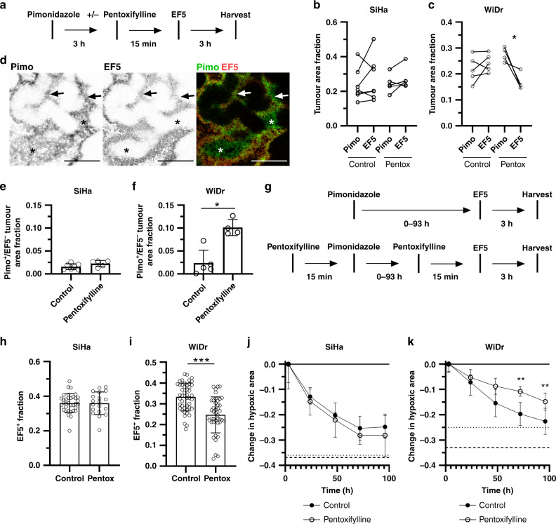
Methods: Hypoxic cells in SiHa and WiDr human tumour xenografts were labelled using pimonidazole and EF5, and turnover was quantified as the loss of labelled cells over time. The perfusion-modifying drug pentoxifylline was used to reoxygenate transiently hypoxic cells prior to hypoxia marker administration or irradiation.

Results: Chronically hypoxic cells constantly turnover in SiHa and WiDr tumours, with half-lives ranging from 42-82 h and significant numbers surviving >96 h. Transiently hypoxic cells constitute 26% of the total hypoxic cells in WiDr tumours. These transiently hypoxic cells survive at least 24 h, but then rapidly turnover with a half-life of 34 h and are undetectable 72 h after labelling. Transiently hypoxic cells are radiation-resistant, although vascular dysfunction induced by 10 Gy of ionising radiation preferentially kills transiently hypoxic cells.

Conclusions: Transiently hypoxic tumour cells survive up to 72 h in WiDr tumours and are radiation-resistant, although transiently hypoxic cells are sensitive to vascular dysfunction induced by high doses of ionising radiation.


Conflict of interest statement

The authors declare no competing interests.

Figures

Fig. 1
Fig. 1. Pimonidazole positive tumour area decreases with time.
a SiHa EF5+ tumour area and b mass versus time post-implant. c WiDr EF5+ tumour area and d mass versus time post-implant. Data points are individual tumours with n = 11–29, mean ± 95% CI, Tukey multiple comparisons test with p < 0.01 for a vs b in c; no significant differences in a. Retrospective data were analyzed for these panels. e Timeline of in vivo experiments. f Representative images of WiDr and SiHa tumours for indicated times between pimonidazole injection and tumour harvest. DAPI (nuclei) is blue, pimonidazole (Pimo) is green, EF5 is red, overlap of Pimo/EF5 is yellow. Sample regions singly labelled by EF5 are indicated by ‘>’, regions of poor tissue integrity (due to tumour necrosis or sectioning issues) are outlined with dashed lines and indicated by ‘#’, scale bars = 500 μm. g Total EF5+ area for SiHa and h WiDr tumours. i SiHa tumour area that was Pimo−EF5+ or j Pimo+EF5− over time. k WiDr tumour area that was Pimo−EF5+ or l Pimo+EF5− over time. m Total Pimo+ area for SiHa and n WiDr tumours. N = 9–12, mean ± 95% CI, Tukey multiple comparisons test with p < 0.05 for a vs b, p < 0.001 for a vs c, p < 0.01 for b vs c. o Decrease in Pimo+ area relative to EF5+ area over time. Total EF5+ hypoxic fractions for SiHa (dashed line) and WiDr (dotted line) shown for reference, n = 9–12, mean ± 95% CI. Data are combined from 3 independent experiments.
Fig. 2
Fig. 2. Pentoxifylline induces 2-nitroimidazole mismatch and reveals chronically and transiently hypoxic cell turnover.
a Experimental timeline. b Paired analysis of Pimo+ and EF5+ tumour area in control and pentoxifylline-treated mice with SiHa tumours or c WiDr tumours. N = 4–5, paired Student’s t-test, *p < 0.05. d Representative images of a WiDr tumour treated with pentoxifylline prior to EF5 injection. Pimo is green, EF5 is red, overlap of Pimo/EF5 is yellow, scale bars = 500 μm. Arrows indicate regions of chronic hypoxia; stars indicate transiently hypoxic regions reoxygenated by pentoxifylline prior to EF5 injection. e Pimo+EF5- area in control or pentoxifylline-treated mice with SiHa tumours or (F) WiDr tumours. N = 4–5, mean ± SD, Mann–Whitney test, *p < 0.05. g Experimental timelines for control and pentoxifylline treatment groups. h EF5+ area in control or pentoxifylline-treated mice with SiHa tumours or i WiDr tumours. N = 19–46, mean ± 95% CI, Mann–Whitney test, ***p < 0.001. j Decrease in Pimo+ area relative to EF5+ area over time in control or pentoxifylline-treated mice with SiHa tumours or k WiDr tumours. Total EF5+ hypoxic fractions for control (dashed line) and pentoxifylline-treated mice (dotted line) shown for reference. N = 9–12, mean ± 95% CI, Sidak multiple comparisons test between control and pentoxifylline time points, **p < 0.01. Data are combined from 3 independent experiments.
Fig. 3
Fig. 3. Hypoxia reduces tumour cell proliferation and loss of pimonidazole staining intensity.
WiDr or SiHa cells were labelled with Pimo at 1% O2 for 3 h and CTY for 20 m before culture in 21% O2, 1% O2 or 0.5% O2. a CTY mean fluorescent intensity (MFI) for WiDr cells and b SiHa cells over time. CTY data are log2-transformed so that a 50% reduction in CTY MFI as cells divide can be more readily observed. N = 3 independent repeats, mean ± SD, Sidak’s multiple comparisons test, *p < 0.05, **p < 0.01, ***p < 0.001. c Pimo MFI for WiDr cells and d SiHa cells over time. N = 3 independent repeats, mean ± SD, Dunnett’s multiple comparison test comparing each time point to the antibody-only (no Pimo) control, *p < 0.05, **p < 0.01, ***p < 0.001. e WiDr cells were labeled with Pimo and CTY prior to treatment with either 10 μM abemaciclib or 50 μM palbociclib at 21% O2 or f 1% O2. Linear regression lines are shown. N = 3 independent repeats, mean ± SEM, Dunnett’s multiple comparison test comparing each group to DMSO control, ***p < 0.001, *p < 0.05. g WiDr and h SiHa cells were cultured in 5% O2, 1% O2, 0.5% O2 or cycles of 25 m at 5% O2 and 25 m at 0.5% O2 for 48 h. Growth rates (exponential change in cell number per hour) were calculated based on cell numbers quantified by automated cell counting. N = 6, mean ± SD, Tukey multiple comparisons test, *p < 0.05, **p < 0.01, ***p < 0.001. Data are from 1 experimental day.
Fig. 4
Fig. 4. Hypoxic cell response to direct and indirect effects of ionising radiation.
a Density of CD31+ blood vessels, b density of perfused Hoechst 33342+ CD31+ blood vessels and c fraction of tumour area positive for EF5 in WiDr tumours 3 or 27 h after irradiation with 5, 2 × 5 or 10 Gy. d Representative images of WiDr tumours 3 or 27 h after 10 Gy. CD31 is red, Hoechst 33342 is blue, EF5 is green, scale bars = 500 μm. Arrow heads are Hoechst negative, EF5+ blood vessels. e Clonogenic survival of cells from WiDr tumours 3 or 27 h after irradiation with 5 Gy, f 2 × 5 Gy or g 10 Gy. Mice were treated with PBS (control; black circles) or pentoxifylline (white circles) 15 m prior to irradiation. Clonogenic data are corrected for plating efficiency from 5 non-irradiated control or pentoxifylline-treated tumours. N = 5, mean ± SD, Tukey multiple comparisons test with a vs b or b vs c p < 0.05, a vs c p < 0.001. Data are from 1 experimental day.

References

    1. Höckel M, Knoop C, Schlenger K, Vorndran B, Baußnann E, Mitze M, et al. Intratumoral pO2 predicts survival in advanced cancer of the uterine cervix. Radiother Oncol. 1993;26:45–50. doi: 10.1016/0167-8140(93)90025-4. - DOI - PubMed
    1. Höckel M, Schlenger K, Aral B, Mitze M, Schäffer U, Vaupel P. Association between tumor hypoxia and malignant progression in advanced cancer of the uterine cervix. Cancer Res. 1996;56:4509–15. - PubMed
    1. Brizel DM, Sibley GS, Prosnitz LR, Scher RL, Dewhirst MW. Tumor hypoxia adversely affects the prognosis of carcinoma of the head and neck. Int J Radiat Oncol Biol Phys. 1997;38:285–9. doi: 10.1016/S0360-3016(97)00101-6. - DOI - PubMed
    1. Nordsmark M, Overgaard M, Overgaard J. Pretreatment oxygenation predicts radiation response in advanced squamous cell carcinoma of the head and neck. Radiother Oncol. 1996;41:31–39. doi: 10.1016/S0167-8140(96)91811-3. - DOI - PubMed
    1. Nordsmark M, Alsner J, Keller J, Nielsen OS, Jensen OM, Horsman MR, et al. Hypoxia in human soft tissue sarcomas: adverse impact on survival and no association with p53 mutations. Br J Cancer. 2001;84:1070–5. doi: 10.1054/bjoc.2001.1728. - DOI - PMC - PubMed

Publication types