. 2022 Jan 15;9(1):2.
doi: 10.1186/s40658-021-00429-9.

Quantitative evaluation of PSMA PET imaging using a realistic anthropomorphic phantom and shell-less radioactive epoxy lesions

Affiliations

Quantitative evaluation of PSMA PET imaging using a realistic anthropomorphic phantom and shell-less radioactive epoxy lesions

Roberto Fedrigo et al. EJNMMI Phys. .

Abstract

Background: Positron emission tomography (PET) with prostate specific membrane antigen (PSMA) have shown superior performance in detecting metastatic prostate cancers. Relative to [18F]fluorodeoxyglucose ([18F]FDG) PET images, PSMA PET images tend to visualize significantly higher-contrast focal lesions. We aim to evaluate segmentation and reconstruction algorithms in this emerging context. Specifically, Bayesian or maximum a posteriori (MAP) image reconstruction, compared to standard ordered subsets expectation maximization (OSEM) reconstruction, has received significant interest for its potential to reach convergence with minimal noise amplifications. However, few phantom studies have evaluated the quantitative accuracy of such reconstructions for high contrast, small lesions (sub-10 mm) that are typically observed in PSMA images. In this study, we cast 3 mm-16-mm spheres using epoxy resin infused with a long half-life positron emitter (sodium-22; 22Na) to simulate prostate cancer metastasis. The anthropomorphic Probe-IQ phantom, which features a liver, bladder, lungs, and ureters, was used to model relevant anatomy. Dynamic PET acquisitions were acquired and images were reconstructed with OSEM (varying subsets and iterations) and BSREM (varying β parameters), and the effects on lesion quantitation were evaluated.

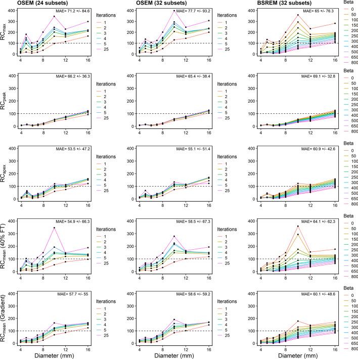
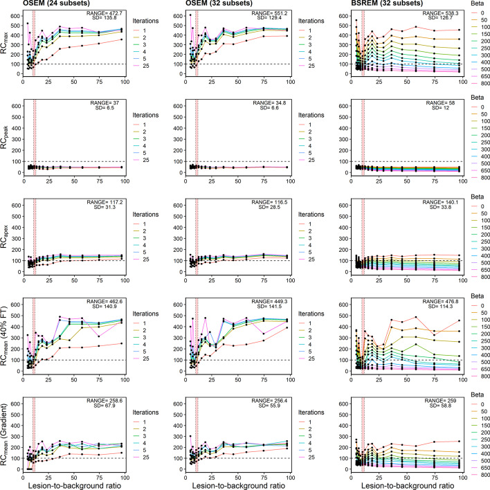
Results: The 22Na lesions were scanned against an aqueous solution containing fluorine-18 (18F) as the background. Regions-of-interest were drawn with MIM Software using 40% fixed threshold (40% FT) and a gradient segmentation algorithm (MIM's PET Edge+). Recovery coefficients (RCs) (max, mean, peak, and newly defined "apex"), metabolic tumour volume (MTV), and total tumour uptake (TTU) were calculated for each sphere. SUVpeak and SUVapex had the most consistent RCs for different lesion-to-background ratios and reconstruction parameters. The gradient-based segmentation algorithm was more accurate than 40% FT for determining MTV and TTU, particularly for lesions [Formula: see text] 6 mm in diameter (R2 = 0.979-0.996 vs. R2 = 0.115-0.527, respectively).

Conclusion: An anthropomorphic phantom was used to evaluate quantitation for PSMA PET imaging of metastatic prostate cancer lesions. BSREM with β = 200-400 and OSEM with 2-5 iterations resulted in the most accurate and robust measurements of SUVmean, MTV, and TTU for imaging conditions in 18F-PSMA PET/CT images. SUVapex, a hybrid metric of SUVmax and SUVpeak, was proposed for robust, accurate, and segmentation-free quantitation of lesions for PSMA PET.

Keywords: PET; PSMA; Phantoms; Segmentation.


Conflict of interest statement

This work depicts compounds pertaining to patent WO 2017/117687 A1, which entitles F. Bénard to royalties upon licensing. No other potential conflicts of interest relevant to this article exist.

Figures

Fig. 1
Fig. 1
Anthropomorphic Probe-IQ Phantom. (Top) Left to Right: medium phantom shell, large phantom shell, pelvis shell with bladder insert. (Bottom) Left to Right: liver, lung inserts, ribs and spine that can be inserted in the large phantom
Fig. 2
Fig. 2
Sodium-22 epoxy spheres. a Schematic of aluminium mould used for casting 3–16-mm spheres. b Radioactive epoxy spheres (3–16 mm) infused with 22Na-NaCl. c Transaxial PET image slices of Probe-IQ pelvis with 22Na spheres inserted into [18F]FDG background, which establishes increased lesion contrast at later times
Fig. 3
Fig. 3
Maximum intensity projection (MIP) images of the anthropomorphic Probe-IQ phantom. The images show increasing lesion contrast as the 18F radioactivity in the background decayed (from image 1 to image 10), and the 22Na lesion radioactivity remained approximately constant
Fig. 4
Fig. 4
PET images comparing real and simulated lesions in PSMA patient and Probe-IQ phantom, respectively. a PSMA PET image of patient with prostate cancer metastasis. b PET image of Probe-IQ phantom with embedded radioactive epoxy spheres. Transaxial slices are shown to highlight the realism of the simulated lesions
Fig. 5
Fig. 5
SUV metrics applied to simulated lesions in the Probe-IQ phantom. Max, peak, apex, 40% FT, and gradient methods applied to PET images of 14-mm, 8-mm, and 6-mm 22Na epoxy spheres reconstructed with OSEM (32 subsets, two iterations)
Fig. 6
Fig. 6
Recovery curves. (Top to bottom) Recovery concentration coefficients measured in Probe-IQ pelvis using Max, Peak, Apex, and Mean (40% FT and gradient). (Left to right) Reconstruction algorithms using OSEM + PSF (24 and 32 subsets, respectively) and BSREM. Mean absolute error (MAE) ± Standard Deviation indicated on each plot
Fig. 7
Fig. 7
Robustness of recovery curves. Recovery concentration coefficient versus lesion-to-background ratio for 10-mm lesion measured in Probe-IQ pelvis. (Top to bottom) Max, Peak, Apex, and Mean (40% FT and gradient). (Left to right) Reconstruction algorithms using OSEM + PSF (24 and 32 subsets, respectively) and BSREM. Range and standard deviation of recovery coefficients annotated on plots. [18F]DCFPyL background activity levels are represented by red shaded region
Fig. 8
Fig. 8
Tumour volume and uptake accuracy using 40% fixed threshold and gradient segmentation. Difference between measured value and ground truth, plotted vs. ground truth, for metabolic tumour volume (MTV) and total tumour uptake (TTU) metrics. (Top to bottom) 40% FT and gradient segmentation. (Left to right) Reconstruction algorithms using OSEM + PSF (24 and 32 subsets respectively) and BSREM. Blue line indicates overall fit
Fig. 9
Fig. 9
Contrast-to-noise ratio (CNR) and signal-to-noise ratio (SNR) metrics. (Top to bottom) CNR and SNR plotted vs. lesion diameter, using RCmean with gradient segmentation. (Left to right) Reconstruction algorithms using OSEM + PSF (24 and 32 subsets, respectively) and BSREM
Fig. 10
Fig. 10
Recovery curves for epoxy spheres with different 22Na activity concentrations. (Top to bottom) Probe-IQ recovery concentration coefficients vs. lesion diameter using Max, Peak, Apex, and Mean (40% FT and gradient). (Left to right)—reconstruction algorithms using OSEM + PSF (24 and 32 subsets, respectively) and BSREM. Each colour represents a different lesion-to-background activity ratio
Fig. 11
Fig. 11
Reconstruction algorithms applied to patient imaged with PSMA PET. (Left to right) [18F]DCFPyL PET images reconstructed with OSEM (24 subsets, two iterations), OSEM (32 subsets, two iterations), and BSREM (32 subsets, 25 iterations, β = 300)
Fig. 12
Fig. 12
Lesion activity concentration, volume, and uptake determined from patient analysis. Activity concentration plotted using max, peak, apex, mean (40% FT and gradient) metrics (top), metabolic tumour volume; MTV using 40% FT and gradient segmentation (middle row), and total tumour uptake; TTU using 40% FT and gradient segmentation (bottom) for ten lesions from [18F]DCFPyL patient images

References

    1. Rawla P. Epidemiology of prostate cancer. World J Oncol. 2019;10(2):63–89. doi: 10.14740/wjon1191. - DOI - PMC - PubMed
    1. Society AC. Survival rates for prostate cancer. cancer facts and figures. 2021 [cited 2021 Apr 6]. Available from: https://www.cancer.org/cancer/prostate-cancer/detection-diagnosis-stagin...
    1. Maurer T, Eiber M, Schwaiger M, Gschwend JE. Current use of PSMA-PET in prostate cancer management. Nat Rev Urol. 2016;13(4):226–235. doi: 10.1038/nrurol.2016.26. - DOI - PubMed
    1. Rousseau E, Wilson D, Lacroix-Poisson F, Krauze A, Chi K, Gleave M, et al. A prospective study on 18F-DCFPYL PSMA PET/CT imaging in biochemical recurrence of prostate cancer. J Nucl Med. 2019;60(11):1587–1593. doi: 10.2967/jnumed.119.226381. - DOI - PMC - PubMed
    1. Rowe SP, Macura KJ, Mena E, Blackford AL, Nadal R, Antonarakis ES, et al. PSMA-based [18F]DCFPyL PET/CT is superior to conventional imaging for lesion detection in patients with metastatic prostate cancer. Mol Imaging Biol. 2016;18(3):411–419. doi: 10.1007/s11307-016-0957-6. - DOI - PMC - PubMed

LinkOut - more resources