Mechanisms of energy dissipation and relationship with tissue composition in human meniscus
- PMID: 35032627
- PMCID: PMC8940718
- DOI: 10.1016/j.joca.2022.01.001
Mechanisms of energy dissipation and relationship with tissue composition in human meniscus
Abstract
Objective: The human meniscus is essential in maintaining proper knee joint function. The meniscus absorbs shock, distributes loads, and stabilizes the knee joint to prevent the onset of osteoarthritis. The extent of its shock-absorbing role can be estimated by measuring the energy dissipated by the meniscus during cyclic mechanical loading.
Methods: Samples were prepared from the central and horn regions of medial and lateral human menisci from 8 donors (both knees for total of 16 samples). Cyclic compression tests at several compression strains and frequencies yielded the energy dissipated per tissue volume. A GEE regression model was used to investigate the effects of compression, meniscal side and region, and water content on energy dissipation in order to account for repeated measures within samples.
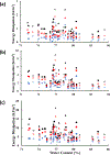
Results: Energy dissipation by the meniscus increased with compressive strain from ∼0.1 kJ/m3 (at 10% strain) to ∼10 kJ/m3 (at 20% strain) and decreased with loading frequency. Samples from the anterior region provided the largest energy dissipation when compared to central and posterior samples (P < 0.05). Water content for the 16 meniscal tissues was 77.9 (C.I. 72.0-83.8%) of the total tissue mass. A negative correlation was found between energy dissipation and water content (P < 0.05).
Conclusion: The extent of energy dissipated by the meniscus is inversely related to loading frequency and meniscal water content.
Keywords: Cyclic compression; Hysteresis; Meniscal dampening; Water content.
Copyright © 2022 Osteoarthritis Research Society International. Published by Elsevier Ltd. All rights reserved.
Conflict of interest statement
CONFLICT OF INTEREST
The authors declare that they have no known competing financial interests or personal relationships that could have appeared to influence the work reported in this paper.
Figures


References
-
- Freutel M, Scholz NB, Seitz AM, Ignatius A, Dürselen L. Mechanical properties and morphological analysis of the transitional zone between meniscal body and ligamentous meniscal attachments. Journal of Biomechanics 2015; 48: 1350–1355. - PubMed
-
- Sweigart MA, Athanasiou KA. Tensile and Compressive Properties of the Medial Rabbit Meniscus. Proceedings of the Institution of Mechanical Engineers, Part H: Journal of Engineering in Medicine 2005; 219: 337–347. - PubMed
-
- Walker PS, Arno S, Bell C, Salvadore G, Borukhov I, Oh C. Function of the medial meniscus in force transmission and stability. Journal of Biomechanics 2015; 48: 1383–1388. - PubMed
-
- Seitz AM, Galbusera F, Krais C, Ignatius A, Dürselen L. Stress-relaxation response of human menisci under confined compression conditions. Journal of the Mechanical Behavior of Biomedical Materials 2013; 26: 68–80. - PubMed
-
- Lohmander LS, Englund PM, Dahl LL, Roos EM. The Long-term Consequence of Anterior Cruciate Ligament and Meniscus Injuries. The American Journal of Sports Medicine 2007; 35: 1756–1769. - PubMed
Publication types
MeSH terms
Substances
Grants and funding
LinkOut - more resources
Full Text Sources

