. 2022 Jan 15;21(1):17.
doi: 10.1186/s12943-022-01494-z.

A nuclear function for an oncogenic microRNA as a modulator of snRNA and splicing

Affiliations

A nuclear function for an oncogenic microRNA as a modulator of snRNA and splicing

Rachid El Fatimy et al. Mol Cancer. .

Abstract

Background: miRNAs are regulatory transcripts established as repressors of mRNA stability and translation that have been functionally implicated in carcinogenesis. miR-10b is one of the key onco-miRs associated with multiple forms of cancer. Malignant gliomas exhibit particularly striking dependence on miR-10b. However, despite the therapeutic potential of miR-10b targeting, this miRNA's poorly investigated and largely unconventional properties hamper the clinical translation.

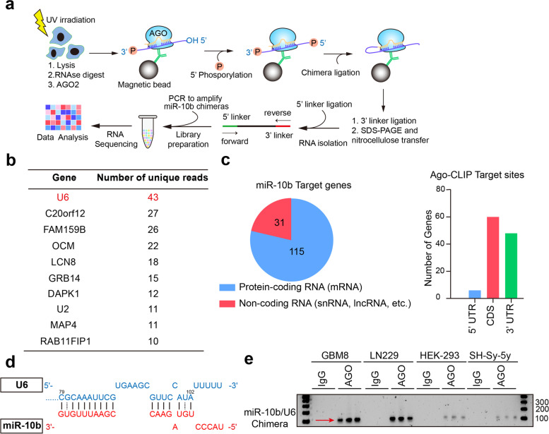
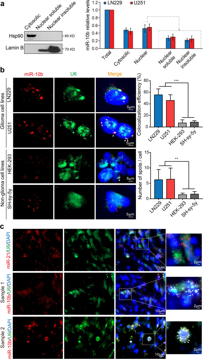
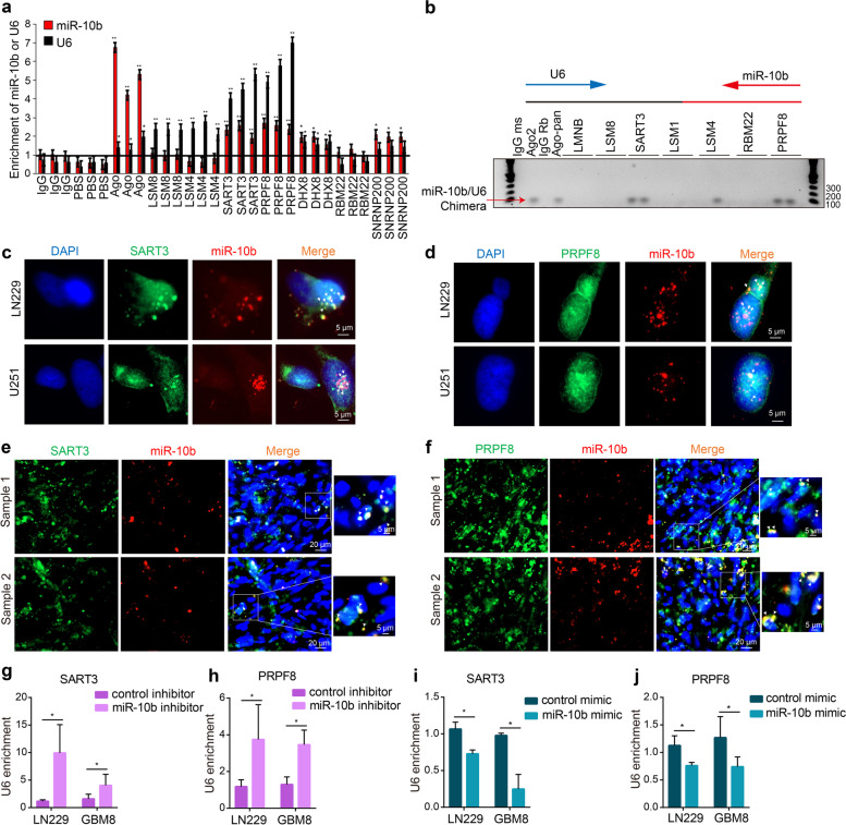
Methods: We utilized Covalent Ligation of Endogenous Argonaute-bound RNAs and their high-throughput RNA sequencing to identify miR-10b interactome and a combination of biochemical and imaging approaches for target validation. They included Crosslinking and RNA immunoprecipitation with spliceosomal proteins, a combination of miRNA FISH with protein immunofluorescence in glioma cells and patient-derived tumors, native Northern blotting, and the transcriptome-wide analysis of alternative splicing.

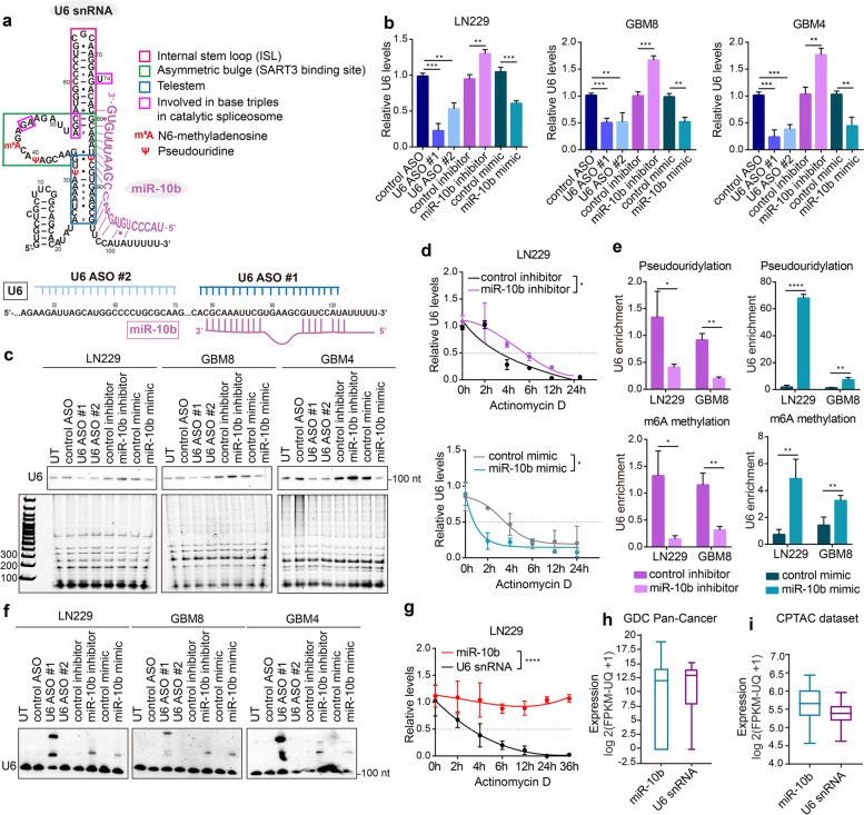
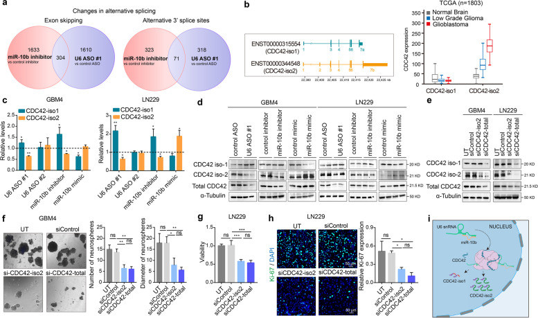
Results: We demonstrate that miR-10b binds to U6 snRNA, a core component of the spliceosomal machinery. We provide evidence of the direct binding between miR-10b and U6, in situ imaging of miR-10b and U6 co-localization in glioma cells and tumors, and biochemical co-isolation of miR-10b with the components of the spliceosome. We further demonstrate that miR-10b modulates U6 N-6-adenosine methylation and pseudouridylation, U6 binding to splicing factors SART3 and PRPF8, and regulates U6 stability, conformation, and levels. These effects on U6 result in global splicing alterations, exemplified by the altered ratio of the isoforms of a small GTPase CDC42, reduced overall CDC42 levels, and downstream CDC42 -mediated effects on cell viability.

Conclusions: We identified U6 snRNA, the key RNA component of the spliceosome, as the top miR-10b target in glioblastoma. We, therefore, present an unexpected intersection of the miRNA and splicing machineries and a new nuclear function for a major cancer-associated miRNA.

Keywords: CDC42; Glioblastoma; Nucleus; Splicing machinery; U6 snRNA; miR-10b.


Conflict of interest statement

All other authors declare no competing financial interests.

Figures

Fig. 1
Fig. 1
CLEAR-CLIP identifies U6 snRNA as the top miR-10b interacting transcript. a Schematics of CLEAR-CLIP procedure, described in details in the Methods. b Top genes represented by the highest number of unique reads identified in chimeric miR-10b libraries in LN229 cells. c Summary of miR-10b chimeras and binding site distribution in mRNAs. More details are provided in Fig. S1, Table S1 and Table S2. d miR-10b binding site within U6 snRNA, at the 3′ end (positions 79 to 102) of U6. e miR-10b-U6 chimera detected by PCR, with one primer corresponding to miR-10b and another to U6, in CLEAR-CLIP libraries produced from glioma LN229 cells, GBM8 stem cells, and non-glioma HEK-293 and SH-Sy-5y cell lines (n = 3)
Fig. 2
Fig. 2
Co-localization of miR-10b and U6 snRNA in human GBM cells and tumors. a Fractionation of glioma cells indicates that miR-10b is distributed in both cytosolic and nuclear compartments. Representative Western blotting of cytoplasmic, nuclear soluble, and nuclear insoluble fractions of glioma LN229 cells for Hsp90 (cytosolic) and Lamin B (nuclear) markers (left). MiR-10b levels in cytoplasmic, nuclear soluble, and nuclear insoluble fractions in LN229 and U251 glioma lines by qRT-PCR (mean ± SD, n = 3) (right). b Representative FISH images of miR-10b (Alexa FluorTM 546, red) and U6 (Alexa FluorTM 488, green) in cultured LN229 and U251 glioma cells and non-glioma HEK-293 and SH-Sy-5y cell lines with a fluorescently labeled probe, and nuclei stained with DAPI. Arrows mark the colocalization of miR-10b with U6. Quantification of the colocalization between miR-10b and U6 is presented in right panels. The percentage of cells with the colocalized miR-10b and U6 signals and the number of colocalization spots per cell are shown. At least 80 cells were analyzed for each cell line. P values were calculated using one-way ANOVA. c Representative FISH images of miR-10b or miR-21 (red) and U6 (green) in patient-derived GBM tissues with the corresponding fluorescently labeled probes, and nuclei stained with DAPI. Arrows mark the colocalization of miR-10b with U6. ** P < 0.01; *** P < 0.001
Fig. 3
Fig. 3
miR-10b is enriched in spliceosomal SART3 and PRPF8 RNPs. a iCLIP of glioma cells for AGO2 and spliceosomal proteins LSM8, LSM4, SART3, PRPF8, DHX8, RBM22, and SNRNP200, followed by qRT-PCR for miR-10b and U6, demonstrates miR-10b enrichment in SART3 and PRPF8 RNPs. The results are expressed as fold-changes relative to the IgG (mean ± SD, n = 3). P values were calculated using two-tail unpaired t-test. b miR-10b-U6 chimeras were detected with a pair of PCR primers, one corresponding to miR-10b and another to U6, using iCLIP assay with AGO2 and Pan-Ago antibodies, and antibodies against LMNB and splieosomal factors LSM8, SART3, LSM1, LSM4, RBM22, PRPF8 in LN229 cells. c, d Representative images of miR-10b FISH (red) and either SART3 (c) or PRPF8 (d) immunofluorescence (green) in glioma cells, and nuclei stained with DAPI (blue) (n = 3). Arrows mark the miR-10b colocalization to SART3 and PRPF8 RNPs. e, f Representative images of the colocalization of miR-10b FISH (red) and either SART3 (e) or PRPF8 (f) immunofluorescence (green) in human GBM tumors, and nuclei stained with DAPI (blue) (n = 3). Arrows mark the miR-10b colocalization to SART3 and PRPF8 RNPs. g-j iCLIP with SART3 and PRPF8 antibodies on LN229 and GBM8 cells, transfected with either miR-10b inhibitor or mimic and the corresponding control oligonucleotides, followed by qRT-PCR detection of U6, demonstrate that U6 is displaced from the RNPs by miR-10b (mean ± SD, n = 3). P values were calculated using two-tail unpaired t-test. * P < 0.05; ** P < 0.01
Fig. 4
Fig. 4
miR-10b regulates U6 snRNA levels, stability, and conformation. a Sequence, putative secondary structure, nucleotide modifications of human U6, and miR-10b binding to U6 (top panel). The structure and modifications of human U6 have not been experimentally determined, and are shown to mimic that of yeast U6 [30]. Positions of U6-targeting ASOs are shown in the lower panel. U6 ASO-1 binds to the 3’end of U6, similarly to miR-10b. b qRT-PCR analysis of U6 levels in glioma cells and GSCs transfected with either U6 ASOs, miR-10b inhibitor, mimic, or corresponding control oligonucletides (mean ± SD, n = 3). P values were calculated using two-tail unpaired t-test. c Northern blot analysis of glioma cells transfected with either U6 ASOs, miR-10b inhibitor, or mimic, with a U6-specific probe, demonstrates that U6 levels are regulated by miR-10b. The lower panel demonstrates that other small RNA species, resolved by denaturing electrophoresis, are not regulated by miR-10b. d qRT-PCR analysis of U6 levels in LN229 cells transfected with miR-10b inhibitor (top) or mimic (bottom), and treated with 5 μg/ml Actinomycin D demonstrates that miR-10b reduces U6 half-life (mean ± SD, n = 3). P values were calculated using two-way ANOVA. e iCLIP with antibodies recognizing pseudouridylation (top) and m6A methylation (bottom) on LN229 and GBM8 cells transfected with either miR-10b inhibitor or mimic, and the corresponding control oligonucleotides, followed by U6 qRT-PCR detection, demonstrate that U6 modification are regulated by miR-10b (mean ± SD, n = 3). P values were calculated using two-tail unpaired t-test. f Representative native Northern blotting with U6-specific probe demonstrates that miR-10b modulation leads to the appearance of an additional U6 structural variant. g Glioma cells treated with Actinomycin D, followed by qRT-PCR for miR-10b and U6 levels, exhibit high stability of miR-10b (mean ± SD, n = 3). P values were calculated using two-way ANOVA. h Expression of miR-10b and U6 in 155 GBM samples representing TCGA/ GDC Pan-Cancer dataset, retrieved from UCSC XENA browser (https://xena.ucsc.edu/). The RNAseq data are FPKM normalized and presented as log2(FPKM-uq + 1). The center line shows the median expression, the box limits indicate the 25th and 75th percentiles, and whiskers extend to the minimum and maximum values. i Expression of miR-10b and U6 in 96 GBM samples retrieved from Clinical Proteomic Tumor Analysis Consortium (CPTAC) (portal.gdc.cancer.gov) [31]. miR-10b and U6 levels were plotted from two separate sequencing libraries generated on the same RNA samples. The data are FPKM normalized using the GDC’s RNA-Seq pipeline and expressed as log2(FPKM-uq + 1). * P < 0.05; ** P < 0.01; *** P < 0.001; **** P < 0.0001
Fig. 5
Fig. 5
miR-10b regulates alternative splicing of CDC42, thereby controlling its total levels via U6 regulation. a Alternative splicing events induced by miR-10b inhibitor and U6 ASO #1, as determined by RNAseq (n = 3). Venn diagrams indicate the numbers of exon skipping (SE) and alternative 3′ splice sites (A3SS) modulation, in the indicated conditions. b Schematic illustration of two major CDC42 mRNA isoforms, with alternative exons encoding alternative 5′ and 3′ UTRs (left panel). Expression analysis of CDC42 isoforms in the normal brain (n = 1141), low grade glioma (LGG) (n = 509) and GBM (n = 153) in TCGA database demonstrates that ENST00000344548 (CDC42-iso2) is the pathologic variant associated with glioma progression, whereas ENST00000315554 (CDC42-iso1) is present at similarly low levels in the normal brain, LGG, and GBM (right panel). c qRT-PCR analysis of CDC42-iso1 or iso2 mRNAs in glioma cells and GSCs transfected with either U6 ASOs, or miR-10b inhibitor or mimic. The results are expressed as the fold-changes relative to the corresponding control groups (mean ± SD, n = 3). P values were calculated using two-tail unpaired t-test. d Western blot analysis of the indicated CDC42 forms in the cells transfected with either U6 ASO, or miR-10b inhibitor or mimic. e Western blot analysis of the cells transfected with either selective siRNA targeting iso2 (siCDC42-iso2) or both CDC42 isoforms (siCDC42-total) demonstrates that KD of iso2 is sufficient for reducing total CDC42 levels. f Analysis of GSC spheroids transfected with either siCDC42-iso2 or siCDC42-total demonstrates that CDC42 KD reduces GSC growth. The number and size of GSC spheres have been monitored using Image J at day 7 after transfection (mean ± SD, n = 3, 8 images per cultured analyzed). P values were calculated using two-tail unpaired t-test. g Analysis of glioma LN229 cells transfected with either siCDC42-iso2 or siCDC42-total demonstrates that CDC42 KD reduces glioma growth. Cell viability was monitored 72 h after transfection by WST-1 assay (mean ± SD, n = 3). P values were calculated using two-tail unpaired t-test. h Representative images of LN229 cells immuno-stained for Ki-67 (green) and DAPI staining (blue) and quantification of the Ki67+ cells (mean ± SD, n = 3). P values were calculated using two-tail unpaired t-test. i Schematic illustration of alternative splicing of CDC42, regulated by miR-10b via its binding to U6 snRNA. * P < 0.05; ** P < 0.01; *** P < 0.001; ns, no significance

References

    1. Lund AH. miR-10 in development and cancer. Cell Death Differ. 2010;17(2):209–214. - PubMed
    1. Tehler D, Hoyland-Kroghsbo NM, Lund AH. The miR-10 microRNA precursor family. RNA Biol. 2011;8(5):728–734. - PMC - PubMed
    1. Ma L, Teruya-Feldstein J, Weinberg RA. Tumour invasion and metastasis initiated by microRNA-10b in breast cancer. Nature. 2007;449(7163):682–688. - PubMed
    1. Sheedy P, Medarova Z. The fundamental role of miR-10b in metastatic cancer. Am J Cancer Res. 2018;8(9):1674. - PMC - PubMed
    1. Kim J, Siverly AN, Chen D, Wang M, Yuan Y, Wang Y, et al. Ablation of miR-10b suppresses oncogene-induced mammary tumorigenesis and metastasis and reactivates tumor-suppressive pathways. Cancer Res. 2016;76(21):6424–6435. - PMC - PubMed

Publication types