. 2022 Apr:54:101060.
doi: 10.1016/j.dcn.2022.101060. Epub 2022 Jan 10.

A survey of protocols from 54 infant and toddler neuroimaging research labs

Affiliations

A survey of protocols from 54 infant and toddler neuroimaging research labs

Cassandra L Hendrix et al. Dev Cogn Neurosci. 2022 Apr.

Abstract

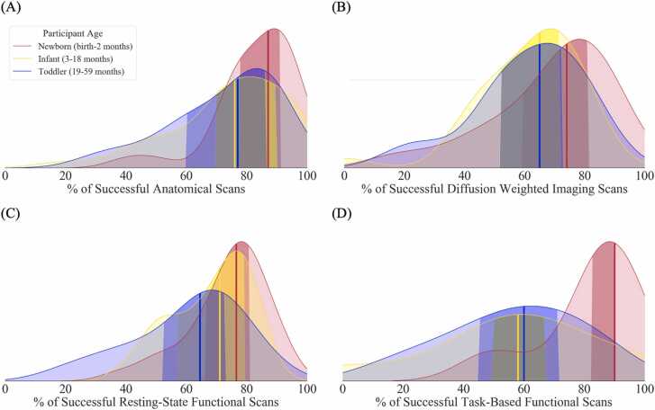
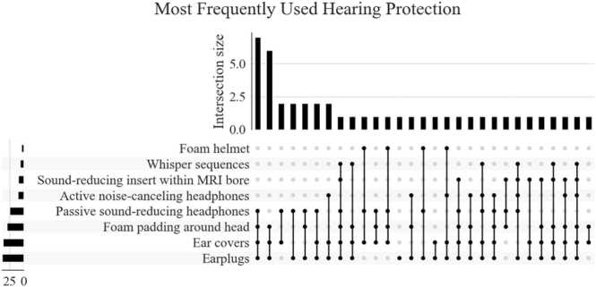
Infant and toddler MRI enables unprecedented insight into the developing brain. However, consensus about optimal data collection practices is lacking, which slows growth of the field and impedes replication efforts. The goal of this study was to collect systematic data across a large number of infant/toddler research laboratories to better understand preferred practices. Survey data addressed MRI acquisition strategies, scan success rates, visit preparations, scanning protocols, accommodations for families, study design, and policies regarding incidental findings. Respondents had on average 8 years' experience in early life neuroimaging and represented more than fifty research laboratories. Areas of consensus across labs included higher success rates among newborns compared to older infants or toddlers, high rates of data loss across age groups, endorsement of multiple layers of hearing protection, and age-specific scan preparation and participant accommodation. Researchers remain divided on decisions in longitudinal study design and practices regarding incidental findings. This study summarizes practices honed over years of work by a large collection of scientists, which may serve as an important resource for those new to the field. The ability to reference data about best practices facilitates future harmonization, data sharing, and reproducibility, all of which advance this important frontier in developmental science.

Keywords: Developmental methods; Infants; Magnetic resonance imaging; Toddlers.


Conflict of interest statement

The authors declare that they have no known competing financial interests or personal relationships that could have appeared to influence the work reported in this paper.

Figures

Fig. 1
Fig. 1
Most common participant ages and scanning sequences in early life neuroimaging. (A) The number of researchers scanning a particular, or combination of, age group(s) is displayed in Fig. 1A. Most researchers in our sample scan at least 2 early life age groups. (B) The number of researchers who report collecting each MRI scan type is displayed on the y-axis of Fig. 1B. Along the x-axis is the number of researchers who report collecting each combination of scans. The most common scan protocol includes field maps, T2w, rsfMRI, DTI, and T1 scans.
Fig. 2
Fig. 2
Scan success rates by participant age and type of scan. Researchers were asked to estimate the percent of scans that result in usable data for each participant age group and type of scan they examine. Across (A) anatomical (i.e., T1 and T2w), (B) diffusion weighted imaging (DWI), (C) rsfMRI, and (D) task-based fMRI scans, researchers consistently reported the highest success rates for newborn participants. The group median for each participant age bracket is displayed by the bolded line, and the group interquartile range is displayed as darker shading within the distribution.
Fig. 3
Fig. 3
Easiest and most difficult ages to scan. Experts were asked “What is the easiest age range to scan?” and “What is the most difficult age range to scan?” in a free response format. Each line represents a single researcher’s response, with dots indicating the lower and upper ranges of their response to each question. The gray shading represents the modal responses for each question. The text block to the right of the chart is a word cloud created from the researcher’s free response answers to questions asking them to explain why the age range they indicated was easy or difficult, respectively. (A) Experts consistently reported that 0–3 months is the easiest age range to scan, largely because they are most likely to fall asleep at the visit. (B) 12–36 months was consistently described as the most difficult age range to scan, secondary to increased child awareness of novelty and difficulty falling sleep in the scanning environment.
Fig. 4
Fig. 4
Most frequently used hearing protection during infant and toddler MRI scans. The number of researchers using each individual form of hearing protection is indicated by the bars on the y-axis. The bar graph along the x axis indicates the number of researchers who report using each combination of hearing protection. nearly all researchers apply at least 3 forms of hearing protection to infants and toddlers during MRI visits.

References

    1. Amaro E., Williams S.C.R., Shergill S.S., Fu C.H.Y., MacSweeney M., Picchioni M.M., Brammer M.J., McGuire P.K. Acoustic noise and functional magnetic resonance imaging: Current strategies and future prospects. J. Magn. Reson. Imaging. 2002;16:497–510. doi: 10.1002/jmri.10186. - DOI - PubMed
    1. Antonov N.K., Ruzal-Shapiro C.B., Morel K.D., Millar W.S., Kashyap S., Lauren C.T., Garzon M.C. Feed and wrap MRI technique in infants. Clin. Pediatr. (Philos. ). 2017;56:1095–1103. doi: 10.1177/0009922816677806. - DOI - PubMed
    1. Carney O., Hughes E., Tusor N., Dimitrova R., Arulkumaran S., Baruteau K.P., Collado A.E., Cordero-Grande L., Chew A., Falconer S., Allsop J.M., Rueckert D., Hajnal J., Edwards A.D., Rutherford M. Incidental findings on brain MR imaging of asymptomatic term neonates in the Developing Human Connectome Project. EClinicalMedicine. 2021;38 doi: 10.1016/j.eclinm.2021.100984. - DOI - PMC - PubMed
    1. Copeland A., Silver E., Korja R., Lehtola S.J., Merisaari H., Saukko E., Sinisalo S., Saunavaara J., Lähdesmäki T., Parkkola R., Nolvi S., Karlsson L., Karlsson H., Tuulari J.J. Infant and child MRI: a review of scanning procedures. Front. Neurosci. 2021;15:1–16. doi: 10.3389/fnins.2021.666020. - DOI - PMC - PubMed
    1. Dubois J., Alison M., Counsell S.J., Lucie H.P., Hüppi P.S., Benders M.J.N.L. MRI of the neonatal brain: a review of methodological challenges and neuroscientific advances. J. Magn. Reson. Imaging. 2020 doi: 10.1002/jmri.27192. - DOI - PMC - PubMed

Publication types