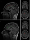
Prenatal gyrification pattern affects age at onset in frontotemporal dementia
- PMID: 35034126
- PMCID: PMC9476616
- DOI: 10.1093/cercor/bhab457
Prenatal gyrification pattern affects age at onset in frontotemporal dementia
Erratum in
-
Correction to: Prenatal gyrification pattern affects age at onset in frontotemporal dementia.Cereb Cortex. 2023 Jun 8;33(12):8087. doi: 10.1093/cercor/bhad188. Cereb Cortex. 2023. PMID: 37183181 Free PMC article. No abstract available.
Abstract
The paracingulate sulcus is a tertiary sulcus formed during the third trimester. In healthy individuals paracingulate sulcation is more prevalent in the left hemisphere. The anterior cingulate and paracingulate gyri are focal points of neurodegeneration in behavioral variant frontotemporal dementia (bvFTD). This study aims to determine the prevalence and impact of paracingulate sulcation in bvFTD. Structural magnetic resonance images of individuals with bvFTD (n = 105, mean age 66.9 years), Alzheimer's disease (n = 92, 73.3), and healthy controls (n = 110, 62.4) were evaluated using standard protocol for hemispheric paracingulate sulcal presence. No difference in left hemisphere paracingulate sulcal frequency was observed between groups; 0.72, 0.79, and 0.70, respectively, in the bvFTD, Alzheimer's disease, and healthy control groups, (P = 0.3). A significant impact of right (but not left) hemispheric paracingulate sulcation on age at disease onset was identified in bvFTD (mean 60.4 years where absent vs. 63.8 where present [P = 0.04, Cohen's d = 0.42]). This relationship was not observed in Alzheimer's disease. These findings demonstrate a relationship between prenatal neuronal development and the expression of a neurodegenerative disease providing a gross morphological example of brain reserve.
Keywords: behavioral variant frontotemporal dementia; cingulate; paracingulate; sulcation.
© The Author(s) 2022. Published by Oxford University Press.
Figures


References
-
- Borroni B, Premi E, Agosti C, Alberici A, Garibotto V, Bellelli G, Paghera B, Lucchini S, Giubbini R, Perani D, et al. 2009. Revisiting brain reserve hypothesis in frontotemporal dementia: evidence from a brain perfusion study. Dement Geriatr Cogn Disord. 28(2):130–135. - PubMed
-
- Chi JG, Dooling EC, Gilles FH. 1977. Gyral development of the human brain. Ann Neurol. 1(1):86–93. - PubMed
-
- Clark GM, Mackay CE, Davidson ME, Iversen SD, Collinson SL, James AC, Roberts N, Crow TJ. 2010. Paracingulate sulcus asymmetry; sex difference, correlation with semantic fluency and change over time in adolescent onset psychosis. Psychiatry Res. 184(1):10–15. - PubMed
-
- Del Maschio N, Sulpizio S, Fedeli D, Ramanujan K, Ding G, Weekes BS, Cachia A, Abutalebi J. 2019. ACC sulcal patterns and their modulation on cognitive control efficiency across lifespan: a neuroanatomical study on bilinguals and monolinguals. Cereb Cortex. 29(7):3091–3101. - PubMed
Publication types
MeSH terms
Grants and funding
LinkOut - more resources
Full Text Sources
Other Literature Sources
Medical

