

Clinical implications of microvascular CT scan signs in COVID-19 patients requiring invasive mechanical ventilation
- PMID: 35034320
- PMCID: PMC8761248
- DOI: 10.1007/s11547-021-01444-7
Clinical implications of microvascular CT scan signs in COVID-19 patients requiring invasive mechanical ventilation
Abstract
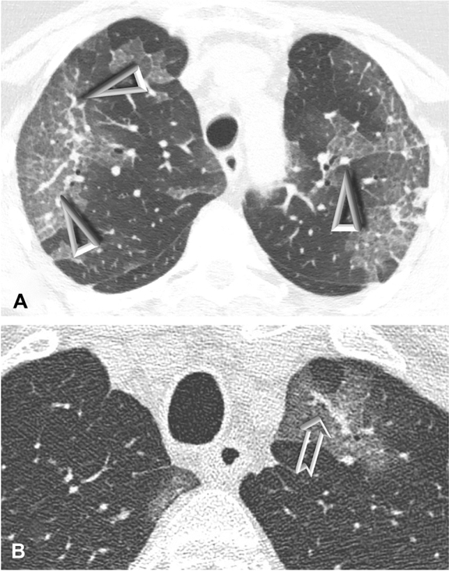
Purpose: COVID-19-related acute respiratory distress syndrome (ARDS) is characterized by the presence of signs of microvascular involvement at the CT scan, such as the vascular tree in bud (TIB) and the vascular enlargement pattern (VEP). Recent evidence suggests that TIB could be associated with an increased duration of invasive mechanical ventilation (IMV) and intensive care unit (ICU) stay. The primary objective of this study was to evaluate whether microvascular involvement signs could have a prognostic significance concerning liberation from IMV.
Material and methods: All the COVID-19 patients requiring IMV admitted to 16 Italian ICUs and having a lung CT scan recorded within 3 days from intubation were enrolled in this secondary analysis. Radiologic, clinical and biochemical data were collected.
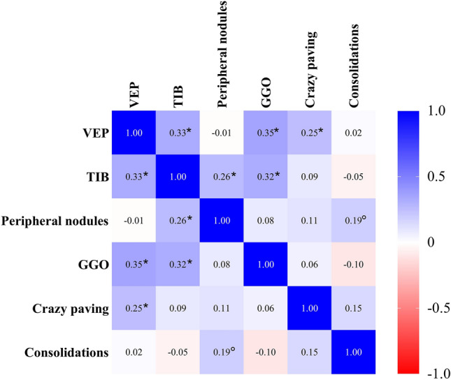
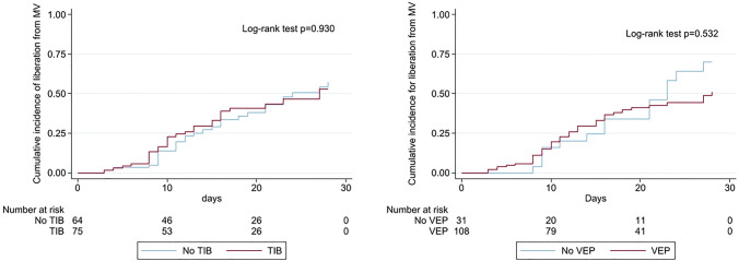
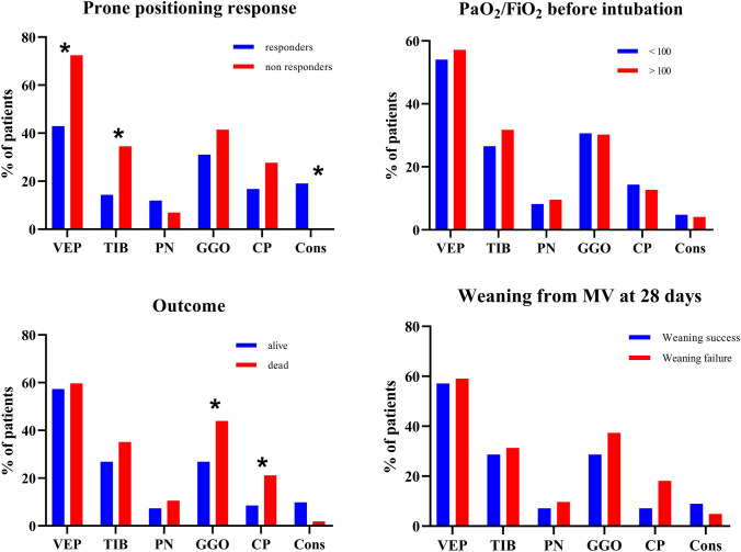
Results: A total of 139 patients affected by COVID-19 related ARDS were enrolled. After grouping based on TIB or VEP detection, we found no differences in terms of duration of IMV and mortality. Extension of VEP and TIB was significantly correlated with ground-glass opacities (GGOs) and crazy paving pattern extension. A parenchymal extent over 50% of GGO and crazy paving pattern was more frequently observed among non-survivors, while a VEP and TIB extent involving 3 or more lobes was significantly more frequent in non-responders to prone positioning.
Conclusions: The presence of early CT scan signs of microvascular involvement in COVID-19 patients does not appear to be associated with differences in duration of IMV and mortality. However, patients with a high extension of VEP and TIB may have a reduced oxygenation response to prone positioning.
Trial registration: NCT04411459.
Keywords: Acute respiratory distress syndrome; Mechanical ventilation; Novel coronavirus disease 2019; Pulmonary perfusion; Thoracic imaging.
© 2022. Italian Society of Medical Radiology.
Conflict of interest statement
The authors have nothing to disclose.
Figures
References
-
- Wu Z, McGoogan JM. Characteristics of and important lessons from the coronavirus disease 2019 (COVID-19) outbreak in China: summary of a report of 72314 cases from the chinese center for disease control and prevention. JAMA J Am Med Assoc. 2020;323:1239–1242. doi: 10.1001/jama.2020.2648. - DOI - PubMed
