. 2021 Dec 3;49(6):534-550.
doi: 10.1080/12298093.2021.1997461. eCollection 2021.

New Species and Eight Undescribed Species Belonging to the Families Aspergillaceae and Trichocomaceae in Korea

Affiliations

New Species and Eight Undescribed Species Belonging to the Families Aspergillaceae and Trichocomaceae in Korea

Thuong T T Nguyen et al. Mycobiology. .

Abstract

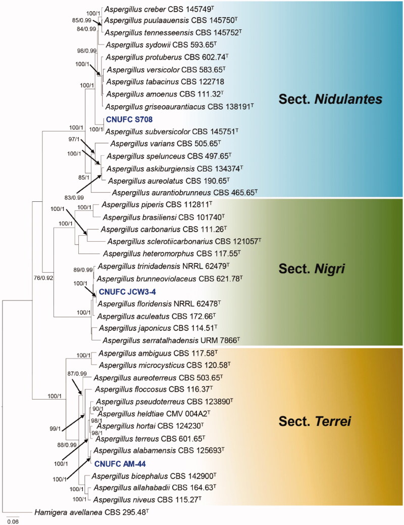
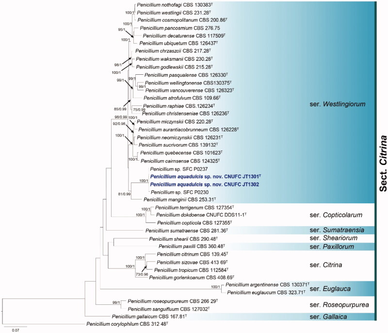
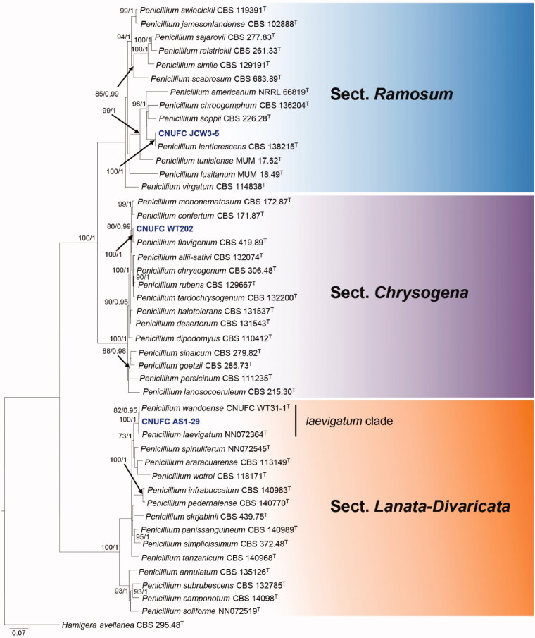
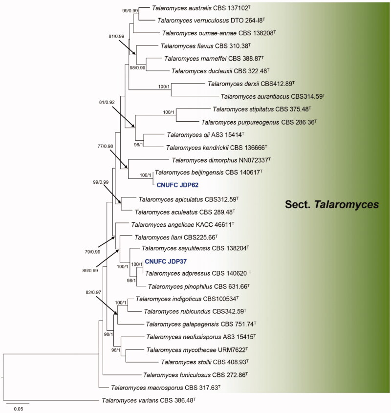
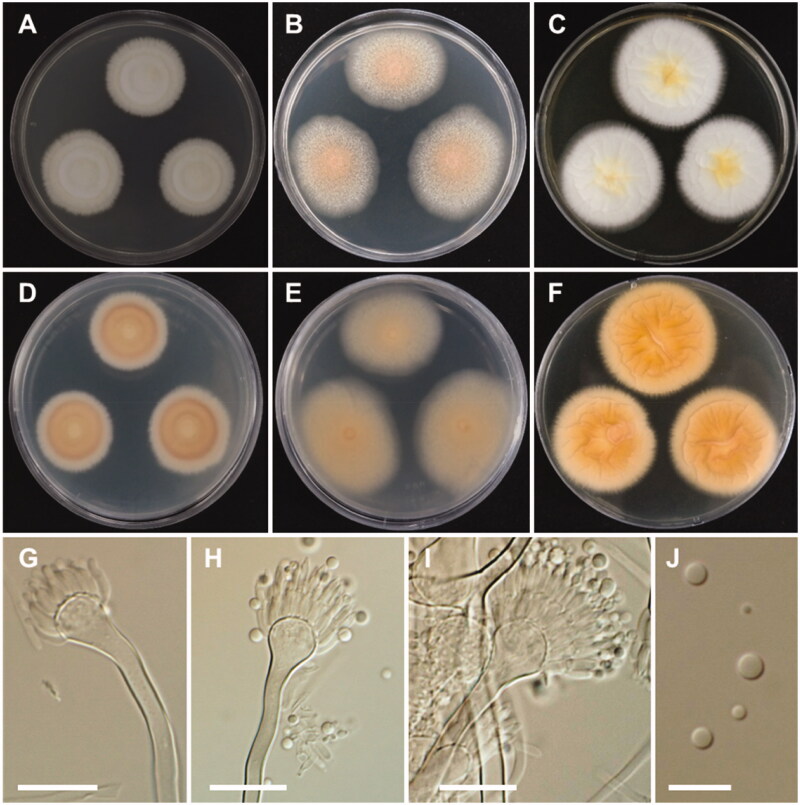
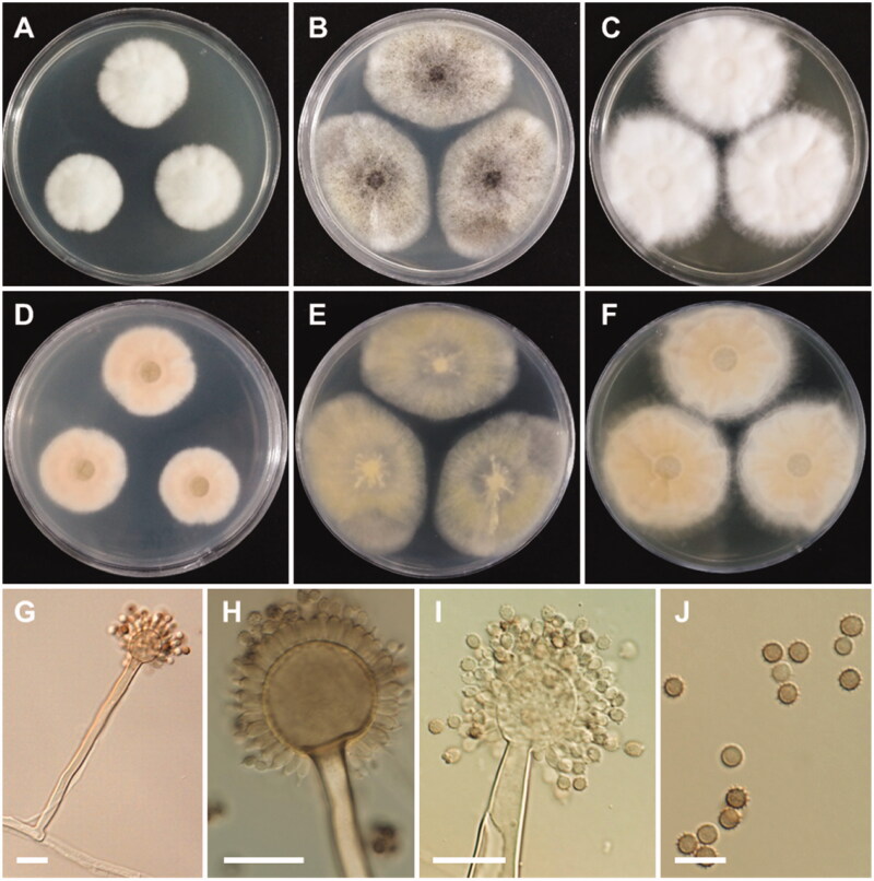
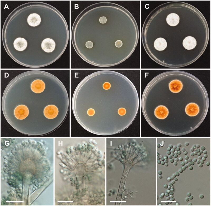
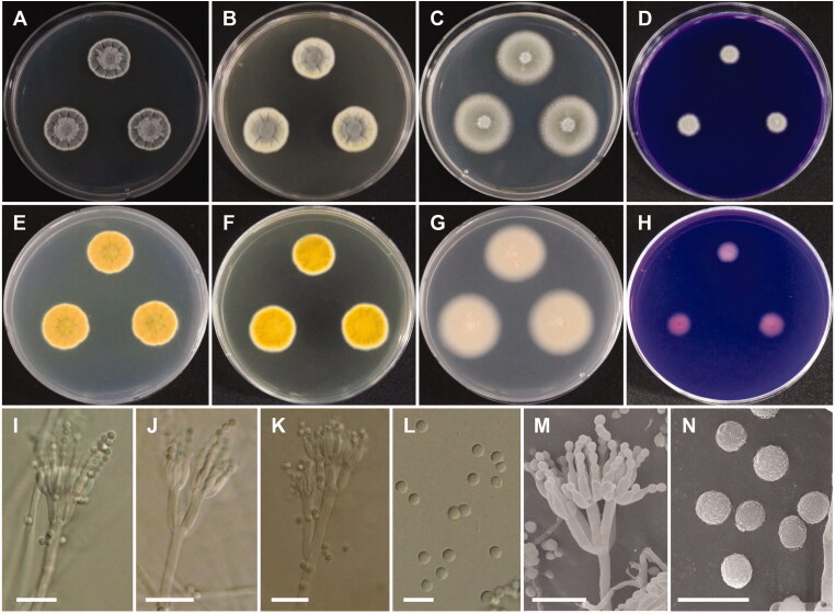
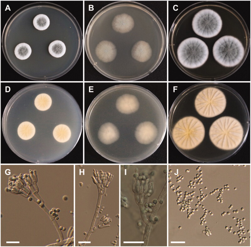
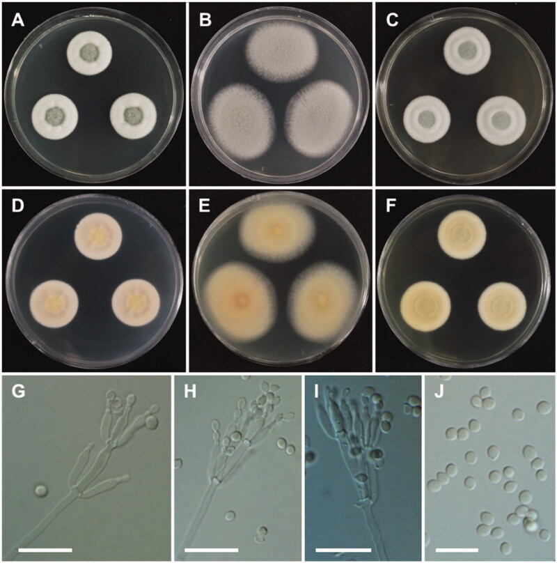
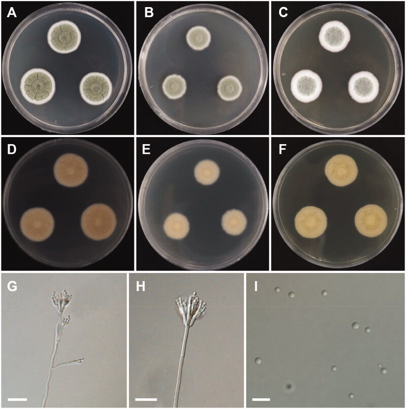
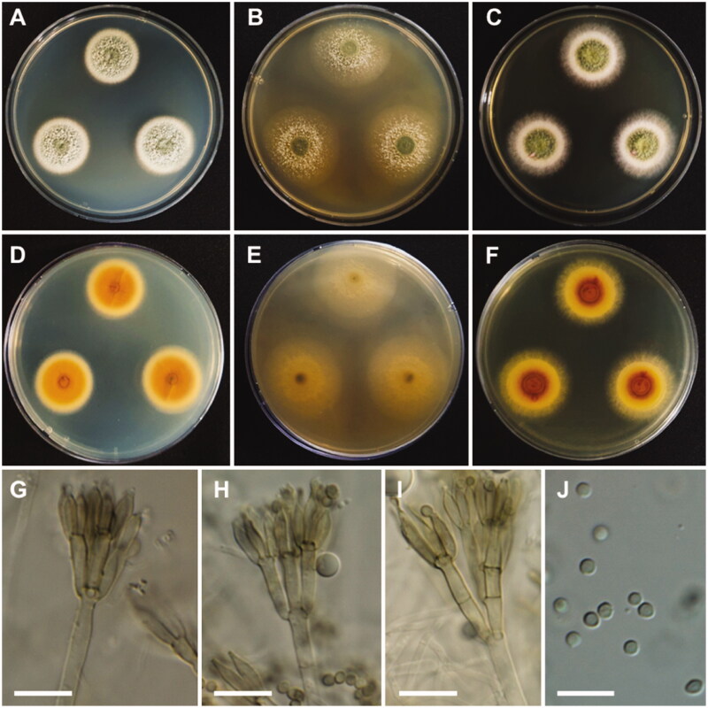
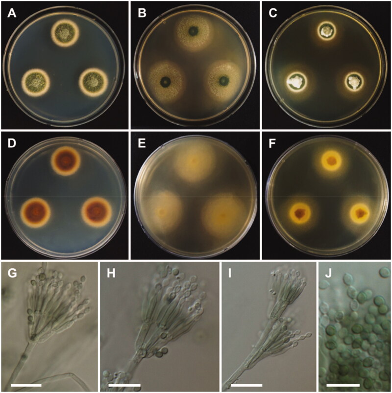
During a survey of fungal diversity associated with insects, mud, soil, and freshwater niches in different areas in Korea, nine interesting fungal strains were isolated. Based on their morphological characteristics and molecular phylogeny analyses, using a combined data set of β-tubulin (BenA), calmodulin (CaM), and second largest subunit of RNA polymerase (RPB2) sequences, the strains CNUFC AM-44, CNUFC JCW3-4, CNUFC S708, CNUFC WT202, CNUFC AS1-29, CNUFC JCW3-5, CNUFC JDP37, and CNUFC JDP62 were identified as Aspergillus alabamensis, A. floridensis, A. subversicolor, Penicillium flavigenum, P. laevigatum, P. lenticrescens, Talaromyces adpressus, and T. beijingensis, respectively. The strain CNUFC JT1301 belongs to series Westlingiorum in section Citrina and is phylogenetically related to P. manginii. However, slow growth when cultivated on CYA, MEA, CREA is observed and the property can be used to easily distinguish the new species from these species. Additionally, P. manginii is known to produce sclerotia, while CNUFC JT1301 strain does not. Herein, the new fungal species is proposed as P. aquadulcis sp. nov. Eight species, A. alabamensis, A. floridensis, A. subversicolor, P. flavigenum, P. laevigatum, P. lenticrescens, T. adpressus, and T. beijingensis, have not been previously reported in Korea. The present study expands the known distribution of fungal species belonging to the families Aspergillaceae and Trichocomaceae in Korea.

Keywords: Freshwater; insect; morphology; mud; phylogeny; soil; taxonomy.


Conflict of interest statement

No potential conflict of interest was reported by the author(s).

Figures

Figure 1.
Figure 1.
Phylogram generated from the Maximum Likelihood (RAxML) analysis based on a combined BenA, CaM and RPB2 sequence data for species classified in the Aspergillus sections Nigri, Terrei and Nidulantes. Bootstrap values ≥70% MLBS and ≥0.90 BPP are indicated above or below branches. Hamigera avellanea CBS 295.48 was used as the outgroup. The newly generated sequences are indicated in blue bold. T = ex type.
Figure 2.
Figure 2.
Phylogram generated from the Maximum Likelihood (RAxML) analysis based on a combined BenA, CaM and RPB2 sequence data for species classified in Penicillium section Citrina. Bootstrap values ≥70% MLBS and ≥0.90 BPP are indicated above or below branches. Penicillium corylophilum CBS 312.48 was used as outgroup. The newly generated sequence is indicated in blue bold. T = ex type.
Figure 3.
Figure 3.
Phylogram generated from the Maximum Likelihood (RAxML) analysis based on a combined BenA, CaM and RPB2 sequence data for species classified in the Penicillium sections Chrysogena, Lanata-Divaricata and Ramosum. Bootstrap values ≥70% MLBS and ≥0.90 BPP are indicated above or below branches. Hamigera avellanea CBS 295.48 was used as the outgroup. The newly generated sequences are indicated in blue bold. T = ex type.
Figure 4.
Figure 4.
Phylogram generated from the Maximum Likelihood (RAxML) analysis based on a combined BenA, CaM and RPB2 sequence data for species classified in the section Talaromyces. Bootstrap values ≥70% MLBS and ≥0.90 BPP are indicated above or below branches. Talaromyces varians CBS 386.48 was used as the outgroup. The newly generated sequences are indicated in blue bold. T = ex type.
Figure 5.
Figure 5.
Morphology of Aspergillus alabamensis. A, D: Colonies on Czapek yeast autolysate agar (CYA). B, E: Colonies on malt extract agar (MEA). C, F: Colonies on Yeast extract sucrose agar (YES). (A–C: obverse view, D–F: reverse view). (G–I) Conidiophores; (J) Conidia (scale bars: G–I = 20 μm, J = 10 μm).
Figure 6.
Figure 6.
Morphology of Aspergillus floridensis. A, D: Colonies on Czapek yeast autolysate agar (CYA). B, E: Colonies on malt extract agar (MEA). C, F: Colonies on yeast extract sucrose agar (YES). (A–C: obverse view, D–F: reverse view). (G–I) Conidiophores. (J) Conidia (scale bars: G–I = 20 μm, J = 10 μm).
Figure 7.
Figure 7.
Morphology of Aspergillus subversicolor. A, D: Colonies on Czapek yeast autolysate agar (CYA). B, E: Colonies on malt extract agar (MEA). C, F: Colonies on yeast extract sucrose agar (YES). (A–C: obverse view, D–F: reverse view). (G–I) Conidiophores; (J) Conidia (scale bars: G–J = 20 μm).
Figure 8.
Figure 8.
Morphology of Penicillium aquadulcis sp. nov. A, E: Colonies on Czapek yeast autolysate agar (CYA). B, F: Colonies on yeast extract sucrose agar (YES). C, G: Colonies on malt extract agar (MEA). D, H: Colonies on creatine sucrose agar (CREA). (A–D: obverse view, E–H: reverse view), (I–L: LM; M, N: SEM). (I–K, M) Conidiophores. (L, N) Conidia (scale bars: I–K, M = 10 μm, L, N = 5 μm).
Figure 9.
Figure 9.
Morphology of Penicillium flavigenum. A, D: Colonies on Czapek yeast autolysate agar (CYA). B, E: Colonies on malt extract agar (MEA). C, F: Colonies on yeast extract sucrose agar (YES). (A–C: obverse view, D–F: reverse view). (G–I) Conidiophores; (J) Conidia (scale bars: G–J = 20 μm).
Figure 10.
Figure 10.
Morphology of Penicillium laevigatum. A, D: Colonies on Czapek yeast autolysate agar (CYA). B, E: Colonies on malt extract agar (MEA). C, F: Colonies on yeast extract sucrose agar (YES). (A–C: obverse view, D–F: reverse view). (G–I) Conidiophores; (J) Conidia (scale bars: G–I = 20 μm, J = 10 μm).
Figure 11.
Figure 11.
Morphology of Penicillium lenticrescens. A, D: Colonies on Czapek yeast autolysate agar (CYA). B, E: Colonies on malt extract agar (MEA). C, F: Colonies on yeast extract sucrose agar (YES). (A–C: obverse view, D–F: reverse view). (G, H) Conidiophores; (I) Conidia (scale bars: G, H = 20, I = 10 μm).
Figure 12.
Figure 12.
Morphology of Talaromyces adpressus. A, D: Colonies on Czapek yeast autolysate agar (CYA). B, E: Colonies on malt extract agar (MEA). C, F: Colonies on yeast extract sucrose agar (YES). (A–C: obverse view, D–F: reverse view). (G–I) Conidiophores; (J) Conidia (scale bars: G–J = 10 μm).
Figure 13.
Figure 13.
Morphology of Talaromyces beijingensis. A, D: Colonies on Czapek yeast autolysate agar (CYA). B, E: Colonies on malt extract agar (MEA). C, F: Colonies on yeast extract sucrose agar (YES). (A–C: obverse view, D–F: reverse view). (G–I) Conidiophores; (J) Conidia (scale bars: G–J = 10 μm).

References

    1. Samson RA, Visagie CM, Houbraken J, et al. Phylogeny, identification and nomenclature of the genus Aspergillus. Stud Mycol. 2014;78:141–173. - PMC - PubMed
    1. Visagie CM, Hirooka Y, Tanney JB, et al. Aspergillus, Penicillium and Talaromyces isolated from house dust samples collected around the world. Stud Mycol. 2014;78:63–139. - PMC - PubMed
    1. Yilmaz N, Visagie CM, Houbraken J, et al. Polyphasic taxonomy of the genus Talaromyces. Stud Mycol. 2014;78:175–341. - PMC - PubMed
    1. Frisvad JC, Smedsgaard J, Larsen TO, et al. Mycotoxins, drugs and other extrolites produced by species in Penicillium subgenus Penicillium. Stud Mycol. 2004;49:201–241.
    1. Frisvad JC. Taxonomy, chemodiversity, and chemoconsistency of Aspergillus, Penicillium, and Talaromyces species. Front Microbiol. 2014;5:773. - PMC - PubMed

LinkOut - more resources