

A case of a small-sized cavernous hemangioma in the right ventricle - an incidental finding
- PMID: 35035650
- PMCID: PMC8753057
- DOI: 10.1016/j.radcr.2021.12.038
A case of a small-sized cavernous hemangioma in the right ventricle - an incidental finding
Erratum in
-
Erratum regarding missing declaration of competing interest statements in previously published articles.Radiol Case Rep. 2023 Jan 24;18(3):1391-1392. doi: 10.1016/j.radcr.2023.01.015. eCollection 2023 Mar. Radiol Case Rep. 2023. PMID: 36818995 Free PMC article.
Abstract
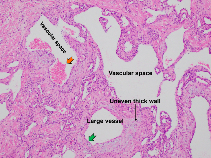
A cardiac cavernous hemangioma is a rare, primary, benign tumor that is usually diagnosed in young or middle-aged patients. In this article, we report the case of a 71-year-old male patient whose doctors incidentally discovered a heart tumor on his transthoracic echocardiography. Triple-phase computed tomography (CT) (pre-contrast, arterial and portal venous) missed the lesion, and magnetic resonance imaging (MRI) revealed a small, oval tumor attached to the wall of the right ventricle. The tumor was successfully removed surgically, and the patient recovered after 2 weeks. A histopathological examination resulted in the diagnosis of a benign cavernous hemangioma.
Keywords: Cardiac tumor; Cavernous hemangioma; Computed tomography (CT); Echocardiography; Magnetic resonance imaging (MRI).
© 2021 The Authors. Published by Elsevier Inc. on behalf of University of Washington.
Figures


Similar articles
-
Dumbbell shaped craniorbital cavernous hemangioma.BMC Neurol. 2020 Apr 22;20(1):149. doi: 10.1186/s12883-020-01734-z. BMC Neurol. 2020. PMID: 32321464 Free PMC article.
-
Exophytic cavernous hemangioma arising from the right ventricle: Report of a rare case.Pathol Int. 2021 Apr;71(4):267-271. doi: 10.1111/pin.13075. Epub 2021 Feb 9. Pathol Int. 2021. PMID: 33559333
-
Adrenal cavernous hemangioma: a case report.BMC Surg. 2018 Nov 20;18(1):103. doi: 10.1186/s12893-018-0429-9. BMC Surg. 2018. PMID: 30458815 Free PMC article.
-
Intraventricular cavernous hemangioma at the foramen of Monro: Case report and literature review.Clin Neurol Neurosurg. 2006 Sep;108(6):604-9. doi: 10.1016/j.clineuro.2005.04.004. Clin Neurol Neurosurg. 2006. PMID: 15916846 Review.
-
Cardiac hemangioma: a report of two cases and review of the literature.Heart Vessels. 2003 Jul;18(3):153-6. doi: 10.1007/s00380-003-0699-7. Heart Vessels. 2003. PMID: 12955432 Review.
Cited by
-
Cardiac Hemangiomas: A Five-Year Systematic Review of Diagnosis, Treatment, and Outcomes.Cancers (Basel). 2025 Apr 30;17(9):1532. doi: 10.3390/cancers17091532. Cancers (Basel). 2025. PMID: 40361457 Free PMC article. Review.
-
Cavernous mediastinal hemangioma presenting with persistent cough: a rare case report and review of literature.J Cardiothorac Surg. 2023 Jan 5;18(1):3. doi: 10.1186/s13019-023-02130-7. J Cardiothorac Surg. 2023. PMID: 36604701 Free PMC article. Review.
-
Epicardial cavernous haemangioma; A case report of a unique incidental finding.Eur Heart J Case Rep. 2024 Mar 21;8(4):ytae146. doi: 10.1093/ehjcr/ytae146. eCollection 2024 Apr. Eur Heart J Case Rep. 2024. PMID: 38660462 Free PMC article.
References
Publication types
LinkOut - more resources
Full Text Sources
