. 2021 Dec 15;21(6):e45.
doi: 10.4110/in.2021.21.e45. eCollection 2021 Dec.

A Potential New Mouse Model of Axial Spondyloarthritis Involving the Complement System

Affiliations

A Potential New Mouse Model of Axial Spondyloarthritis Involving the Complement System

V Michael Holers et al. Immune Netw. .

Abstract

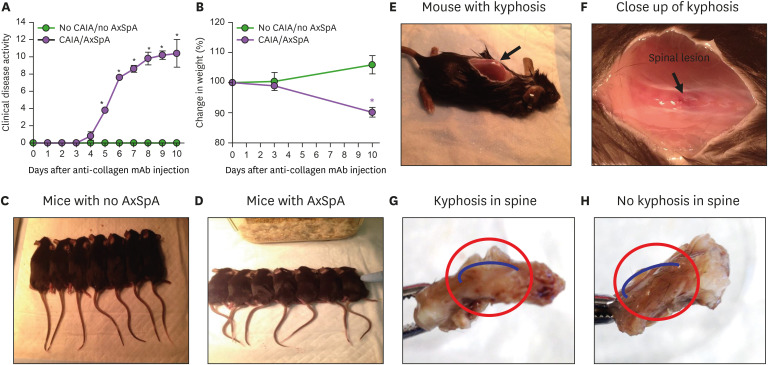
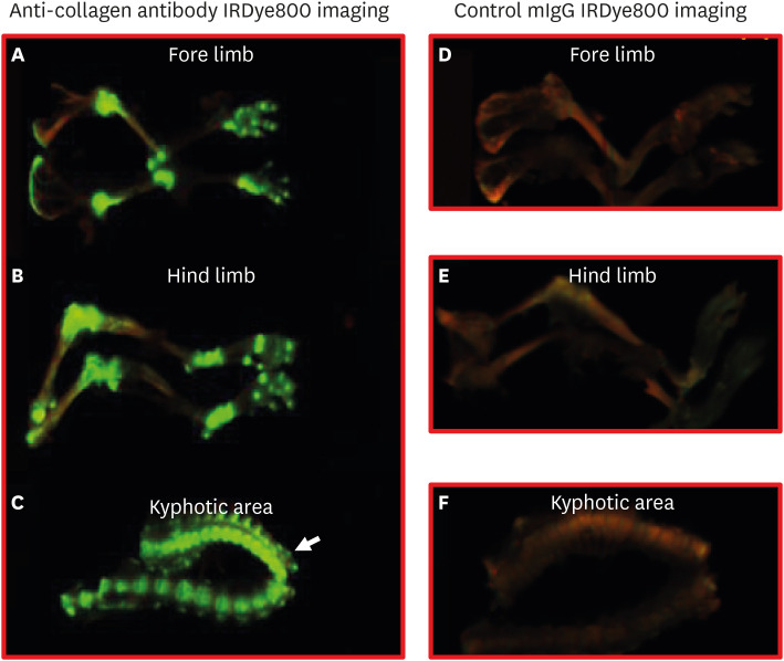
Many mouse models of rheumatoid arthritis have been identified, but only a limited number are present for axial spondyloarthritis (AxSpA). Collagen Ab-induced arthritis (CAIA) is one of the most widely used mouse models of arthritis, and it is complement-dependent. We found that mice developing CAIA also developed spinal lesions similar to those found in AxSpA. To induce CAIA, mice were injected intraperitoneally at day 0 with anti-collagen Abs, followed by LPS injection at day 3. CAIA mice demonstrated a significant kyphosis through the spine, as well as hypertrophic cartilage and osseous damage of the intravertebral joints. Immunohistochemical staining of the kyphotic area revealed increased complement C3 deposition and macrophage infiltration, with localization to the intravertebral joint margins. Near Infrared (NIR) in vivo imaging showed that anti-collagen Abs conjugated with IRDye® 800CW not only localized to cartilage surface in the joints but also to the spine in arthritic mice. We report here a novel preclinical mouse model in which, associated with the induction of CAIA, mice also exhibited salient features of AxSpA; this new experimental model of AxSpA may allow investigators to shed light on the local causal mechanisms of AxSpA bone and soft tissue changes as well as treatment.

Keywords: Arthritis; Axial spondyloarthritis; Complement.


Conflict of interest statement

Conflict of Interest: The authors declare no potential conflicts of interest.

Figures

Figure 1
Figure 1. Mice with CAIA developed AxSpA-like phenotype and mice with no CAIA developed no AxSpA-like phenotype. All WT mice with arthritis consistently developed kyphosis and spine lesions indicating the development of AxSpA. (A) CDA in WT injected with anti-collagen Ab developed arthritis and AxSpA compared with control mice. (B) Percent change in weight in mice with CAIA/AxSpA compared with control mice without CAIA/AxSpA. (C) Representative gross images of mice with normal posture without kyphosis in a cohort of WT with no arthritis and no AxSpA. (D) Representative gross images of mice with bended backbone posture showing kyphosis in a cohort of arthritic mice co-developed AxSpA. (E) The kyphotic area in mice with CAIA/AxSpA was clearly visible at autopsy after making an incision on the overlying skin. (F) Close up view of an incision made to show the morphology of the spine lesion similar to AxSpA on a mice with induced arthritis (black arrow). (G) After dissection of the spine with arthritis the curved reddish spine area was exposed indicating AxSpA (curved blue line within red circle). (H) The same exam was performed in a mouse without arthritis; no reddish spine area was seen, and it was straight (curved blue line in the red circle). Data expressed as mean±SD.
*p <0.05 considered significant. CDA shown in WT mice with CAIA/AxSpA (n=5) and WT control mice with no CAIA/no AxSpA (n=5). A separate cohort of WT mice with CAIA/AxSpA (n=7) or control WT mice without CAIA/AxSpA (n=7).
Figure 2
Figure 2. Histopathology, C3 deposition and macrophage infiltration of kyphotic area of spine. (A) A large and reactive chondrocyte proliferation in the kyphotic area of the vertebrae of mice with arthritis and AxSpA specifically in IVD area. (B) A normal chondrocyte growth in mice in inter vertebral disc area with no arthritis and no kyphosis. (C) Histopathological scores for inflammation, IVD destruction, cartilage damage, bone erosion, excess tissue formation and ectopic chondrocyte presence on the cartilage in the kyphotic area in mice with or without CAIA/AxSpA. (D) Increased C3 deposition score in the spine lesion and surrounding area of a treated mouse with spine lesion. (E) Minimal or no C3 deposition in spine areas of mice without arthritis and AxSpA. (F) C3 deposition in the spinal kyphotic and surrounding area as well as on the IVD. (G) Score of macrophages (red color) marked by a block arrow seen in the kyphotic area of mice with arthritis and AxSpA. (H) Few macrophages present in the spine areas of mice without arthritis and AxSpA. (I) Scores for F4/80 positivity in the spine lesion area and vertebral peripheral areas. Data expressed for histopathology scores as mean±SD (n=5 in each group). Data expressed for C3 deposition and macrophage as mean±SD. All microphotographs at 10× magnification.
BM, bone marrow. *p<0.05 considered significant (n=4 in each group).
Figure 3
Figure 3. Comparative histological examination of H&E sections of mice with and without CAIA/AxSpA and spine lesions. (A) Knee joint of mice with CAIA/AxSpA showing multilayered synovium (pannus) and damaged cartilage (black arrow). (B) Knee joint of mice without CAIA/AxSpA with normal synovium and no cartilage damage (black arrow). (C) Interior surface of the hunched pouch showing significant cartilage growth of the vertebrae in mice with arthritis and AxSpA (marked by a black arrow in a blue circle). (D) Interior surface of the spine showing no cartilage growth of the vertebrae in a mouse with no arthritis and no AxSpA (marked by a black arrow in a blue circle). (E) Exterior surface of the kyphotic area, confirming large cartilage growth of the vertebrae in mice with arthritis and AxSpA. Enthesitis shown by a black arrow on one side of inter vertebral disc. (F) Exterior surface of the backbone in mice without arthritis and AxSpA showing no cartilage growth of the vertebrae. (G) Exterior surface of the kyphosis confirming huge spine damage in mice with arthritis and AxSpA (marked by a black arrow). (H) Exterior surface showing no bone damage in mice without arthritis and AxSpA (marked by a black arrow). CAIA/AxSpA (n=3); no CAIA/no AxSpA (n=4). (A, B) Pictures at 20× magnification (200 µm scale bar); (C, D) Pictures at 2× magnification (500 µm scale bar); and (E-H) Pictures at 10× magnification (100 µm scale bar). Scale bar shown at the bottom of right hand corner.
C, cartilage; MS, muscle; BM, bone marrow.
Figure 4
Figure 4. In vivo imaging of IRDye 800 labelled anti-CII mAbs and control mIgG at day 14. In vivo imaging of IRDye 800 labelled anti-CII mAbs actually not only reached joints but also to the spine during the development of AxSpA lesions. (A) IRDye800 labelled anti-CII mAbs localized in forelimbs (green color). (B) IRDye800 labelled anti-CII mAbs localized in hind limbs (green color). (C) Localization of the labelled anti-CII mAbs localized in spine (green color) indicating specifically anti-CII mAbs caused spine lesion similar to the AxSpA (white arrow). (D) IRDye800 labelled control mIgG not localized in the forelimbs (no green color). (E) IRDye800 labelled control mIgG not localized in hind limbs (no green color). (F) No localization of the IRDye800 labelled control mIgG in spine (no green color). Only fluorescence background is seen in mice injected with IRDye800 mIgG. One of the representative images of each from 2 independent, in vivo, experiments have been shown.

References

    1. Walsh J, Hunter T, Schroeder K, Sandoval D, Bolce R. Trends in diagnostic prevalence and treatment patterns of male and female ankylosing spondylitis patients in the United States, 2006-2016. BMC Rheumatol. 2019;3:39. - PMC - PubMed
    1. Braun J, Bollow M, Remlinger G, Eggens U, Rudwaleit M, Distler A, Sieper J. Prevalence of spondylarthropathies in HLA-B27 positive and negative blood donors. Arthritis Rheum. 1998;41:58–67. - PubMed
    1. Tran TM, Dorris ML, Satumtira N, Richardson JA, Hammer RE, Shang J, Taurog JD. Additional human beta2-microglobulin curbs HLA-B27 misfolding and promotes arthritis and spondylitis without colitis in male HLA-B27-transgenic rats. Arthritis Rheum. 2006;54:1317–1327. - PubMed
    1. Baeten D, Breban M, Lories R, Schett G, Sieper J. Are spondylarthritides related but distinct conditions or a single disease with a heterogeneous phenotype? Arthritis Rheum. 2013;65:12–20. - PubMed
    1. Sherlock JP, Joyce-Shaikh B, Turner SP, Chao CC, Sathe M, Grein J, Gorman DM, Bowman EP, McClanahan TK, Yearley JH, et al. IL-23 induces spondyloarthropathy by acting on ROR-γt+ CD3+CD4−CD8− entheseal resident T cells. Nat Med. 2012;18:1069–1076. - PubMed