Anomalous Branch of the Left Hepatic Artery With Pericardial, Diaphragmatic, Splenic and Gastric Supply During Selective Internal Radiotherapy (SIRT)
- PMID: 35036206
- PMCID: PMC8752408
- DOI: 10.7759/cureus.20373
Anomalous Branch of the Left Hepatic Artery With Pericardial, Diaphragmatic, Splenic and Gastric Supply During Selective Internal Radiotherapy (SIRT)
Abstract
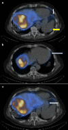
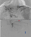
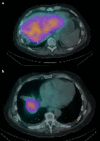
Selective internal radiotherapy (SIRT) is an established modality for the treatment of hepatic malignancy. The procedure is normally carried out in two parts. The first part involves a planning or "work-up" angiogram to delineate anatomy and plan safe yttrium-90 (Y90) delivery, and the second part for the administration of the Y90 microspheres. The work-up angiogram has three main purposes including delineation of hepatic and tumor vascular anatomy, which might influence the administration of the microbeads, identification, and embolization of blood vessels, which may complicate treatment or contribute to non-target Y90 microsphere deposition and administration of technetium 99 (metastable) labeled macroaggregated albumin (99mTcMAA) at the planned administration points prior to the same day single-photon emission computed tomography (SPECT) or planar SPECT to identify sites of 99mTcMAA uptake. We present the case of a SIRT procedure that demonstrated an anomalous artery arising from the left hepatic artery with supply to the pericardium, diaphragm, fundus of the stomach, and spleen. This is a rare vascular variant that highlights the importance of thorough assessment of both the planning angiograms and SPECT CT for the presence of anatomical variants and abnormal extrahepatic 99mTcMAA uptake to help reduce the need to recall patients for repeat work-up procedures.
Keywords: anomalous left hepatic artery branch; diaphragm; pericardium; selective internal radiotherapy (sirt); single photon emission tomography (spect); spleen; stomach.
Copyright © 2021, Karim et al.
Conflict of interest statement
The authors have declared that no competing interests exist.
Figures


References
-
- Same-day yttrium-90 radioembolization: feasibility with resin microspheres. Li MD, Chu KF, DePietro A, et al. J Vasc Interv Radiol. 2019;30:314–319. - PubMed
-
- Anatomy of liver arteries for interventional radiology. Favelier S, Germain T, Genson PY, Cercueil JP, Denys A, Krausé D, Guiu B. Diagn Interv Imaging. 2015;96:537–546. - PubMed
-
- Vascular redistribution for SIRT—a quantitative assessment of treatment success and long-term analysis of recurrence and survival outcomes. Borg P, Wong JJ, Lawrance N, et al. J Clin Interv Radiol ISVIR. 2019;03:89–97.
-
- Flow-directed catheters in hepatic embolization therapy—a review with clinical cases. Iqbal S, Breyfogle LJ, Flacke S. J Clin Interv Radiol ISVIR. 2021;5:99–105.
Publication types
LinkOut - more resources
Full Text Sources
