

Metastatic paraganglioma presenting as ajunctional scotoma
- PMID: 35036631
- PMCID: PMC8749452
- DOI: 10.1016/j.ajoc.2021.101253
Metastatic paraganglioma presenting as ajunctional scotoma
Abstract
Purpose: To report a unique case of metastatic paraganglioma presenting as a junctional scotoma.
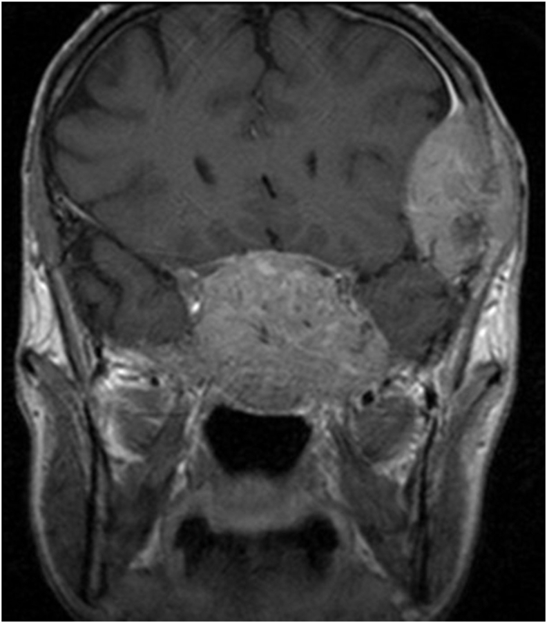
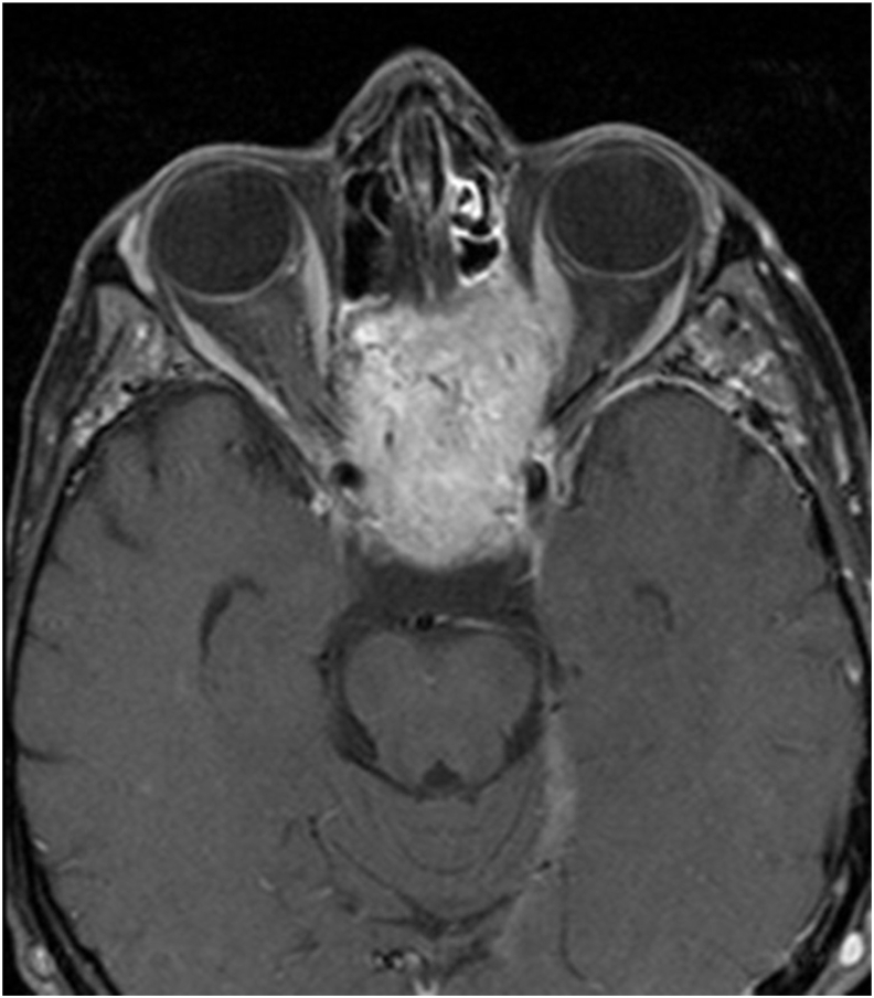
Observations: A 38-year-old Caucasian man with a history of abdominal paraganglioma presented with minimally blurred vision 20/25 visual acuity in the left eye. The patient was found to have a junctional scotoma upon visual field testing. Cranial MRI revealed a large peri-clival mass compressing the pre-chiasmal optic nerves and other loci of metastatic disease. Intracranial masses, including metastases, can present with a relatively intact central acuity and nonspecific visual symptoms.
Conclusions and importance: To the best of our knowledge, this is the first report of metastatic paraganglioma causing a junctional scotoma. In cases with junctional scotoma, careful neuro-ophthalmic assessment and imaging are of paramount importance, even in patients with excellent visual acuity.
Keywords: Junctional scotoma; Optic chiasm; Paraganglioma; Visual field defect.
© 2021 Published by Elsevier Inc.
Conflict of interest statement
The authors have no relevant conflicts of interest to disclose with this manuscript.
Figures

References
-
- Shin W.-J., Song B.-J., Kim J.-M. Junctional scotoma in giant cerebral aneurysm. Kor J Ophthalmol. 2002;16(2):124–129. - PubMed
-
- Borgman C.J. Atypical junctional scotoma secondary to optic chiasm atrophy: a case report. Clin Exp Optom. 2019;102(6):627–630. - PubMed
-
- Lee J.H., Tobias S., Kwon J.-T., Sade B., Kosmorsky G. Wilbrand's knee: does it exist? Surg Neurol. 2006;66(1):11–17. - PubMed
Publication types
LinkOut - more resources
Full Text Sources
