. 2021 Dec 20;7(1):70-84.
doi: 10.1021/acsomega.1c03216. eCollection 2022 Jan 11.

Characterization of Cyclophilin from Thaumarchaeota Nitrosopumilus maritimus: Implications on the Diversity of Chaperone-like Activity in the Archaeal Domain

Affiliations

Characterization of Cyclophilin from Thaumarchaeota Nitrosopumilus maritimus: Implications on the Diversity of Chaperone-like Activity in the Archaeal Domain

Vineeta Kaushik et al. ACS Omega. .

Abstract

The Archaea constitute separate domain of life and show resemblance with bacteria in their metabolic pathways while showing similarity with eukaryotes at the level of molecular processes such as cell division, DNA replication, protein synthesis, and proteostasis. However, the molecular machinery of archaea can be considered a simpler version of that found in eukaryotes because of the absence of multiple paralogs for any given molecular factor. Therefore, archaeal systems can possibly be used as a model system for understanding the eukaryotic protein folding machinery and thereby may help to address the molecular mechanism of various protein (mis)foldings and diseases. In the process of protein folding, the cis-trans isomerization of the peptide-prolyl bond is a rate-limiting step for the correct folding of proteins. Different types of peptidyl-prolyl cis-trans isomerases can accelerate this reaction, e.g., cyclophilin, FKBP, and parvulin. Among the five phyla of the archaeal domain, homologs of the cyclophilin protein are found only in two. Here we have characterized a cyclophilin from an archaeal organism, Nitrosopumilus maritimus (NmCyp), belonging to the phylum Thaumarchaeota. Like other known cyclophilins, NmCyp also possesses PPIase activity that can be inhibited by cyclosporine A. Generally, archaeal proteins are expected to possess differential thermal stability due to their adaptation to extreme environmental niche conditions. However, NmCyp exhibits low thermal stability and starts to aggregate beyond 40 °C. The properties of NmCyp are compared to those reported for the cyclophilin from another archaeal organism, Methanobrevibacter ruminantium. The current study sheds light on the differential behavior of cyclophilin proteins from two different phyla of archaea.


Conflict of interest statement

The authors declare no competing financial interest.

Figures

Figure 1
Figure 1
Schematic representation of the NmCyp protein. NmCyp has 158 amino acids with one PPiB domain. Binding sites as predicted in comparison to other known cyclophilins and the sites conserved among all archaeal cyclophilins are represented by the red dot and blue star, respectively.
Figure 2
Figure 2
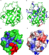
Modeled structure of NmCyp. (A) Cartoon representation of NmCyp using PyMOL. (B) Active site residues and an extra stretch of eight amino acid residues are marked on the cartoon structure of NmCyp in blue and red, respectively. (C) Surface representation of NmCyp with active site residues and an extra stretch of eight amino acid residues in blue and red, respectively. (D) Electrostatic potential surface diagram of NmCyp, where red, blue, and white symbolize negative, positive, and neutral charge.
Figure 3
Figure 3
PPIase activity of NmCyp. The PPIase activity of purified recombinant NmCyp was determined using the synthetic peptide N-succinyl-ala-ala-pro-phe-p-nitroaniline. In the absence of NmCyp, the reaction proceeds to completion based on the intrinsic rate of the cis-to-trans isomerization of the peptide only (black). The addition of NmCyp to the reaction mixture results in an increase of the initial rate of cis–trans isomerization reaction (red). The addition of CsA, a cyclophilin family inhibitor, again lowers the rate of reaction (blue). The PPIase activity of NmCyp was recorded for 20 min at 390 nm.
Figure 4
Figure 4
Thermal aggregation prevention of BCAII and citrate synthase by NmCyp. (A) The aggregation prevention of BCAII with the NmCyp protein using 1:1 molar concentration. (B) The aggregation prevention of citrate synthase with varying concentrations of the NmCyp protein. The zoom-in view shows the spectra of citrate synthase at 45 °C. In the figure, A.U. stands for ″arbitrary units″.
Figure 5
Figure 5
CD and fluorescence spectra of native NmCyp. (A) Far-UV CD spectrum of native NmCyp from 200 to 250 nm. (B) Near-UV CD spectrum of native NmCyp from 260 to 350 nm. (C) Fluorescence emission spectrum of NmCyp on excitation at 280 nm. In the figure, A.U. stands for ″arbitrary units″.
Figure 6
Figure 6
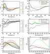
Thermostability of NmCyp. (A) Far-UV CD spectra of NmCyp at different temperatures (25–90 °C). (B) Far-UV CD spectra of the native NmCyp (25 °C), unfolded NmCyp (90 °C), and irreversible denatured NmCyp (cooled from 90 to 25 °C). (C) Near-UV CD spectra of NmCyp at different temperatures (25–90 °C). (D) Near-UV CD spectra of the native NmCyp (25 °C), unfolded NmCyp (90 °C), and irreversible denatured NmCyp (cooled from 90 to 25 °C). (E) Fluorescence emission spectra of NmCyp at different temperatures. (F) Emission spectra of the native NmCyp (25 °C), unfolded NmCyp (90 °C), and irreversible denatured NmCyp (cooled from 90 to 25 °C). In the figure, A.U. stands for ″arbitrary units″.
Figure 7
Figure 7
Equilibrium unfolding and refolding of NmCyp with different concentrations of urea. (A) Far-UV CD spectra of NmCyp at different urea concentrations. (B) Near-UV CD spectra of the native NmCyp at different urea concentrations. (C) Unfolding data (222 nm) of NmCyp at different concentrations of urea fitted into a two-state model. (D) Refolding of NmCyp after diluting the urea.
Figure 8
Figure 8
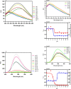
Equilibrium unfolding and refolding of NmCyp with different concentrations of urea using fluorescence spectroscopy. (A) Emission spectra of NmCyp at different concentrations of urea. (B) Emission spectra of the refolded protein on diluting the denaturant concentration till 0.2 M. (C) Comparison of λmax of the unfolded and refolded NmCyp protein in the presence of urea. (D) The pattern of the unfolding of NmCyp monitored by the change in fluorescence emission intensity. The data were fitted to a three-state equation, and thermodynamic parameters were calculated (Table S4). In the figure, A.U. stands for ″arbitrary units″.
Figure 9
Figure 9
ANS-based extrinsic fluorescence studies of NmCyp at different urea concentrations. (A) ANS-based emission spectra of the protein in the presence of an increasing concentration of urea. (B) A change in emission maxima (λmax) on increasing concentration of urea.
Figure 10
Figure 10
Equilibrium unfolding and refolding of NmCyp in the presence of GdnHCl. (A) Far-UV CD spectra from 200 to 250 nm at different concentrations of GdnHCl (0–7 M). (B) Near-UV CD spectra from 260 to 350 nm at different concentrations of GdnHCl (0–6 M). (C) Far-UV CD spectra of refolded proteins on diluting the denaturant. (D) GdnHCl dependent unfolding of NmCyp monitored by the change in mdeg values by CD spectroscopy. The data were fitted to a two-state equation, and thermodynamic parameters were calculated. (E) Normalized data of the NmCyp unfolded and refolded protein at different concentrations of GdnHCl.
Figure 11
Figure 11
Equilibrium unfolding of NmCyp in the presence of GdnHCl using fluorescence spectroscopy. (A) Emission spectra of NmCyp in the presence of different concentrations of GdnHCl (0–6 M). (B) GdnHCl-dependent unfolding of NmCyp monitored by the change in λmax of emission via fluorescence spectroscopy. The data were fitted to a three-state equation, and thermodynamic parameters were calculated. (C) The emission spectra of refolded proteins upon diluting the denaturant. (D) λmax of the unfolded and refolded NmCyp protein at different concentrations of GdnHCl. In the figure, A.U. stands for ″arbitrary units″.
Figure 12
Figure 12
ANS-based extrinsic fluorescence studies of NmCyp at different GdnHCl concentrations. (A) Emission spectra of the protein in the presence of increasing concentrations of GdnHCl. The concentration of ANS used was 50 μM. (B) Change in emission maxima (λmax) on increasing concentration of GdnHCl. In the figure, A.U. stands for ″arbitrary units″.
Figure 13
Figure 13
Effect of pH on the secondary and tertiary structure of NmCyp. (A) Far-UV CD spectra of NmCyp at different pHs (1–11). (B) A zoom-in view of the regions clustered close together in the pH range 5–11 for better visualization.(C) Near-UV CD spectra of NmCyp at different pHs (1–11). (D) A zoom-in view of the regions clustered close together in the pH range 5–11 for better visualization.
Figure 14
Figure 14
(A) The intrinsic fluorescence of NmCyp at different pHs from 1 to 11. Panel 1 shows a zoom-in view of the spectra recorded at pH 5–11 (for better visualization), and panel 2 shows the change in λmax and emission intensity at different pHs. (B) The ANS-mediated extrinsic fluorescence of NmCyp at different pHs ranging from 1 to 11. Panel 3 shows a zoom-in view of the spectra recorded at pH 6–11, and panel 4 shows the change in λmax and intensity of NmCyp in the pH range 1–11 after incubating with 50 μM of ANS. In the figure, A.U. stands for ″arbitrary units″.

Similar articles

References

    1. Wang P.; Heitman J. The cyclophilins. Genome Biol. 2005, 6, 226.10.1186/gb-2005-6-7-226. - DOI - PMC - PubMed
    1. Maruyama T.; Furutani M. Archaealpeptidylprolylcis-trans isomerases (PPIases). Front Biosci. 2000, 5, 821–836. 10.2741/maruyama. - DOI - PubMed
    1. Nath P. R.; Isakov N. Insights into peptidyl-prolyl cis-trans isomerase structure and function in immunocytes. ImmunolLett. 2015, 163, 120–131. 10.1016/j.imlet.2014.11.002. - DOI - PubMed
    1. Marks A. R. Cellular functions of immunophilins. Physiol Rev. 1996, 76, 631–649. 10.1152/physrev.1996.76.3.631. - DOI - PubMed
    1. Arevalo-Rodriguez M.; Wu X.; Hanes S. D.; Heitman J. Prolyl isomerases in yeast. Front Biosci. 2004, 9, 2420–2446. 10.2741/1405. - DOI - PubMed