. 2021 Dec 8:7:62-67.
doi: 10.1016/j.sopen.2021.11.007. eCollection 2022 Jan.

Whipple pancreatoduodenectomy: A technical illustration

Affiliations

Whipple pancreatoduodenectomy: A technical illustration

Shankar I Logarajah et al. Surg Open Sci. .

Abstract

Background: The Whipple procedure in its current form owes its evolution to the groundbreaking and innovative work of giants in the field of surgery. From being a multistep procedure with high morbidity and mortality, it is now ubiquitously performed in a single setting, often offered via minimally invasive approaches. Training to perform this procedure is an arduous task, and different training paradigms vary significantly.

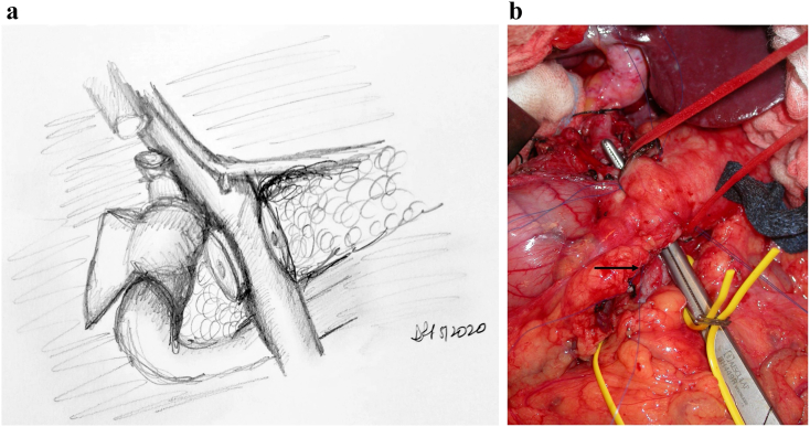
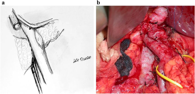
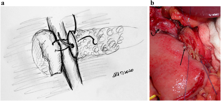
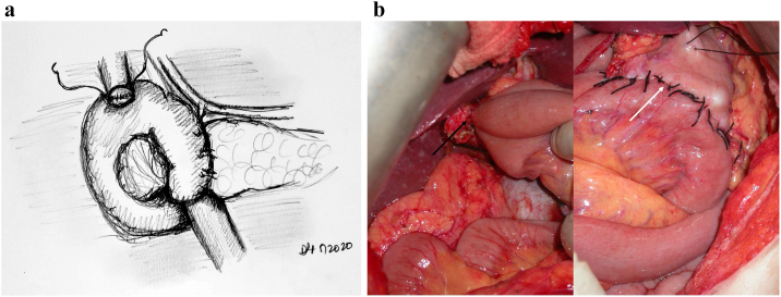
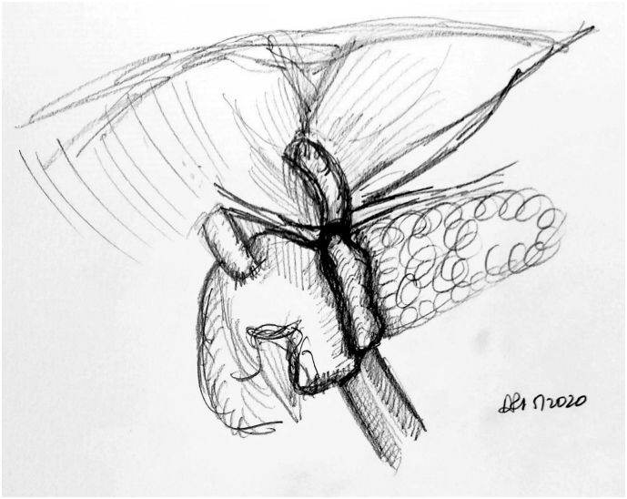
Objectives/methods: The purpose of this paper is to share a standard method by which the surgeon can perform the Whipple procedure in a systematic manner. Using illustrations to make the steps clearer, the authors will postulate that an improvement in mean operative time can be realistically achieved by most pancreatic surgeons. The focus is also on presenting this complex procedure as reproducible and teachable techniques for trainees.

Conclusion: This illustrated review of the Whipple procedure as performed at our institution is intended to help facilitate a streamlined and stepwise progression through what is undoubtedly a challenging surgical procedure. Although the procedure described will not apply to all Whipple operations given the heterogeneity in anatomy and circumstances, our hope is that this will lead to a more efficient procedure and decreased operating room time and costs as well as provide a framework to teach and measure technical progress for surgical trainees.


Figures

Fig 1
Fig 1
Step 1: Right to left medial visceral rotation and dissection of the right mesocolon. Note. Following a right to left medial visceral rotation (the Cattell–Braasch maneuver), the right mesocolon is dissected off the duodenum and head of pancreas. In this process, the SMV is identified early in the dissection, with the gastrocolic trunk of Henle.
Fig 2
Fig 2
A, Step 2: Dissection of the right gastroepiploic vein and superior mesenteric vein. Note. The right gastroepiploic vein is now identified, dissected, and ligated. This allows further cephalad dissection of the SMV leading into the PV and inferior end of the retropancreatic tunnel. B, Operative exposure of SMV which is encircled with vessel loop. Thick arrow points to ligated stump of right gastroepiploic vein; thin arrow points to intersection between the SMV and inferior end of the retropancreatic tunnel.
Fig 3
Fig 3
A, Step 3: Entering the lesser sac and dissecting the station 8 lymph node. Note. Enter the lesser sac via pars flaccida. Identify the station 8 lymph node or hepatic artery lymph node and dissect it off the CHA underneath. Following identification of the CHA, dissect just caudal to it to identify the portal vein between the CHA and the superior border of the pancreas. B, Intraoperative picture demonstrating identification of the hepatoduodenal ligament (black arrow).
Fig 4
Fig 4
Step 4: Identification and dissection of the gastroduodenal artery and exposure of the portal vein. Note. Follow the CHA to identify the GDA and proper hepatic artery. Dissect and ligate the GDA, allowing further exposure of the PV and access to the superior end of the retropancreatic tunnel.
Fig 5
Fig 5
A, Step 5: Transection of the pancreas, bile duct, stomach, and jejunum. Note. Having identified the PV, the pancreas and bile duct can now be safely transected. The stomach and jejunum are also transected, leaving behind the uncinate attached to the PV/SMV and the SMA. B, Intraoperative photograph demonstrating peon and umbilical tape placed through retropancreatic tunnel; division of the pancreas can now proceed. Arrow highlighting figure-of-8 Prolene used to control bleeding arcade vessels, which is the alternative method of controlling the bleeding to electrocautery.
Fig 6
Fig 6
A, Step 6: Transecting the uncinate and skeletonizing the SMA distally. Note. Dissection is then performed through the mesopancreas and the uncinate from the aorta, skeletonizing the SMA distally. A harmonic scalpel is then used to transect the uncinate off the SMA and SMV. B, Intraoperative photograph following pancreatic transection; black arrow pointing to pancreatic duct.
Fig 7
Fig 7
A, Step 7: Modified Blumgart technique to create the pancreatojejunostomy. Note. Modified Blumgart technique using an outer buttressing layer of 2-0 silk and inner duct to mucosa using 5-0 Prolene is performed to create the pancreatojejunostomy. B, Intraoperative photograph demonstrating the alternative method of pancreatojejunostomy utilizing a two-layer anastomosis; arrow noting pediatric feeding tube cannulating the pancreatic duct.
Fig 8
Fig 8
A, Step 8: Creation of the HJ and GJ.Note. HJ is created using a single-layered, running 5-0 polydioxanone suture. A stapled, antecolic, retrogastric, isoperistaltic GJ is then created. B, Intraoperative photographs demonstrating completed HJ (black arrow) and GJ (white arrow).
Fig 9
Fig 9
Step 9: Using the falciform ligament to cover the pancreatojejunostomy. Note. The falciform ligament is passed posterior to the pancreatojejunostomy and sutured to itself, forming a layer of buttress and coverage for the pancreatojejunostomy.

References

    1. Howard J.M. History of pancreatic head resection—the evaluation of surgical technique. Am J Surg. 2007;194(4 SUPPL):S6–S10. doi: 10.1016/j.amjsurg.2007.05.029. - DOI
    1. Cameron J.L., Riall T.S., Coleman J., Belcher K.A. One thousand consecutive pancreaticoduodenectomies. Ann Surg. 2006;244(1):10–15. doi: 10.1097/01.sla.0000217673.04165.ea. - DOI - PMC - PubMed
    1. Fernández-Del Castillo C., Morales-Oyarvide V., McGrath D., et al. Evolution of the Whipple procedure at the Massachusetts General Hospital. Surg (United States) 2012;152(3 SUPPL):S56–S63. doi: 10.1016/j.surg.2012.05.022. - DOI - PMC - PubMed
    1. Sheetz K.H., Dimick J.B., Nathan H. Centralization of high-risk cancer surgery within existing hospital systems. J Clin Oncol. 2019;37(34):3234–3242. doi: 10.1200/JCO.18.02035. - DOI - PMC - PubMed
    1. Strasberg S.M., Sanchez L.A., Hawkins W.G., Fields R.C., et al. Resection of tumors of the neck of the pancreas with venous invasion: the “Whipple at the Splenic Artery (WATSA)” procedure. J Gastrintest Surg. 2012;16(5):1048–1054. - PubMed

LinkOut - more resources